

- Department of Neurosurgery, São Paulo Federal University, São Paulo, Brasil
Correspondence Address:
Franz J. Onishi
Department of Neurosurgery, São Paulo Federal University, São Paulo, Brasil
DOI:10.4103/sni.sni_74_17
Copyright: © 2017 Surgical Neurology International This is an open access article distributed under the terms of the Creative Commons Attribution-NonCommercial-ShareAlike 3.0 License, which allows others to remix, tweak, and build upon the work non-commercially, as long as the author is credited and the new creations are licensed under the identical terms.How to cite this article: Franz J. Onishi, Mirto N. Prandini, Sergio Cavalheiro. Delayed presentation of spinal foreign body – Case report and review of literature. 11-Jul-2017;8:143
How to cite this URL: Franz J. Onishi, Mirto N. Prandini, Sergio Cavalheiro. Delayed presentation of spinal foreign body – Case report and review of literature. 11-Jul-2017;8:143. Available from: http://surgicalneurologyint.com/surgicalint-articles/delayed-presentation-of-spinal-foreign-body-case-report-and-review-of-literature/
Date of Submission
20-Feb-2017
Date of Acceptance
04-Apr-2017
Date of Web Publication
11-Jul-2017
Abstract
Background:Although spinal cord injuries are frequent causes of myelopathy in young patients, stab wounds of the spinal cord rarely occur and are typically maximal symptomatic immediately after the trauma.
Case Description:A 31-year-old male developed delayed onset of symptoms 4 years after a stab wound to the cervical spinal cord attributed to a plant needle (plant called Mandacaru). Following removal of the foreign body and decompression/excision of scarring at the C34 level, the patient's symptoms resolved.
Conclusion:Surgical excision should be encouraged to remove chronic penetrating foreign bodies to both decompress and untether the spinal cord.
Keywords: Delayed myelopathy, spinal cord injury, stab injury, surgical treatment
INTRODUCTION
Spinal stab wounds account for 8–11% of all penetrating spinal injuries and almost all injuries are acute.[
CASE REPORT
A 31-year-old white male presented with a progressive spastic quadriparesis over 1-year duration. Four years previously, he had fallen from a horse into a tree called Mandacaru (Cereus jamaracu) in northeast Brazil. At the time of the injury, the patient had several skin wounds but no neurological symptoms or signs. Four years later, he presented with new onset of spastic tetraparesis characterized by motor 4/5 strength in the upper and lower limbs; he could only ambulate with assistance.
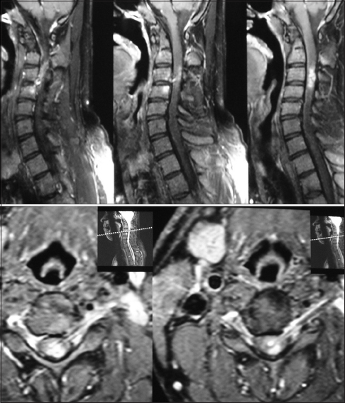
While plain radiographs of cervical spine revealed upper cervical kyphosis, computed tomography (CT) scan [
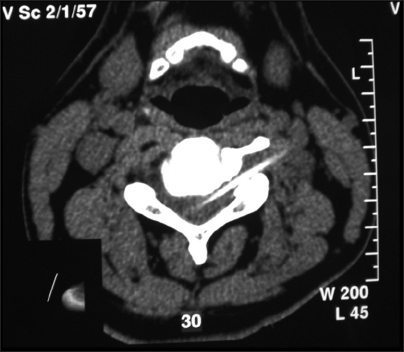
The needle was removed utilizing a left side approach to the left C3-C4 foramen. (Hodgson-like approach[
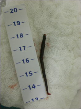
A 5-cm thorn was removed [
DISCUSSION
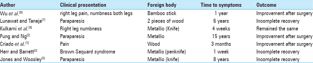
There are seven reported[
Mechanism of injury
The foreign body usually enters the spinal canal via the interlaminar space or through the intervertebral foramen. The delayed onset of neurological deficits are typically attributed to retained fragments within the spinal canal. These lesions may transfix the spinal cord causing slow progressive damage due to tethering[
CONCLUSION
Few patients exhibit delayed onset of symptoms following penetrating injuries to the spinal cord. If delayed neurological deterioration occurs, patients should be reassessed with X-ray, MRI, and CT to demonstrate the site, location, and type of penetrating lesion and whether it has been adequately excised, or whether there is a residual foreign body. Repeated surgical intervention, offering decompression and untethering, may or may not result in neurological recovery.
Financial support and sponsorship
Nil.
Conflicts of interest
There are no conflicts of interest.
References
1. Criado E, Oller D, Fulghum J. Delayed diagnosis of a foreign body in the spinal canal. South Med J. 1990. 83: 332-3
2. Fung CF, Ng TH. Delayed myelopathy after a stab wound with a retained intraspinal foreign body: Case report. J Trauma. 1992. 32: 539-41
3. Herr RD, Barrett J. An unusual presentation of Brown-Sequard syndrome. Ann Emerg Med. 1987. 16: 1285-8
4. Hodgson AR. Approach to the cervical spine C3-C7. Clin Orthop. 1996. 39: 129-34
5. Jones FD, Woosley RE. Delayed myelopathy secondary to retained intraspinal metallic fragment. Case report. J Neurosurg. 1981. 55: 979-82
6. Kulkarni AV, Bhandari M, Stiver S, Reddy K. Delayed presentation of spinal stab wound: Case report and review of the literature. J Emerg Med. 2000. 18: 209-13
7. Lunawat SK, Taneja DK. A foreign body in the spinal canal. A case report. J Bone Joint Surg Br. 2000. 82: 267-8
8. Skadorwa T, Ciszek B. Pediatric arrow shot injury to cervical spinal cord-sagittal cord transection with no neurological deficit and good outcome: Case report and review of literature. Childs Nerv Syst. 2013. 29: 1933-9
9. Wu QH, Chen WS, Chen QX. Delayed myelopathy after stab injury with intraspinal non metal foreign body granuloma. Chin J Traumatol. 2008. 11: 126-8

