

- Department of Neurosurgery, Marshall University Joan C. Edwards School of Medicine, West Virginia, USA
Correspondence Address:
Rida Mazagri
Department of Neurosurgery, Marshall University Joan C. Edwards School of Medicine, West Virginia, USA
DOI:10.4103/sni.sni_148_17
Copyright: © 2017 Surgical Neurology International This is an open access article distributed under the terms of the Creative Commons Attribution-NonCommercial-ShareAlike 3.0 License, which allows others to remix, tweak, and build upon the work non-commercially, as long as the author is credited and the new creations are licensed under the identical terms.How to cite this article: Yusif Mohammed, Shahed Elhamdani, Mobeen Farooq, Rida Mazagri. A case report and review of thoracic spinal angiolipoma. 13-Jun-2017;8:113
How to cite this URL: Yusif Mohammed, Shahed Elhamdani, Mobeen Farooq, Rida Mazagri. A case report and review of thoracic spinal angiolipoma. 13-Jun-2017;8:113. Available from: http://surgicalneurologyint.com/surgicalint-articles/a-case-report-and-review-of-thoracic-spinal-angiolipoma/
Date of Submission
16-Apr-2017
Date of Acceptance
05-May-2017
Date of Web Publication
13-Jun-2017
Abstract
Background:While it is a rare entity, spinal angiolipomas are well-defined benign tumors that have been described sporadically in the literature starting from the late 1800s. Composed of mature lipomatous and angiomatous elements, these tumors manifest neurological symptoms due to progressive spinal cord or root compression. We present a case of a thoracic spinal angiolipoma and review the relevant literature.
Case Description:A 68-year-old male with ongoing bilateral lower extremity weakness was found on enhanced magnetic resonance imaging to have an extradural mass in the thoracic spine causing cord compression. A T4–T8 laminectomy and complete excision of the epidural mass resulted in reversal of the patient's neurological symptoms. Histopathology identified the mass as a thoracic spinal angiolipoma.
Conclusion:Given its uncommon occurrence and excellent prognosis, our report serves as a reminder to always consider spinal angiolipoma in the differential diagnosis of epidural masses.
Keywords: Epidural mass, magnetic resonance imaging, spinal angiolipomas, spinal tumor
INTRODUCTION
Spinal angiolipomas are benign epidural neoplasms that comprise approximately 1% of all spinal axis tumors and up to 3% of all primary extradural spinal tumors.[
CASE HISTORY
A 68-year-old white male presented complaining of tingling in both feet and a “funny feeling” in the lower abdomen since 1 year. He reported weakness in his left leg along with balance problems (e.g., stumbling but not falling). The neurological exam revealed mild weakness of the hip flexors bilaterally, left side greater than right, hyperactive patellar and Achilles responses, and bilateral Babinski signs. The thoracic magnetic resonance imaging (MRI) revealed an extra-axial T2 hyperintense and T1 isointense soft tissue mass at the T5–T7 levels resulting in extrinsic compression of the spinal cord [Figure
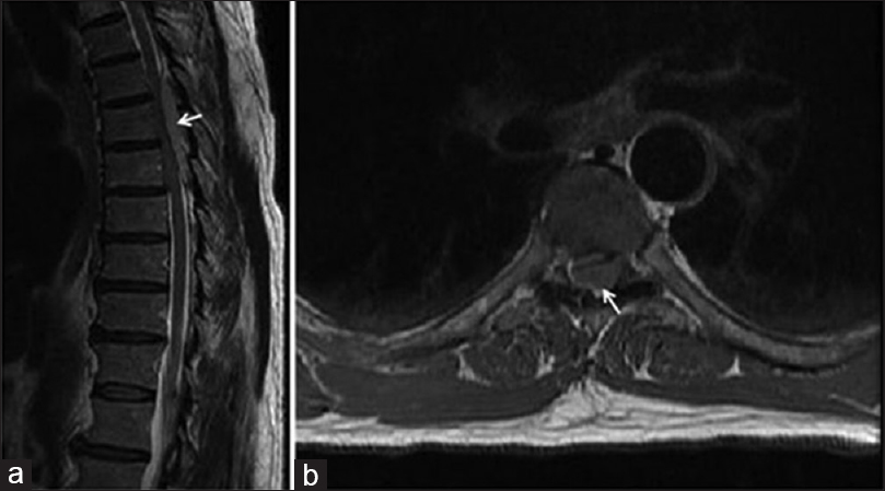
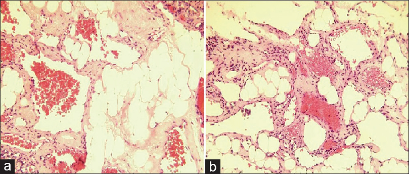
The patient was taken to the operation room and a laminectomy was performed at the T4–T8 levels. Excessive epidural fatty tissue with mild vascular elements was encountered and uneventfully removed. The dura was opened but no intradural lesion was present. Histopathology revealed a lipovascular proliferation with fat and myxoid tissue without malignancy [Figure
DISCUSSION
Angiolipomas are benign tumors that most likely originate from the same progenitor tissue as lipomas and hemangiomas as they appear to have features common to both tumor subgroups.[
Histologically, spinal angiolipomas are composed of mature adipose tissue as well as blood vessels that may or may not be surrounded by a thin capsule.[
CONCLUSION
This case report highlights the importance of considering spinal angiolipoma in the differential diagnosis of a long-standing, slowly progressive paraparesis in the presence of an extradural spinal mass. Ultimately, histology is still necessary to correctly establish the diagnosis.
Financial support and sponsorship
Nil.
Conflicts of interest
There are no conflicts of interest.
References
1. Benvenutti-Regato M, De la Garza-Ramos R, Caro-Osorio E. Thoracic epidural spinal angiolipoma with coexisting lumbar spinal stenosis: Case report and review of the literature. Int J Spine Surg. 2015. 9: 67-
2. Fourney DR, Tong KA, Macaulay RJ, Griebel RW. Spinal angiolipoma. Can J Neurol Sci. 2001. 28: 82-8
3. Leu NH, Chen CY, Shy CG, Lu CY, Wu CS, Chen DC. MR imaging of an infiltrating spinal epidural angiolipoma. AJNR Am J Neuroradiol. 2003. 24: 1008-11
4. Meng J, Du Y, Yang HF, Hu FB, Huang YY, Li B. Thoracic epidural angiolipoma: A case report and review of the literature. World J Radiol. 2013. 5: 187-92
5. Si Y, Wang Z, Pan Y, Lin G, Yu T. Spinal angiolipoma: Etiology, imaging findings, classification, treatment, and prognosis. Eur Spine J. 2014. 23: 417-25

