

- Departament of Surgery, Federal University of Ceara, Ceará, Brazil
- Division of Neurosurgery, General Hospital of Fortaleza, Fortaleza, Ceará, Brazil
Correspondence Address:
Anderson Alexsander Rodrigues Teixeira
Division of Neurosurgery, General Hospital of Fortaleza, Fortaleza, Ceará, Brazil
DOI:10.4103/sni.sni_256_18
Copyright: © 2018 Surgical Neurology International This is an open access journal, and articles are distributed under the terms of the Creative Commons Attribution-NonCommercial-ShareAlike 4.0 License, which allows others to remix, tweak, and build upon the work non-commercially, as long as appropriate credit is given and the new creations are licensed under the identical terms.How to cite this article: Anderson Alexsander Rodrigues Teixeira, Lucas Fernandes Ferreira, Bruno Nunes Ferraz De Abreu, Euler Nicolau Sauaia Filho, Francisco Ramos Junior. A rare cause of thoracic cord compression. 21-Sep-2018;9:194
How to cite this URL: Anderson Alexsander Rodrigues Teixeira, Lucas Fernandes Ferreira, Bruno Nunes Ferraz De Abreu, Euler Nicolau Sauaia Filho, Francisco Ramos Junior. A rare cause of thoracic cord compression. 21-Sep-2018;9:194. Available from: http://surgicalneurologyint.com/surgicalint-articles/9015/
Date of Submission
26-Jul-2018
Date of Acceptance
14-Aug-2018
Date of Web Publication
21-Sep-2018
Abstract
Background:The posterior longitudinal ligament (PLL) extends from the foramen magnum to the sacrum. In some cases, it becomes calcified/ossified; the term for this is ossification of the PLL (OPLL).
Case Description:A 50-year-old female presented with acute sphincter dysfunction and paraparesis attributed to T2–T4 OPLL. The patient underwent a C7-T5 laminectomy to decompress the spinal cord. After 1 postoperative week, and certainly by 6 months postoperatively, the patient's motor and sensory deficits showed improvement.
Conclusion:Surgery for thoracic OPLL includes laminoplasty, laminectomy with/without fusion, anterior decompression through a posterior approach (transpedicular, costotransversectomy), and circumferential decompression (e.g. combined anterior/posterior approaches). In cases like the one presented, patients who originally present with acute paraparesis/sphincter dysfunction may demonstrate postoperative improvement.
Keywords: Ossification of posterior longitudinal ligament, spinal cord compression, Thoracic vertebrae
INTRODUCTION
Ossification of the posterior longitudinal ligament (OPLL) occurs most frequently in Asian patients, but may also present in other populations. It involves the thoracic spinal cord (T-OPLL) in 15% of cases, while 70% appears in the cervical region.[
CASE REPORT
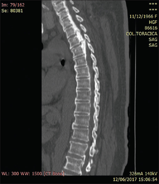
A 50-year-old Brazilian female presented with 6 months of lower extremity paresis (not walking for the past 2 months) accompanied by 1 week of urinary and fecal incontinence. On examination, the patient had diffuse lower extremity weakness, hyperactive reflexes with bilateral Babinski responses, and absent vibratory/proprioception at the T10 level. A computed tomography (CT) scan showed extensive thoracic OPLL extending from the T2–T4 levels, with some fractures within the OPLL mass [
The patient underwent an emergent C7-T5 laminectomy to decompress the spinal cord. One week postoperatively, her paraparesis improved (e.g., regarding motor/sensory findings): the patient started to walk again. Six months later, the patient exhibited continued improvement in her motor deficit and sphincter dysfunction.
DISCUSSION
OPLL is almost exclusively found in the Japanese, Chinese, and Korean patients; the prevalence is between 1.9% and 4.8% in the Japanese literature, but it also occurs in the Caucasian American population (e.g., 0.12%).[
Thoracic ossification of the posterior longitudinal ligament types
T-OPLL is subclassified into flat or nozzle types; The flat type is continuous or mixed T-OPLL and presents as sharp protrusions behind the disk space.[
Surgery for thoracic ossification of the posterior longitudinal ligament
Surgical for T-OPLL may be warranted to relieve spinal cord compression.[
CONCLUSION
About 10% of patients with T-OPLL may present with acute paraparesis and sphincter dysfunction. The diagnostic work-up should include both MR and CT studies. Surgical management should depend on the type/location of the T-OPLL and may include anterior, posterior, and/or circumferential surgery with/without fusion.
Declaration of patient consent
The authors certify that they have obtained all appropriate patient consent forms. In the form the patient(s) has/have given his/her/their consent for his/her/their images and other clinical information to be reported in the journal. The patients understand that their names and initials will not be published and due efforts will be made to conceal their identity, but anonymity cannot be guaranteed.
Financial support and sponsorship
Nil.
Conflicts of interest
There are no conflicts of interest.
References
1. Abiola R, Rubery P, Mesfin A. Ossification of the posterior longitudinal ligament: Etiology, diagnosis, and outcomes of nonoperative and operative management. Global Spine J. 2016. 6: 195-204
2. Choi JY, Sung KH. Complete removal of ossification of the posterior longitudinal ligament in the mid-thoracic spine. Acta Neurochir (Wien). 2005. 147: 675-7
3. Kawaguchi Y, Nakano M, Yasuda T, Seki S, Suzuki K, Yahara Y. Serum biomarkers in patients with ossification of the posterior longitudinal ligament (OPLL): Inflammation in OPLL. PLoS One. 2017. 12: e0174881-
4. Kim KH, Kuh SU, Park JY, Lee SJ, Park HS, Chin DK. Association between BMP-2 and COL6A1 gene polymorphisms with susceptibility to ossification of the posterior longitudinal ligament of the cervical spine in Korean patients and family members. Genet Mol Res. 2014. 13: 2240-7
5. Matsumoto M, Chiba K, Toyama Y, Takeshita K, Seichi A, Nakamura K. Surgical results and related factors for ossification of posterior longitudinal ligament of the thoracic spine: A multi-institutional retrospective study. Spine (Phila Pa 1976). 2008. 33: 1034-41
6. Matsuyama Y, Sakai Y, Katayama Y, Imagama S, Ito Z, Wakao N. Indirect posterior decompression with corrective fusion for ossification of the posterior longitudinal ligament of the thoracic spine: Is it possible to predict the surgical results?. Eur Spine J. 2009. 18: 943-8
7. Smith ZA, Buchanan CC, Raphael D, Khoo LT. Ossification of the posterior longitudinal ligament: Pathogenesis, management, and current surgical approaches. A review. Neurosurg Focus. 2011. 30: E10-
8. Wang P, Liu X, Zhu B, Ma Y, Yong L, Teng Z. Association of IL17RC and COL6A1 genetic polymorphisms with susceptibility to ossification of the thoracic posterior longitudinal ligament in Chinese patients. J Orthop Surg Res. 2018. 13: 109-
9. Yang C, Bi Z, Fu C, Zhang Z. A modified decompression surgery for thoracic myelopathy caused by ossification of posterior longitudinal ligament: A case report and literature review. Spine (Phila Pa 1976). 2010. 35: E609-13

