

- Department of Neurosurgery, Iwate Prefectural Chubu Hospital, Kitakami, Iwate, Japan
Correspondence Address:
Hiroshi Kashimura
Department of Neurosurgery, Iwate Prefectural Chubu Hospital, Kitakami, Iwate, Japan
DOI:10.4103/2152-7806.183499
Copyright: © 2016 Surgical Neurology International This is an open access article distributed under the terms of the Creative Commons Attribution-NonCommercial-ShareAlike 3.0 License, which allows others to remix, tweak, and build upon the work non-commercially, as long as the author is credited and the new creations are licensed under the identical terms.How to cite this article: Yoshida J, Kashimura H, Takeda M, Aso K. An unusual variant of the callosomarginal artery from the A1 segment of the anterior cerebral artery. Surg Neurol Int 03-Jun-2016;7:
How to cite this URL: Yoshida J, Kashimura H, Takeda M, Aso K. An unusual variant of the callosomarginal artery from the A1 segment of the anterior cerebral artery. Surg Neurol Int 03-Jun-2016;7:. Available from: http://surgicalneurologyint.com/surgicalint_articles/an-unusual-variant-of-the-callosomarginal-artery-from-the-a1-segment-of-the-anterior-cerebral-artery/
Abstract
Background:Although the anatomy of the A1 segment of the anterior cerebral artery (ACA) is highly variable, a callosomarginal artery (CMA) arising from the A1 segment of the ACA is rare.
Case Description:A 27-year-old man presented with severe headache and was admitted to our hospital. Initial computed tomography (CT) showed subarachnoid hemorrhage in the basal cistern. Three-dimensional CT angiography revealed a saccular aneurysm arising from the left internal carotid bifurcation and showed an anomalous cortical branch originating from the left A1 segment of the ACA. The anomalous artery was interpreted as a CMA.
Conclusions:Recognizing this variant preoperatively could be helpful in preventing complications of surgery. Careful follow-up studies are necessary in the present case to monitor the development of another aneurysm at the junction between the left CMA and the left A1 segment of the ACA.
Keywords: Aneurysm, anterior cerebral artery, callosomarginal artery, variant
INTRODUCTION
The anatomy of the A1 segment of the anterior cerebral artery (ACA) is highly variable.[
CASE REPORT
A 27-year-old man presented with severe headache and subsequent loss of consciousness and was admitted to our hospital. No focal neurological abnormality was noted. Initial computed tomography (CT) showed typical findings of subarachnoid hemorrhage in the basal cistern. Three-dimensional CT angiography revealed a saccular aneurysm arising from the left internal carotid bifurcation, fenestration of the right P1 segment of the posterior cerebral artery, and an anomalous cortical branch originating from the left A1 segment of the ACA [
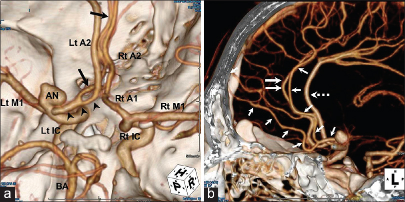
Figure 1
(a) Superoinferior view of the three-dimensional computed tomography angiogram, showing a saccular aneurysm arising from the left internal carotid artery bifurcation and an anomalous cortical branch originating from the left A1 segment of the anterior cerebral artery (black arrowheads). The anomalous artery runs anteromedially and then ascends superiorly and parallel to the right A2 segment of the anterior cerebral artery (black arrows). (b) Left lateral view of the three-dimensional computed tomography angiogram, showing the arterial course of an anomalous cortical branch originating from the left A1 segment of the anterior cerebral artery (white arrows) and its relationship to the bilateral A2 segment of the anterior cerebral artery. The artery runs parallel to the right A2 segment of the anterior cerebral artery (white double arrows). The left A2 segment of the anterior cerebral artery supplies the bihemispheric branches (white dotted arrow). A1: A1 segment of the anterior cerebral artery, A2: A2 segment of the anterior cerebral artery, BA: Basilar artery, IC: Internal carotid artery, Lt: Left, M1: M1 segment of the middle cerebral artery, Rt: Right, AN: Aneurysm
Left frontotemporal craniotomy was performed, and the aneurysm was successfully obliterated with clipping with a bayonet-shaped Yasargil titanium clip (No. FT727T). Postoperative digital subtraction angiography confirmed complete aneurysm occlusion, showed the right A2 segment, and showed that the anomalous cortical branch originating from the left A1 segment of the ACA terminated at the medial internal frontal artery [
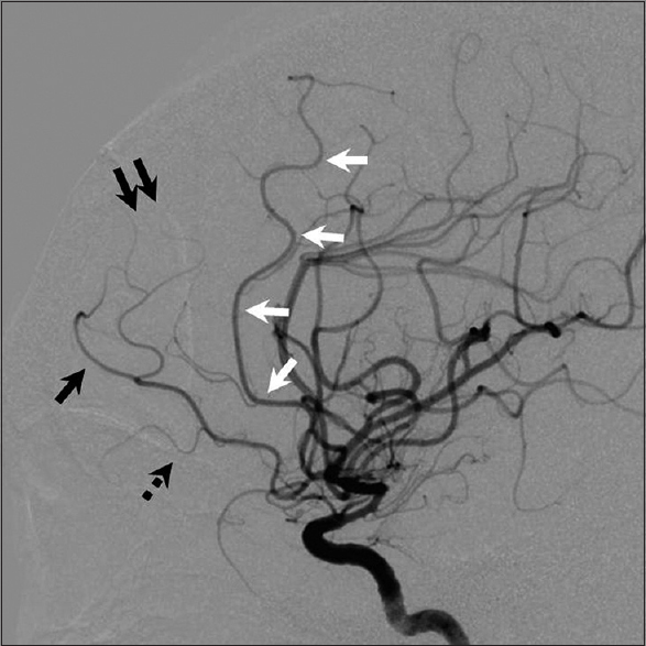
Figure 2
Lateral view of the right carotid arterial digital subtraction angiogram showing the arterial course of an anomalous cortical branch originating from the left A1 segment of the anterior cerebral artery. The artery has two main branches: The inferior branch forming the common trunk of the fronto-orbital (black dotted arrow), frontopolor (black arrow) and the anterior internal frontal arteries (black double arrows), and superior branch forming the callosomarginal branch of the anterior cerebral artery. The artery terminates in in medial internal frontal artery (white arrows)
DISCUSSION
The FOA is the first cortical branch of the ACA and normally arises from the ipsilateral pericallosal artery. It may uncommonly arise from the A1 segment just proximal to the anterior communicating artery.[
The CMA is defined as the artery that courses in or near the cingulated sulcus and gives origin to two or more cortical branches. This artery runs parallel to the pericallosal artery and gives origin to the three internal frontal arteries, even though the most consistent branch to originate from it is the middle internal frontal artery.[
Anomalous origin of a cortical branch from the A1 segment of the ACA predisposes to the formation of aneurysm. The pathogenesis of associated aneurysms has not been fully clarified. Mechanisms contributing to aneurysm formation may involve either increased local hemodynamic forces or structural weakness of the arterial wall. Structural anomalies, such as persistent trigeminal artery, azygous ACA, and fenestration of the intracranial arteries, show higher rates of aneurysm formation than other vessels. Therefore, careful follow-up studies are necessary in the present case to monitor for the development of another aneurysm at the junction between the left CMA and the left A1 segment of the ACA.
Financial support and sponsorship
Nil.
Conflicts of interest
There are no conflicts of interest.
References
1. Aso K, Kashimura H, Takeda M, Chida K. An unusual variant of the common trunk of the fronto-orbital and frontopolar arteries associated with a ruptured aneurysm of the A1 segment of the anterior cerebral artery. Surg Neurol Int. 2015. 6: S418-20
2. Hong SK. Ruptured proximal anterior cerebral artery (A1) aneurysm located at an anomalous branching of the fronto-orbital artery – A case report. J Korean Med Sci. 1997. 12: 576-80
3. Horie N, Morikawa M, Fukuda S, Hayashi K, Suyama K, Nagata I. New variant of persistent primitive olfactory artery associated with a ruptured aneurysm. J Neurosurg. 2012. 117: 26-8
4. Krayenbuhl HA, Yasargil MG.editors. Cerebral Angiography. New York: Thieme Medical Publishers; 1968. p. 20-85
5. Krishnamoorthy T, Gupta AK, Bhattacharya RN, Rajesh BJ, Purkayastha S. Anomalous origin of the callosomarginal artery from the A1 segment with an associated saccular aneurysm. AJNR Am J Neuroradiol. 2006. 27: 2075-7
6. Lee ER, Eastwood JD. An unusual variant of the fronto-orbital artery. AJNR Am J Neuroradiol. 2000. 21: 939-40
7. Marinkovic S, Milisavljevic M, Kovacevic M. Anatomical bases for surgical approach to the initial segment of the anterior cerebral artery. Microanatomy of Heubner's artery and perforating branches of the anterior cerebral artery. Surg Radiol Anat. 1986. 8: 7-18
8. Rhoton AL. The supratentorial arteries. Neurosurgery. 2002. 51: S53-120
9. Sato Y, Kashimura H, Takeda M, Chida K, Kubo Y, Ogasawara K. Aneurysm of the A1 segment of the anterior cerebral artery associated with the persistent primitive olfactory artery. World Neurosurg. 2015. 84: 2079.e7-9
10. Yasargil MG, Yasargil MG.editors. Anterior communicating artery aneurysm. Microneurosurgery. Stuttgart: Georg Thieme Verlag; 1984. I: 113-6

