Tools

Chompunut Asavaaree, Cara Doyle, Saksith Smithason
  1. Section of Vascular Medicine, Cardiovascular Center, Lumberton, NC, USA
  2. School of Osteopathic Medicine, Campbell University, Lumberton, NC, USA
  3. Neurosurgeon, Neurological Center, Lumberton, NC, USA

Correspondence Address:
Saksith Smithason
Neurosurgeon, Neurological Center, Lumberton, NC, USA

DOI:10.4103/sni.sni_254_18

Copyright: © 2018 Surgical Neurology International This is an open access journal, and articles are distributed under the terms of the Creative Commons Attribution-NonCommercial-ShareAlike 4.0 License, which allows others to remix, tweak, and build upon the work non-commercially, as long as appropriate credit is given and the new creations are licensed under the identical terms.

How to cite this article: Chompunut Asavaaree, Cara Doyle, Saksith Smithason. Artery of Percheron infarction results in severe bradycardia: A case report. 19-Nov-2018;9:230

How to cite this URL: Chompunut Asavaaree, Cara Doyle, Saksith Smithason. Artery of Percheron infarction results in severe bradycardia: A case report. 19-Nov-2018;9:230. Available from: http://surgicalneurologyint.com/surgicalint-articles/9071/

Date of Submission
25-Jul-2018

Date of Acceptance
25-Sep-2018

Date of Web Publication
19-Nov-2018

Abstract

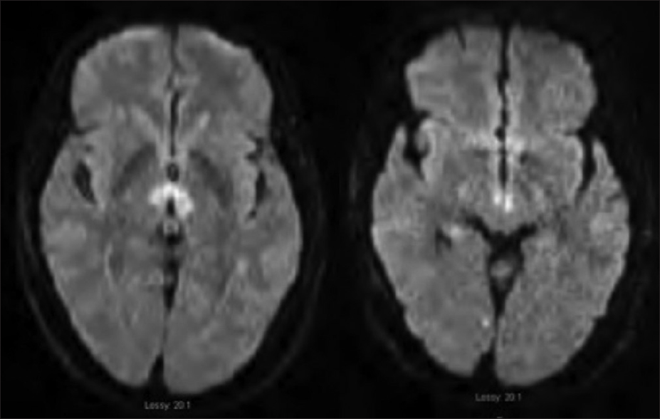
Background:The thalamus is normally supplied by each posterior cerebral artery (PCA). The artery of percheron is a variant of this anatomy as it arises as a single trunk unilaterally from the PCA to supply the thalamus bilaterally. Occlusion of this artery is rare, and the diagnosis is usually missed without obtaining an MRI.

Case Description:We illustrate the case of a 68-year-old male who presented with coma, ocular gaze palsy, and severe bradycardia from bilateral thalamic nuclei and midbrain infarction, as described as an artery of Percheron infarction. The patient recovered neurologically under conservative treatment with a residual vertical diplopia from downward gaze palsy. He underwent cardiac pacer implantation for severe bradycardia at the end of his admission. The thalamic pathway associated with cardiac rhythm, especially the zona inserta, is discussed. Publications related to the artery of Percheron are reviewed.

Conclusion:Coma and ocular gaze palsy are the most common presentations following thalamic and midbrain ischemia from artery of Percheron infarction. To our knowledge, only a single case of artery of Percheron infarction with severe bradycardia has been reported in the past. Our case attested the role of thalamic nuclei controlling cardiac rhythm.

Keywords: Artery of Percheron, coma, severe bradycardia, thalamic infarct, Zonainserta

INTRODUCTION

The artery of Percheron arises as a single common trunk from one of the posterior cerebral arteries (PCA), and provides a bilateral arterial blood supply to the paramedian thalamic and rostral midbrain.[ 6 14 ] Artery of Percheron variance is rare, with an incidence of 11.7% from cadaveric study.[ 6 ] The most common clinical presentations are coma and ocular movement disorders.[ 1 2 3 ] There are four patterns of magnetic resonance imaging (MRI) diffusion study, (1) bilateral paramedian thalamic with midbrain infarction (43%), (2) bilateral paramedian thalamic without midbrain infarction (38%), (3) bilateral paramedian thalamic with anterior thalamic and midbrain infarction (14%), and (4) bilateral paramedian thalamic with anterior thalamus without midbrain infarction (5%).[ 7 ] From a neurophysiology stand point, the thalamus is the part of Reticular Activating System (RAS) that mediates consciousness by connecting the brainstem, thalamus, and cerebral cortex.[ 6 ] The thalamus not only functions to maintain consciousness through the RAS but it also controls the cardiovascular response center at the zona incerta area.[ 12 ] Regarding the cardiovascular connection, zona inserta receives afferent fibers from the anterior insular cortex before sending efferent fibers to the cardiovascular center in medulla.

Alterations of consciousness from a bilateral thalamic infarction is prevalent in the literature,[ 13 14 ] whereas only one case has been reported having severe bradycardia.[ 12 ] We demonstrate a case with coma, ocular gaze palsy, and severe bradycardia following infarction of the artery of Percheron.

CASE

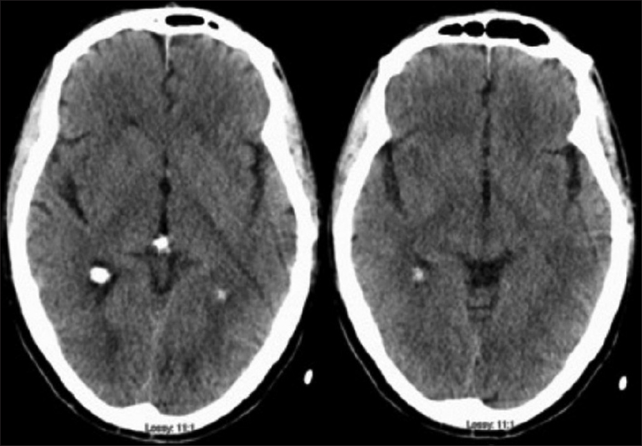
A 68-year-old, right-handed Native American male with a past medical history of hypertension and Gleason grade 3 prostate cancer presented to the emergency room with alterations of consciousness and severe bradycardia. His past surgical history included prostate cancer surgery and degenerative lumbar spine surgery. He is thin, athletic build with a BMI of 18.8. He is a non-smoker and has a baseline heart rate of 50 beats per minute. On the day of admission, he suddenly became unconscious while talking to his wife and fell from the chair. Less than five minutes later, he woke up and was confused. He was found incoherent upon arrival of the emergency medical team. His vitals were normal, and he was able to maintain his airway en route to the hospital. At the emergency room, his heart rate was 30–40 beats/min, blood pressure 120/78 mmHg, respiratory rate 16 breaths/min, and temperature 36.8° C. His oxygen saturation was above 92% on room air. Neurologically, his eyes were closed but opened in response to painful stimuli (E2, Glassgow Coma Score). He made noises and spoke incoherent words sporadically (V3, Glassgow Coma Score). He was not able to follow commands but was able to localize painful stimuli to his extremities equally bilaterally (M5, Glassgow Coma Score). His pupils were 3 mm in diameter and reactive to light. He was not alert enough to follow the extraocular muscle testing. His face was symmetric, and his reflexes were normal. Neither Babinski nor clonus sign were present. Given his initial Glasgow coma score of 10, he was intubated in the emergency room for airway protection. Code stroke was activated at the same time as a computer tomography (CT) head was obtained [ Figure 1 ]. Subsequently, a stroke was ruled out after a normal head CT. This misled to an initial diagnosis of a coma from metabolic causes. Emergency blood chemistry was sent which resulted normal. A spot electroencephalogram was negative for seizure activity or any post ictal changes. He received respiratory support in the intensive care unit. At 24 hours after admission, an MRI was obtained which showed bilateral thalamic and midbrain infarction [ Figure 2 ]. Considering the etiology of this cerebral infarction, a Computed tomography angiography (CTA) of the brain and neck was obtained. This showed patency of all intra and extra cranial vasculature without evidence of stenosis [ Figure 3 ]. Given these MRI findings and his extensive negative laboratory workup, his diagnosis was changed to an infarction of the artery of Percheron. His heart rate was 30–40 beats/min with a systolic blood pressure ranging from 100–110 mmHg. His condition continued to improve, and he was successfully extubated the next day. At 48 h after admission, his heart rate ranged from 30–40 bpm with a systolic blood pressure ranging from 100–110 mmHg. He did not receive any inotropic or chronotropic medications during this admission. The continuous electrocardiography monitoring demonstrated sinus bradycardia. There were no cardiac wall motion abnormalities demonstrated by echocardiography, and the patient had an ejection fraction of 55%. His neurological examination was positive for dysarthria and downward gaze palsy. He was then transferred to the regular nursing floor where he received both physical and speech therapy with improvement. He did not suffer from any syncopal episodes nor did he have symptoms of cerebral hypo-perfusion for the duration of his admission. He stayed in the hospital for 10 days, and later underwent cardiac pacer placement [ Figure 4 ] before he was discharged to home. His home medications included aspirin, atorvastatin, diltiazem, seroquel, and asotalol. Follow-up appointment at 3 months revealed that his only complaint pertaining to a neurological deficit was vertical diplopia from slight downward gaze palsy.


Figure 1

Normal non-contrast head CT during initial work up upon arrival to the emergency room


Figure 2

Diffusion weighted image 48 h after admission demonstrating a bilateral thalamic (left) and midbrain (right) infarction


Figure 3

Follow-up CTA demonstrating patency of the basilar artery, the right-sided dominance of the vertebral artery, and patency of both posterior cerebral arteries


Figure 4

AP CXR status post pacemaker placement


DISCUSSION

Although myocardial contraction is mainly controlled by the intrinsic sinoatrium and atrioventricular nodes, it also receives extrinsic neural input from anterior insula, posterior hypothalamus, rostral ventrolateral medulla, and the zona incerta.[ 17 12 ] Episodes of cardiovascular changes after neural triggers are well known in medicine. For example, the vasovagal response has been described as a known cause of syncope or fainting. Stimulation of the Herring nerve, a branch of the Vagus nerve that innervates the carotid bulb, produces bradycardia. This is known as the carotid reflex. Myocardial damage following ischemia, as known as Takotsubo syndrome results from sympathetic hyperactivity.[ 11 16 18 ] Moreover, psychological stressors upregulate sympathetic neural input with resultant cardiac arrhythmia generation.[ 13 ] The above evidence supports the rich and complex interplay among the nervous and cardiac systems. Unlike the sympathetic system, the knowledge of thalamic visceral pathway is still limited. Thalamic nuclei, specifically the zona incerta is the center of the diencephalon that controls visceral response and receives input from multiple areas of the neural axis.[ 12 ] The zona incerta is initially described as zone of uncertainty.[ 10 ] Stimulation of the zona incerta and ventrolateral diencephalon lower blood pressure and heart rate in rats.[ 17 ] The proposed mechanism is interrupting the sympathetic efferent from posterior thalamus, which received an afferent from anterior insular cortex.[ 13 19 ] The zona incerta is part of the thalamic nuclei that normally receives its vascular supply from independent arteries, the left and right PCA. In certain instance, both thalamic nuclei are supplied by a single end perforator called artery of Percheron.[ 14 ] We confidently diagnose this case with an artery of Percheron infarction as a cause of severe bradycardia and coma for a number of reasons. One, the MRI pattern of bilateral paramedian thalamic and midbrain infarction is the most common finding in this type of infarction.[ 7 ] Two, the simultaneous onset of coma and bradycardia resulted from bilateral thalamic infarction. Three, we could not find another cause of bradycardia such as myocardial ischemia, thyroid disease, or atrial fibrillation.[ 15 ] To be specific and better clarify our hypothesis, the RAS system along with the zona incerta pathway inside thalamic nuclei were both disrupted from ischemia. From the literature search, a single case of severe bradycardia after bilateral thalamic infarction has been previously reported.[ 12 ] More research is needed to explore the pathway that connects the thalamic nuclei to the cardiovascular system. With a rapid growing knowledge in neuromodulation, such as deep brain stimulation for Parkinson disease and depression,[ 8 ] thalamic nuclei could potentially be the next target for cardiovascular disease.

Consciousness is mediated by the RAS. Its nuclei are spread throughout the thalamus and brainstem, sending reciprocal fibers bilaterally to multiple areas of the cerebral cortex.[ 21 ] The thalamic and brain stem RAS nuclei receive blood supply from the vertebral, basilar, and proximal posterior cerebellar artery. Disruption of the vascular supply to the RAS can alter consciousness ranging from dizziness, syncope, and coma.[ 5 ] Bilateral thalamic nuclei along with midbrain infarction are typical for the artery of Percheron.[ 7 ] The artery of Percheron is rarely visualized on magnetic resonance angiography, with a reported incidence of 0.4% of all strokes.[ 13 ] No literature has reported the sensitivity of CTA to detect the artery of Percheron. Coma and ophthalmoplegia are the most common presentations. An infarction in this location results in a supranuclear vertical gaze palsy, which is related to the rostral interstitial and Edinger-Westphal nuclei.[ 20 ] Considering the fact that CT can easily miss an early thalamic infarct, basilar artery occlusion, and venous sinus thrombosis,[ 20 ] it is important to not immediately rule these pathologies out as evidenced by us missing the thalamic infarction on initial head CT. At that time, code stroke was subsequently deactivated, and we shifted our approach toward other causes of coma. We followed the algorithmic approach to the comatose patient that targeted metabolic causes, seizure, central nervous system infection, and poisoning in most cases.[ 4 ] In our case, we did not find any common etiology of his coma within the first 24 h, hence why we pursued a brain MRI and a CTA of the head and neck to rule out any pathology that could have been missed. Besides a bilateral thalamic nuclei and midbrain infarction on brain MRI, there was no other pathology observed. Neither head CTA nor neck CTA revealed any vascular pathology. To optimize treatment for the patient, we searched extensively for the etiology of a bilateral thalamic nuclei and midbrain infarction. Thromboembolism, vascular dissection, and vascular steal phenomenon were ruled out given a normal appearing of intra and extracranial CTA. Cardiac embolism was ruled out given a normal echocardiography and emboli monitoring. Global cerebral hypoperfusion was ruled out given the infarction is outside the watershed area. In addition, considering the fact that small artery atherosclerosis and cardioembolism are the most frequent stroke mechanisms,[ 3 ] this further supported the conclusion that our patient was having an artery of Percheron infarction from atherosclerosis. Other rare reports including an artery of Percheron infarction following transpheniod pituitary surgery[ 2 ] and cardiac catheterization.[ 9 ] Given the rarity of this case, physicians must be aware of bilateral thalamic infarctions and the artery of Percheron variant when approaching the comatose patient.

Declaration of patient consent

The authors certify that they have obtained all appropriate patient consent forms. In the form the patient(s) has/have given his/her/their consent for his/her/their images and other clinical information to be reported in the journal. The patients understand that their names and initials will not be published and due efforts will be made to conceal their identity, but anonymity cannot be guaranteed.

Financial support and sponsorship

Nil.

Conflicts of interest

There are no conflicts of interest.

References

1. Arauz A, Patino-Rodriguez HM, Vargas-Gonzalez JC, Arguelles-Morales N, Silos H, Ruiz-Franco A. Clinical spectrum of artery of Percheron infarct: Clinical-radiological correlations. J Stroke Cerebrovasc Dis. 2014. 23: 1083-8

2. Aryan S, Thakar S, Hegde AS. Artery of Percheron infarction after endoscopic pituitary surgery. Acta Neurochir (Wien). 2016. 158: 1973-5

3. Bjornstad B, Goodman SH, Sirven JI, Dodick DW. Paroxysmal sleep as a presenting symptom of bilateral paramedian thalamic infarctions. Mayo Clin Proc. 2003. 78: 347-9

4. Edlow JA, Rabinstein A, Traub SJ, Wijdicks EFM. Diagnosis of reversible causes of coma. Lancet. 2014. 384: 2064-76

5. Jumean K, Arqoub AA, Al Hadidi MA, Hawatmeh A, Shaaban H. Bilateral thalamic stroke due to occlusion of the artery of Percheron in a patient with a patent foramen ovale. J Nat Sci Biol Med. 2016. 7: 109-12

6. Kocaeli H, Yilmazlar S, Kuytu T, Korfali E. The artery of Percheron revisited: Acadaveric anatomical study. Acta Neurochir (Wien). 2013. 155: 533-9

7. Lazzaro NA, Wright B, Castillo M, Fischbein NJ, Glastonbury CM, Hildenbrand PG. Artery of percheron infarction: Imaging patterns and clinical spectrum. AJNR Am J Neuroradiol. 2010. 31: 1283-9

8. Mayberg HS, Lozano AM, Voon V, McNeely HE, Seminowicz D, Hamani C. Deep brain stimulation for treatment-resistant depression. Neuron. 2005. 45: 651-60

9. Mazek H, Sherif K, Suarez J, Wischmeyer J. The artery of Percheron infarction after coronary angiography. Case Rep Cardiol 2016. 2016. p.

10. Mitrofanis J. Some certainty for the “zone of uncertainty”. Exploring the function of the zona incerta?. Neuroscience. 2005. 130: 1-15

11. Oppenheimer S. Cerebrogenic cardiac arrhythmias: Cortical lateralization and clinical significance. Clin Auton Res. 2006. 16: 6-11

12. Peruzzotti-Jametti L, Bacigaluppi M, Giacalone G, Strambo D, Comi G, Sessa M. Life-threatening bradycardia after bilateral paramedian thalamic and midbrain infarction. J Neurol. 2011. 258: 1895-7

13. Pitts-Tucker T, Small J. Artery of Percheron: An unusual stroke presentation. BMJ Case Rep. 2018. p.

14. Qian J, Wu C, Peng J, Liu H. Bilateral paramedian thalamic and midbrain infarction due to occlusion of the artery of percheron in an elderly male: Acase report. Neurol Sci. 2017. 38: 1123-6

15. Richardson L. Sick sinus syndrome. JAAPA. 2017. 30: 50-1

16. S Y-H. Acute cardiac sympathetic disruption in the pathogenesis of the takotsubo syndrome: Asystematic review of the literature to date. Cardiovasc Revasc Med. 2014. 15: 35-42

17. Spencer SE, SawyerWB , LoewyAD . L-glutamate stimulation of the zonaincerta in the rat decreases heart rate and blood pressure. Brain Res. 1988. 458: 72-81

18. Thayer JF, Lane RD. Claude Bernard and the heart-brain connection: Further elaboration of a model of neurovisceral integration. Neurosci Biobehav Rev. 2009. 33: 81-8

19. Thurtell MJ, Halmagyi GM. Complete ophthalmoplegia: An unusual sign of bilateral paramedian midbrain-thalamic infarction. Stroke. 2008. 39: 1355-7

20. Wong ML, Edlow JA. Artery of Percheron stroke. J Emerg Med. 2018. 55: 114-7

21. Yeo SS, Chang PH, Jang SH. The ascending reticular activating system from pontine reticular formation to the thalamus in the human brain. Front Hum Neurosci. 2013. 7: 416-

Leave a Reply

Your email address will not be published. Required fields are marked *