

- Departments of Neurosurgery, All India Institute of Medical Sciences, Jodhpur, Rajasthan, India.
- Departments of Radiodiagnosis, All India Institute of Medical Sciences, Jodhpur, Rajasthan, India.
Correspondence Address:
Jaskaran Singh Gosal
Departments of Radiodiagnosis, All India Institute of Medical Sciences, Jodhpur, Rajasthan, India.
DOI:10.25259/SNI_467_2019
Copyright: © 2019 Surgical Neurology International This is an open-access article distributed under the terms of the Creative Commons Attribution-Non Commercial-Share Alike 4.0 License, which allows others to remix, tweak, and build upon the work non-commercially, as long as the author is credited and the new creations are licensed under the identical terms.How to cite this article: Kartikeya Shukla, Jaskaran Singh Gosal, Mayank Garg, Suryanarayanan Bhaskar, Deepak Kumar Jha, Sarbesh Tiwari. Atypical variant of Baastrup’s disease with lumbar stenosis and cauda equina syndrome. 11-Oct-2019;10:198
How to cite this URL: Kartikeya Shukla, Jaskaran Singh Gosal, Mayank Garg, Suryanarayanan Bhaskar, Deepak Kumar Jha, Sarbesh Tiwari. Atypical variant of Baastrup’s disease with lumbar stenosis and cauda equina syndrome. 11-Oct-2019;10:198. Available from: http://surgicalneurologyint.com/surgicalint-articles/9698/
Date of Submission
15-Sep-2019
Date of Acceptance
20-Sep-2019
Date of Web Publication
11-Oct-2019
Abstract
Background: Classical Baastrup’s disease is a degenerative disorder of the lumbar spine characterized by the approximation of adjacent spinous processes due to excessive lordosis. This results in edema, sclerosis, cyst, bursitis, and midline epidural fibrosis and is often overlooked as a cause of low back pain. Here, we report a patient with atypical Baastrup’s disease and lumbar spinal stenosis who presented with a cauda equina syndrome.
Case Description: A 67-year-old male presented with low back pain of 1 year’s duration. This exacerbated over the past 3 weeks, becoming associated with the left lower limb numbness/weakness and bladder dysfunction. The lumbar magnetic resonance (MR) showed atypical Baastrup’s disease characterized by multiple ill-defined areas of contrast enhancement in the paraspinal region in conjunction with lumbar canal stenosis. The patient underwent lumbar decompression and exhibited improvement in his neurological deficits postoperatively.
Conclusion: This case highlights the atypical MR features of lumbar Baastrup’s disease in conjunction with stenosis. Atypical Baastrup’s disease should be differentiated from classical Baastrup’s disease or other infectious pathologies (e.g., Pott’s disease of the spine) and appropriately treated with timely spinal decompression.
Keywords: Atypical Baastrup’s disease, Baastrup’s disease variant, Cauda equina syndrome, Lumbar canal stenosis, Pott’s spine
INTRODUCTION
Baastrup’s disease, described as lumbar interspinous bursitis, is a degenerative disorder affecting the lumbar spine, involving mostly the L4-L5 level.[
CASE DESCRIPTION
A 67-year-old male presented with 1 year of low back pain, neurogenic claudication for 6 months, 3 weeks of worsening low back/radiating pain to both lower extremities (L5 distribution), and 1 week of voiding difficulty (residual postvoid 230 ml). On examination, he had weakness involving left hip extension, left foot dorsiflexion (e.g., extensor hallucis longus 3/5), decreased pin appreciation in the left L5-S4 dermatomes, and decreased deep tendon reflexes throughout both lower extremities. Rectal sphincter tone was intact. These findings were consistent with the clinical diagnosis of a cauda equina syndrome.
MR findings
The noncontrast MR imaging of the lumbar spine showed severe lumbar canal stenosis at the L4-L5 level attributed to a prolapsed intervertebral disc and hypertrophied ligamentum flavum (HLF). The contrast MR revealed soft tissue enhancement of the paraspinal musculature, and dorsal to the thecal sac, an inflammatory and/or infectious compressive process [
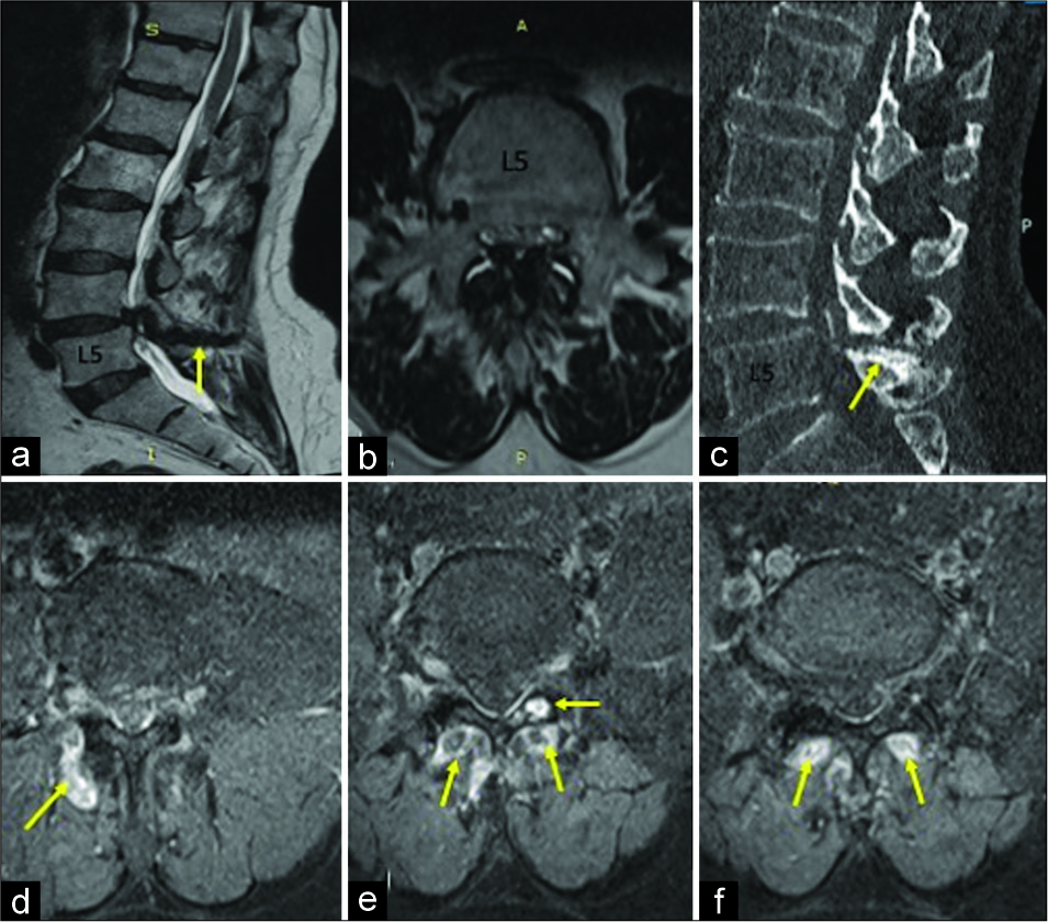
Figure 1:
(a) Sagittal T2-weighted magnetic resonance imaging (MRI): L4-L5 interspinous ligament degeneration/sclerosis (e.g., hypointense streak (arrow) and severe canal stenosis. (b) Axial T2-weighted MRI upper L5 level: lumbar canal stenosis with severe thecal sac compression/hypertrophied ligamentum flavum/facet arthrosis. (c) Sagittal noncontrast computed tomography section lumbosacral spine: sclerosis/flattening of spinous processes of L4/L5 (arrow). (d-f) Axial T1 contrast MR: abnormal ill-defined enhancement – “atypical” Baastrup’s disease (arrows).
Surgery
The patient underwent an L4 laminectomy, L4-L5 discectomy, bilateral foraminotomy, excision of HLF, and biopsy of the paraspinal soft tissues. There was marked L4- L5 thecal sac and nerve root compression. The enhancing dorsal epidural soft tissue (Baastrup’s disease) demonstrated fibrocollagenous changes but no granulomatous, infectious, or neoplastic pathology. Three months later, the patient’s neurological examination normalized.
DISCUSSION
Baastrup’s disease typically occurs in the elderly; 81.3% of patients are above 80 years of age.[
CONCLUSION
This case highlights the clinical and radiological atypical features of Baastrup’s disease involving the lumbar spine. Although rare, it should be recognized and distinguished from other pathologies such as Pott’s disease of the spine.
Declaration of patient consent
The authors certify that they have obtained all appropriate patient consent forms.
Financial support and sponsorship
Nil.
Conflicts of interest
There are no conflicts of interest.
References
1. Alonso F, Bryant E, Iwanaga J, Chapman JR, Oskouian RJ, Tubbs RS. Baastrup’s disease: A comprehensive review of the extant literature. World Neurosurg. 2017. 101: 331-4
2. Farinha F, Raínho C, Cunha I, Barcelos A. Baastrup’s disease: A poorly recognised cause of back pain. Acta Reumatol Port. 2015. 40: 302-3
3. Filippiadis DK, Mazioti A, Argentos S, Anselmetti G, Papakonstantinou O, Kelekis N. Baastrup’s disease (kissing spines syndrome): A pictorial review. Insights Imaging. 2015. 6: 123-8
4. Ghuman MS, Kaur S, Singh G, Saggar K. An unusual cause of low backache: Lumbar interspinous bursitis. Neurol India. 2014. 62: 711-2
5. Johnsson KE, Sass M. Cauda equina syndrome in lumbar spinal stenosis: Case report and incidence in Jutland, Denmark. J Spinal Disord Tech. 2004. 17: 334-5
6. Kwong Y, Rao N, Latief K. MDCT findings in baastrup disease: Disease or normal feature of the aging spine?. AJR Am J Roentgenol. 2011. 196: 1156-9

