Tools

Sandi Lam, Kathryn M. Wagner, Emily Middlebrook, Thomas G. Luerssen
  1. Department of Neurosurgery, Baylor College of Medicine, Texas Children's Hospital, Houston, Texas, USA

Correspondence Address:
Sandi Lam
Department of Neurosurgery, Baylor College of Medicine, Texas Children's Hospital, Houston, Texas, USA

DOI:10.4103/2152-7806.172532

Copyright: © 2015 Surgical Neurology International This is an open access article distributed under the terms of the Creative Commons Attribution-NonCommercial-ShareAlike 3.0 License, which allows others to remix, tweak, and build upon the work non-commercially, as long as the author is credited and the new creations are licensed under the identical terms.

How to cite this article: Lam S, Wagner KM, Middlebrook E, Luerssen TG. Delayed intracranial hypertension after surgery for nonsyndromic craniosynostosis. Surg Neurol Int 23-Dec-2015;6:187

How to cite this URL: Lam S, Wagner KM, Middlebrook E, Luerssen TG. Delayed intracranial hypertension after surgery for nonsyndromic craniosynostosis. Surg Neurol Int 23-Dec-2015;6:187. Available from: http://surgicalneurologyint.com/surgicalint_articles/delayed-intracranial-hypertension-after-surgery-for-nonsyndromic-craniosynostosis/

Date of Submission
11-Jul-2015

Date of Acceptance
07-Oct-2015

Date of Web Publication
23-Dec-2015

Abstract

Keywords: Cranial synostosis, cranial vault, craniofacial, craniosynostosis, intracranial hypertension, intracranial pressure

CASE EXAMPLE

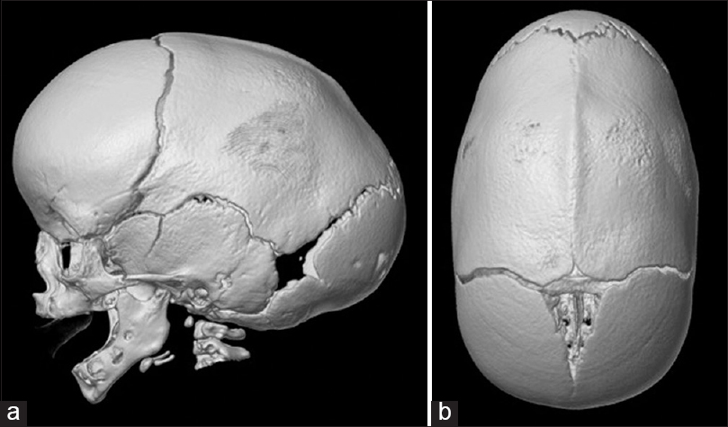
A 2-month-old boy presented with an elongated (anterior-posterior) head shape, prominent wide forehead, and bitemporal narrowing. There was a visible and palpable bony keel along the sagittal suture that was present since birth [ Figure 1 ]. He was the first child in the family, born at 40 weeks’ gestation, with no other obstetric, perinatal, or family history. His parents wished to pursue surgical correction of the sagittal synostosis, which he underwent with an unremarkable postoperative course. He continued to be followed annually as he enrolled in school. His parents have asked about the chances of requiring another surgery.


Figure 1

(a and b) Three-dimensional reconstructions of computed tomography head showing sagittal synostosis


SUMMARY OF POSTOPERATIVE INTRACRANIAL HYPERTENSION AFTER SURGERY ON NONSYNDROMIC CRANIOSYNOSTOSIS

Single-suture synostosis is reported to be associated with elevated intracranial pressure (ICP) in up to 15–20% of cases.[ 2 8 ] Studies have also cited elevated ICP developing after surgical repair of single-suture synostosis, even when intracranial hypertension (IH) was not present preoperatively. Cranial restenosis and delayed IH is known to occur in up to one-third of children after primary repair of syndromic cranial synostosis, though in a much smaller number after surgery on nonsyndromic, single-suture cranial synostosis. This entity is difficult to define, as there is substantial variation among institutions in the screening, diagnosis, and definition of elevated ICP after craniosynostosis surgery. Figures 2 4 conceptually illustrate some variations of craniosynostosis surgery techniques mentioned in the following literature review, including midline sagittal craniotomy and barrel-staves [ Figure 2 ], calvarial vault reconstruction [Figure 3a and b ], and fronto-orbital advancement [ Figure 4 ]. There are a myriad of ways to perform craniofacial reconstructions.


Figure 2

Midline sagittal craniotomy with wedge osteotomies/ barrel-staves. Cuts can be made with endoscopic technique or via open approach


Figure 3

(a and b) Different techniques of calvarial vault reconstruction


Figure 4

Fronto-orbital advancement


RETROSPECTIVE CASE SERIES

Cetas et al. reported that 6.2% of patients who underwent remodeling surgery for single-suture synostosis, and who were followed for at least 3 years, had postoperative elevated ICP. They followed 81 patients from a total of 156 consecutive patients, with average age at operation of 9.1 months.[ 3 ] All of the affected patients were males who had sagittal suture synostosis at ≤5 months of age. These 5 patients presented with delayed clinical and ophthalmological signs and symptoms of high ICP supported by computed tomography (CT) findings and ICP monitoring (three with headaches, three with papilledema, and one with microcephaly). The average time between the first and second surgeries was 30.8 months (range 19.8–35.5 months). Of these 5 affected patients, 3 originally had undergone anterior two-third cranial vault remodeling, while 2 originally had undergone midline sagittal synostectomy with barrel-stave osteotomies. The authors commented on a significant difference in the observed incidence of IH and restenosis depending on the method used for the primary cranial vault reconstruction. They reported a much higher rate after anterior two-third cranial vault reconstruction than after midline craniotomy and barrel-stave reconstruction.

Adamo and Pollack reviewed 164 patients who underwent surgery for nonsyndromic sagittal suture synostosis. One hundred forty three patients had at least 2 years’ follow-up and were followed up for an average of 43.8 months. A total of 1.5% of these patients had to undergo a second surgery for growth restriction leading to an elevation of ICP:[ 1 ] One patient was noted to have deceleration of calvarial growth on growth charts, while the other developed bicoronal synostosis. Both patients presented with headaches and were found to have papilledema; the working diagnosis of IH was corroborated with elevated opening pressure on lumbar puncture (around 30 cm H2O) at the time of reoperation. CT scans showed regrowth over the sagittal synostectomy site. Similar to Cetas’ study, both of these patients were male and had their original surgeries at or prior to 5 months of age.

Thomas et al. reported on 217 children with nonsyndromic sagittal synostosis followed for a mean of 86 months. The overall rate of raised ICP following sagittal synostosis surgery was 6.9%, found at an average of 51 months after initial surgery. Two types of surgery had different outcomes at this British institution: 1.6% (2 out of 128 patients) who underwent calvarial remodeling versus 14.6% (13 out of 89 patients) who underwent modified sagittal strip craniectomy developed raised ICP.[ 10 ] Children who underwent modified strip craniectomy (MSC) were younger at the time of operation, and younger age may have been a factor, as 18.8% of children under 6 months developed elevated ICP versus 9.1% of those with surgery between 6 and 12 months and 0.9% of children over 12 months at the time of surgery.[ 10 ] However, within the MSC group, age at surgery was the same between patients who later developed raised ICP and patients who did not (mean 6.0 ± 1.4 months, median 5.6 months versus mean 6.0 ± 1.4 months, median 6.0 months). It is not known if insufficient bone removal played a role. Unfortunately, the authors were not able to determine the specific technique of MSC for each patient due to limitations in their retrospective chart review spanning over a decade with procedures performed by multiple surgeons.[ 7 9 ] Most patients who underwent ICP monitoring presented with radiographic findings on CT scan or, clinically, with headache/irritability/psychomotor delay, and/or with deterioration in calvarial shape. Elevated ICP was defined by ICP monitoring:[ 1 3 ] The authors defined elevated ICP as baseline above 15 mmHg or the presence of 3 B-waves in a 24-h period. This definition is not one of universal consensus.[ 6 ] The authors found that not one particular sign, symptom, or radiographic finding was more closely associated with the presence of confirmed high ICP on monitoring. A minority (7%) of patients in this group had papilledema on fundoscopic examination.

SYSTEMATIC REVIEW

In a systematic review, Christian et al. found that 5% of patients with sagittal synostosis and 4% of patients with any nonsyndromic craniosynostosis (single- or multiple-suture involvement) developed IH postoperatively.[ 4 ] They found much variance in the way groups defined, screened, and diagnosed postoperative elevated ICP, which made the study of this topic by meta-analysis difficult. The search parameters for this systematic review identified only five studies that met inclusion criteria, with elevated ICP documented by invasive ICP monitoring. Other studies not included in the calculations reported IH diagnosed by lumbar puncture, papilledema findings, and/or clinical symptoms. Signs and symptoms suggesting raised ICP in this population include decreasing head circumference percentiles, worsening head shape deformity, bulging fontanelle/craniectomy defects, headaches, irritability, and developmental delay. In an attempt to sort by surgical technique, the authors found a variety of procedures reported; they grouped surgery technique type into two general categories of cranial remodeling procedures without frontal orbital advancement versus cranial remodeling procedures with advancement. In postoperative nonsyndromic craniosynostosis patients, 23 out of 471 (5%) patients without craniofacial advancement were found to have IH after surgery, and 3 out of 255 (1%) patients developed IH after craniofacial advancement surgery. These results of IH are slightly lower than listed in the previous studies, however, there is substantial variation in the incidence noted in different studies. This review only included studies where IH was diagnosed by invasive ICP monitoring, but there exist other studies using symptoms or lumbar puncture to determine ICP. This review is consistent with Cetas’ series, citing varying rates of restenosis with different procedures.[ 3 ] Measuring rates of restenosis in a review is again made difficult by differences in surgical technique and lack of standardization of definitions.

MECHANISMS AND RISK FACTORS

A possible mechanism for elevated ICP in children who have undergone suturectomy is stenosis of other sutures.[ 11 ] Secondary craniosynostosis may occur as a result of the continued influence of congenitally abnormal skull growth even after surgery or resulting from a detrimental effect of surgery on subsequent growth. Children may have abnormal skull growth postoperatively, leading to decreased intracranial volume and synostosis of other sutures, both of which could lead to increased ICP.[ 3 ] Techniques used for sagittal synostosis repair may involve sagittal plane shortening, which has also been linked with subsequent synostosis and reduced intracranial volume.

Possible risk factors for the development of elevated ICP after surgery gleaned from various case series include male sex, sagittal suture synostosis, and progressive synostosis of sutures that was not present at birth.[ 1 3 11 ] A number of studies have reported that affected patients tended to be younger at the time of primary synostosis surgery <6 months of age. The mechanism by which younger age at primary synostosis repair can be related to the development of postoperative elevated ICP is not understood. Proposed possibilities include a consequence of detaching the periosteum at a young age or the relatively small proportion of cranial volume achieved by the time of repair and subsequent healing in younger children. As well, unexpectedly exuberant ossification, reduced brain growth, or other physiologic imbalance could contribute to insufficient intracranial volume development.

Another cause may be progression of the underlying pathology that led to synostosis. There are a variety of genetic variations that have been implicated in syndromic synostosis: TWIST1 is a transcription factor involved in mesodermal patterning, transforming growth factor beta is involved in cranial suture fusion, and bone morphogenic protein may be part of a signaling cascade involved in restenosis.[ 11 ] Fibroblast growth factor receptor (FGFR) mutations are implicated in the restenosis of sutures.[ 1 ] Although progressive craniosynostosis of sutures are more common in syndromic cases, it is possible that a group of nonsyndromic patients have FGFR mutations leading to postoperative restriction in calvarial growth and raised ICP. FGFR mutations are also implicated in the restenosis of sutures.[ 1 ]

IN PRACTICE

At Texas Children's Hospital, we have a dedicated multidisciplinary craniosynostosis surgery program, where care is integrated for patients and families. Multidisciplinary care includes neurosurgery, plastic surgery, social work, neuropsychology, developmental pediatrics, otolaryngology, and ophthalmology involvement. We follow patients postoperatively on an annual basis until they reach their teenage years. Cetas et al. have recommended screening until the child is at least 6 years old.[ 3 ] We extend this time frame further to follow long-term outcomes. In addition to clinical assessment and tracking of head circumference growth, research quantification of morphometric and volumetric outcomes includes two-dimensional laser surface scanning, traditional photographs, and three-dimensional “3DMD” photographs with volumetric reconstructions. We aim to minimize radiation exposure through CT unless clinically indicated, so CT scanning is not part of our routine follow-up in the absence of symptoms. Fundoscopic exam is performed to assess for papilledema, a sign that becomes increasingly useful to elevated ICP as the child ages.[ 4 ] As no one sign or symptom is 100% sensitive and specific for detecting elevated ICP, a multidisciplinary approach is essential. In addition, monitoring neuropsychological development can help detect cognitive difficulties if present, and our program can help families arrange for appropriate social and educational support services.

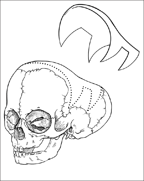
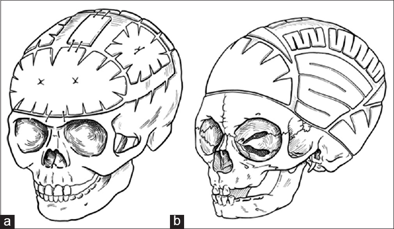
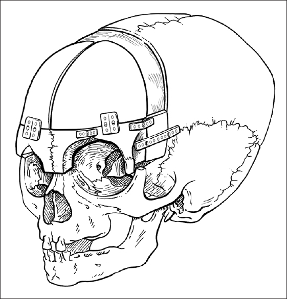
For the child in the above case example, we typically offer endoscopic sagittal synostectomy with bilateral wedge-shaped craniectomies and barrel-stave cuts at 2 months of age requiring overnight observation after surgery, followed by postoperative helmeting. Serial follow-up has shown excellent results at our center, though we remain vigilant in long-term follow-up. Children presenting with sagittal synostosis after 3 months of age are typically offered open surgical correction with modified pi technique or other calvarial vault remodeling techniques tailored to age at surgery.

While the cases of restenosis in the literature tend to be male patients who had surgery at or younger than 5 months of age, Patel et al. found that children who undergo corrective surgery for sagittal synostosis prior to 6 months of age have better long-term neuropsychological outcomes than children who have surgery after 6 months of age.[ 5 ] The best timing of intervention and optimal surgical technique are thus not fully understood at this time. These topics are an active focus of research in the craniofacial surgery community worldwide.

Resource-limited environments may present additional challenges. Surgeons may not have access to nor training for minimally invasive techniques; postoperative helmeting also requires additional expenditure. Open reconstructive surgery is typically the most viable option in certain settings. Experienced surgical teams working with limited resources need careful patient selection, surgical planning, and efficient technical execution to minimize morbidity and optimize outcomes. There is ongoing exploration in determining the most appropriate treatment.

Financial support and sponsorship

Nil.

Conflicts of interest

There are no conflicts of interest.

References

1. Adamo MA, Pollack IF. A single-center experience with symptomatic postoperative calvarial growth restriction after extended strip craniectomy for sagittal craniosynostosis. J Neurosurg Pediatr. 2010. 5: 131-5

2. Bristol RE, Lekovic GP, Rekate HL. The effects of craniosynostosis on the brain with respect to intracranial pressure. Semin Pediatr Neurol. 2004. 11: 262-7

3. Cetas JS, Nasseri M, Saedi T, Kuang AA, Selden NR. Delayed intracranial hypertension after cranial vault remodeling for nonsyndromic single-suture synostosis. J Neurosurg Pediatr. 2013. 11: 661-6

4. Christian EA, Imahiyerobo TA, Nallapa S, Urata M, McComb JG, Krieger MD. Intracranial hypertension after surgical correction for craniosynostosis: A systematic review. Neurosurg Focus. 2015. 38: E6-

5. Patel A, Yang JF, Hashim PW, Travieso R, Terner J, Mayes LC. The impact of age at surgery on long-term neuropsychological outcomes in sagittal craniosynostosis. Plast Reconstr Surg. 2014. 134: 608e-17e

6. Sood S, Marupudi N, Haridas A, Ham SD. Letter to the editor: Intracranial pressure and sagittal craniosynostosis. J Neurosurg Pediatr. 2015. 16: 351-5

7. Souweidane MM. Periodical shifts in the surgical correction of sagittal craniosynostosis. J Neurosurg Pediatr. 2015. 15: 347-8

8. Tamburrini G, Caldarelli M, Massimi L, Santini P, Di Rocco C. Intracranial pressure monitoring in children with single suture and complex craniosynostosis: A review. Childs Nerv Syst. 2005. 21: 913-21

9. Thomas GP, Johnson D, Byren JC, Judge AD, Jayamohan J, Magdum SA. The incidence of raised intracranial pressure in nonsyndromic sagittal craniosynostosis following primary surgery. J Neurosurg Pediatr. 2015. 15: 350-60

10. Thomas GP, Johnson D, Byren JC, Judge AD, Jayamohan J, Magdum SA. Periodical shifts in the surgical correction of sagittal craniosynostosis. J Neurosurg Pediatr. 2015. 15: 348-9

11. Yarbrough CK, Smyth MD, Holekamp TF, Ranalli NJ, Huang AH, Patel KB. Delayed synostoses of uninvolved sutures after surgical treatment of nonsyndromic craniosynostosis. J Craniofac Surg. 2014. 25: 119-23

Leave a Reply

Your email address will not be published. Required fields are marked *