

- Department of Neurosurgery, National Institute of Neurology and Neurosurgery, Guanajuato, Mexico.
- Department of Internal Medicine, Instituto Mexicano del Seguro Social, León, Guanajuato, Mexico.
Correspondence Address:
Marcos Sangrador
Department of Internal Medicine, Instituto Mexicano del Seguro Social, León, Guanajuato, Mexico.
DOI:10.25259/SNI_458_2019
Copyright: © 2019 Surgical Neurology International This is an open-access article distributed under the terms of the Creative Commons Attribution-Non Commercial-Share Alike 4.0 License, which allows others to remix, tweak, and build upon the work non-commercially, as long as the author is credited and the new creations are licensed under the identical terms.How to cite this article: Marcos Sangrador, Jimena González Olvera, Valeria Mendoza Ortiz. Dermatofibrosarcoma of the scalp. 15-Nov-2019;10:221
How to cite this URL: Marcos Sangrador, Jimena González Olvera, Valeria Mendoza Ortiz. Dermatofibrosarcoma of the scalp. 15-Nov-2019;10:221. Available from: http://surgicalneurologyint.com/surgicalint-articles/9754/
Date of Submission
17-Oct-2019
Date of Acceptance
22-Oct-2019
Date of Web Publication
15-Nov-2019
Abstract
A 25-year-old woman presented with a 4 months history of progressive pain and tumefaction in the right parietal region. Deformity of the scalp was evident, and a biopsy was taken, reporting a high- grade dermatofibrosarcoma. She underwent surgical management, achieving a gross total resection. Dermatofibrosarcoma protuberans is a rare tumor arising from the dermis. It tends to have an indolent course and local recurrence after excision.
Keywords: Dermatofibrosarcoma, Protuberans, Scalp
A 25-year-old woman presented with a 4 months history of tumefaction in the right parietal region. Magnetic resonance imaging was performed [
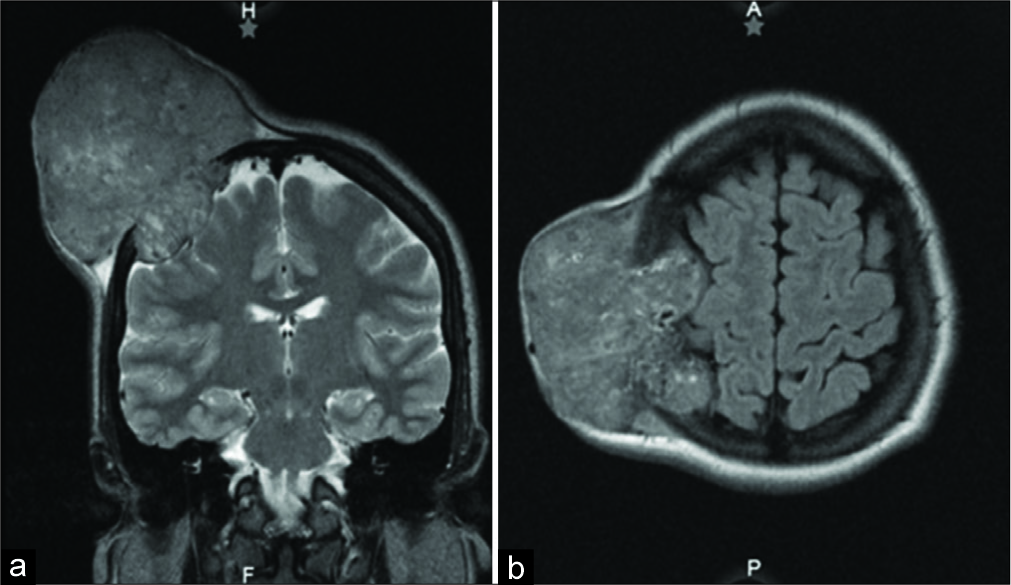
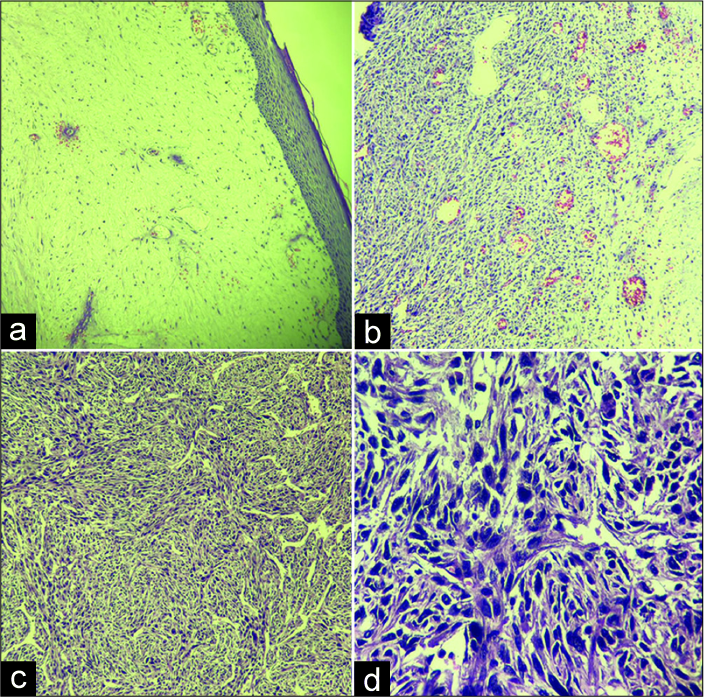
Figure 3:
Histopathologic findings. (a) Dermatofibrosarcoma protuberans expands and infiltrates the dermis, while epidermis shows important thinning. H and E stain ×10. (b) Spindle cells with scant eosinophilic cytoplasm usually are centered on a small vessel. H and E stain ×10. (c) Classic storiform pattern: cells radiating in spokes at right angles around a central point that often contains a vessel. H and E stain ×10 (d) H and E ×100 magnification in which the storiform or cartwheel pattern can be better appreciated.
Declaration of patient consent
The authors certify that they have obtained all appropriate patient consent forms.
Financial support and sponsorship
Nil.
Conflicts of interest
There are no conflicts of interest.
References
1. Gatlin JL, Hosch R, Khan M. Dermatofibrosarcoma protuberans of the scalp with fibrosarcomatous degeneration and pulmonary metastasis. J Clin Imaging Sci. 2011. 1: 55-
2. Gloster HM Jr. Dermatofibrosarcoma protuberans. J Am Acad Dermatol. 1996. 35: 355-74
3. Loss L, Zeitouni NC. Management of scalp dermatofibrosarcoma protuberans. Dermatol Surg. 2005. 31: 1428-33
4. Marakovic J, Vilendecic M, Marinovic T, Lambasa S, Grahovac G. Intracranial recurrence and distant metastasis of scalp dermatofibrosarcoma protuberans. J Neurooncol. 2008. 88: 305-8
5. McLoughlin PM, Girach M, Wood GA. Dermatofibrosarcoma protuberans of the scalp. Br J Oral Maxillofac Surg. 1992. 30: 401-3
6. Rockley PF, Robinson JK, Magid M, Goldblatt D. Dermatofibrosarcoma protuberans of the scalp: A series of cases. J Am Acad Dermatol. 1989. 21: 278-83
7. Weiss S, Goldblum J.editorsEnzinger and Weiss’s Soft Tissue Tumors. Philadelphia, PA: Mosby Elsevier; 2008. p.

