Tools

Domenico Policicchio, Artan Doda, Giampiero Muggianu, Giosuè Dipellegrini, Riccardo Boccaletti
  1. Department of Neurosurgery, Azienda Ospedaliero Universitaria di Sassari, Via Enrico De Nicola 1, 07100 Sassari, Italy
  2. Department of Neurosurgery, Regina Elena National Cancer Institute, Via Elio Chianesi 53, 00144 Rome, Italy

DOI:10.25259/SNI-86-2019

Copyright: © 2019 Surgical Neurology International This is an open-access article distributed under the terms of the Creative Commons Attribution-Non Commercial-Share Alike 4.0 License, which allows others to remix, tweak, and build upon the work non-commercially, as long as the author is credited and the new creations are licensed under the identical terms.

How to cite this article: Domenico Policicchio, Artan Doda, Giampiero Muggianu, Giosuè Dipellegrini, Riccardo Boccaletti. Ethical and therapeutic dilemmas in glioblastoma management during pregnancy: Two case reports and review of the literature. 26-Mar-2019;10:41

How to cite this URL: Domenico Policicchio, Artan Doda, Giampiero Muggianu, Giosuè Dipellegrini, Riccardo Boccaletti. Ethical and therapeutic dilemmas in glioblastoma management during pregnancy: Two case reports and review of the literature. 26-Mar-2019;10:41. Available from: http://surgicalneurologyint.com/surgicalint-articles/9243/

Date of Submission
20-Oct-2018

Date of Acceptance
04-Feb-2019

Date of Web Publication
26-Mar-2019

Abstract

Introduction: There are no guidelines about the management of glioblastoma multiforme (GBM) during pregnancy: treatment of these patients presents therapeutic and ethical challenges.

Case Description: Two patients, respectively, 28 years old at the 14th week of gestation with a thalamic GBM and 38 years old at the 28th week of gestation with fronto-mesial GBM. Patients and their relatives were deeply informed about the natural history of GBM and potential risks and benefits of surgery, radiotherapy (XRT), and chemotherapy (CTX) for both, mother and fetus. The first patient’s will was to preserve her fetus from any related, even minimal, risk of XRT, and CTX until safe delivery despite progression of GBM, accepting only surgery (tumor debulking and shunting of hydrocephalus). The second one asked to deliver the baby as soon as possible (despite the risks of prematurity) to receive the standard treatments of GBM. The two patients survived, respectively, 16 and 46 months after delivery. The first patient’s son is in good clinical conditions; the second one suffered problems linked to prematurity.

Conclusions: Standard treatment of GBM in a pregnant woman could improve the mother’s survival but can expose the fetus to several potential risks. Ethically, relatives should understand that mother has anyway a poor prognosis and, at the same time, fetus prognosis depends on mother’s condition and therapy. It is not possible to warrant absence of risk for both. Considering the absence of guidelines and the relatively poor current data available about management of GBM in a pregnant woman, after a deep explanation of the situation, we think that the will of the mother and her relatives should prevail.

Keywords: Chemotherapy, ethics, glioblastoma, neurosurgery, pregnancy, radiotherapy

INTRODUCTION

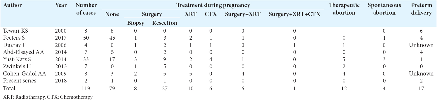
Glioblastoma multiforme (GBM) is the most commonly diagnosed and malignant primary brain tumor. In a pregnant woman, however, it is an unusual and dramatic event because mother, family and physicians must deal with a hard challenge. As far as no current treatment of GBM is curative and there are no clear guidelines about its management in a pregnant woman, the potential benefits in terms of survival to the mother offered by the standard treatment of GBM must be accurately balanced against the potential risks to the fetus. Herein, we present two cases of GBM occurred in pregnant women in different stages of gestational age, review the existing literature and discuss the therapeutic and ethical aspects.

CASE REPORT

Case one

A 28-years-old female at the 14th week of gestation was admitted in April 2016 due to headache, vomiting, and progressive asthenia in the previous 3 weeks. A brain magnetic resonance imaging (MRI) demonstrated a large right thalamic tumor [ Figure 1 ], and the MRI-spectroscopic study showed a high level of choline and low level of N-AcetylAspartate consistent with a high-grade glioma. She presented fully awake, with a slight left hemiparesis and headache. Later on, due to progressive neurological deterioration, she underwent in May 2 external ventricular drainage (EVD) in local anesthesia and 1 week later craniotomy and partial removing of the tumor under general anesthesia, with continuous fetal heart rate (FHR) monitoring, maintaining good fetal conditions during the operation. At the end of the month, she was discharged following the gynecological evaluation who revealed good clinical conditions of both, mother and fetus. 2 weeks later, she deteriorated neurologically presenting vomiting, stupor, and severe hemiparesis. A brain computer tomography scan showed hydrocephalus and she underwent on the left side, a ventricular-peritoneal shunt. The brain-MRI performed 2 days later showed a light improving the hydrocephalus. The abdomen ultrasonography was normal and she was discharged in good clinical conditions except a moderate left arm paresis. In July 5, a brain MRI showed a large thalamic tumor. In July 7, she had elective cesarean section and the day later, underwent a gross debulking of the tumor. Her clinical conditions improved progressively and in August she began CTX with temozolomide (TMZ) and XRT in a standard way.[ 20 , 21 ] 12 months after surgery the patient had a moderate left arm paresis with small residual tumor showed on the brain-MRI [ Figure 2 ]. The baby was born with a retinopathy and bronco dysplasia due to the premature birth. He was treated with retinal laser therapy bilaterally and Lucentis intravitreous on the right side successfully. He was also treated with Synagis 15 mg/kg in 1/month and is growing normally under surveillance by pediatricians. The patient died 16 months after delivery.


Figure 1

Brain magnetic resonance imaging preoperative showing a large right thalamic lesion.


Figure 2

Magnetic resonance imaging with gad, 12 months after the operation.


Case two

A 38-years-old female, on the 28th week of gestation, presented in November 15, 2014, at the emergency department due to general asthenia associated with a headache, as well as left arm paresis and dysesthesia and a slight left facial deficit. A brain MRI showed a large right frontal tumor [ Figure 3 ]. She moved to the department of obstetrics-gynecology and after team counseling (oncologist, neurosurgeon, gynecologist, and neonatologist), 2 days later, she underwent an elective cesarean section. 5 days later she underwent craniotomy and exeresis of the tumor with intraoperative neuronavigation and ultrasound. The histology revealed GBM. The patient was evaluated by the oncologists and radiotherapists and began chemotherapy (CTX) and radiotherapy (XRT) (Stupp protocol). 12 months after surgery her clinical conditions progressively deteriorate with signs of regrowth on MRI [ Figure 4 ]. The baby was born with low birth weight and needed 2 months to stay in the neonatal intensive care unit; he was also treated with Synagis for 4 months. At the age of 4 months he was operated for hypertrophic pyloric stenosis; at present, he suffers epilepsy and sometimes tremor but has a normal psychomotor development. The patient survived 46 months after delivery.


Figure 3

Magnetic resonance imaging with gad on admission, showing a gross frontal glioblastoma multiforme on the right side.


Figure 4

Magnetic resonance imaging with gad 14 months after the operation.


DISCUSSION

GBM is the most aggressive and malignant primary brain tumor with a poor prognosis. Its standard treatment consists of maximal surgical resection, XRT, and concomitant and adjuvant CTX with TMZ.[ 9 , 19 , 20 ] GBM in a pregnant woman is a rare and a dramatic condition because mother, family, and physicians must face a difficult challenge. No current treatment of GBM is curative and there are neither guidelines nor enough evidence about its management in pregnancy; therapeutic strategies are based only on a few case reports and a small case series in literature [ Table 1 ].[ 1 , 6 , 7 , 17 , 21 , 23 - 25 ] The potential benefits in terms of survival to the mother by the standard treatment of GBM must be accurately balanced against the potential risks to the fetus. Treatment will depend on clinical-radiological presentation, histology, gestational age, and the patients’ will.


Table 1

Management of glioblastoma multiforme in pregnancy in different case series.


THERAPEUTIC CONSIDERATIONS

Surgery

General considerations for anesthesia in pregnant patients undergoing nonobstetric operations have already been covered in several review articles;[ 15 ] specific considerations on anesthetic management of pregnant patients treated for brain tumors were published by Abd-Elsayed et al. in 2014.[ 1 ] A thorough evaluation of the management of pregnant patient during surgery is extremely complex and is beyond the purpose of this article. Many aspects such as maternal and fetal physiology, altered drug pharmacodynamics and pharmacokinetics (including placental transfer of drugs), gestational age, and nature of the brain pathology and fetal conditions should be considered. The ultimate goal is to provide safe anesthesia to the mother while simultaneously minimizing the risk to the fetus (including preterm labor or fetal demise). Until date, no anesthetic drug has been proven to be clearly hazardous to the human fetus; particularly, most anesthetic medications, including barbiturates, volatile anesthetic, propofol, opioids, muscle relaxants, and local anesthetics have been widely used during pregnancy with a good safety record.[ 15 ] Both, propofol and thiopental have favorable effects in terms of preservation of cerebral autoregulation and reduction in cerebral metabolic rate and intracranial pressure. General anesthesia may consist of induction with either propofol or thiopental plus a volatile anesthetic for maintenance or induction and maintenance with intravenous propofol.[ 11 ] Volatile anesthetics such as halothane, sevoflurane, desflurane, and isoflurane are shown to inhibit the uterine contractility, which may prove beneficial in preventing preterm contractions.[ 11 , 15 ] One of the major concerns with surgery during pregnancy is the risk of maternal hypotension (due to blood loss and/or anesthesia), which may reduce placental perfusion precipitating fetal ischemia; careful maintenance of stable maternal hemodynamic parameters and oxygenation is mandatory. Many authors advocate continuous FHR monitoring during surgery as maternal hemodynamic stability alone is not an adequate indicator of fetal well-being. The importance of intraoperative FHR monitoring consists in detecting early alterations, allowing optimization of maternal hemodynamics and oxygenation with appropriate fluid therapy, vasopressors, blood product administration, hyperventilation, or position adjustment.[ 15 , 22 ] Optimal timing of surgery in pregnant patients is an argument of debate. Surgery during the first trimester is associated with an increased risk of miscarriage; during second and third trimester, due to greater uterine irritability, surgery increases the risk of preterm labor;[ 11 , 15 , 22 ] and during the third trimester, according to Tewari, increased maternal intravascular volume, carries a high risk of intracranial hemorrhage.[ 21 ] In recent years, several authors agree on the high degree of safety of the neurosurgical intervention and anesthesia during pregnancy. Delaying surgery often resulted in maternal deterioration and urgent intervention. Thus, pregnancy by itself should not be considered a major contraindication for performing a neurosurgical procedure, which should be considered early rather than late in most patients.[ 6 , 11 , 13 , 16 ] According to American College of Obstetricians and Gynecologists’ Committee on Obstetric Practice, regardless of trimester, pregnant woman should not be denied indicated surgery. The choice of anesthetic technique(s), and the selection of appropriate drugs of anesthesia should be guided by maternal indications for surgery and the location of the surgical procedure.

XRT

The major potential complications of fetal radiation exposure include the death of a developing embryo, teratogenesis, growth retardation, central nervous system (CNS) effects, and induction of malignancy. Developing embryos pass different stages (preimplantation, organogenesis, and fetal growth), with specific sensitivities to the effects of radiation. During the first stage of development, the embryo is sensitive to lethal effects of 0.10 Gy of radiation or less, but it is resistant to teratogenic and growth retarding effects of radiation.[ 5 ] This means that overexposure in this stage could be lethal, and embryos that survive are generally free from any abnormalities.[ 4 , 11 ] Teratogenesis is a major risk only during the period of early organogenesis, i.e. during the 3rd and 4th weeks of gestation, with doses over about 0.05 Gy.[ 5 ] Unlike other organ systems, the CNS remains sensitive to ionizing radiation throughout gestation and into the neonatal period. After the 4th week of gestation, radiation doses over about 0.50–1.00 Gy may cause growth retardation and CNS effects such as microcephaly and eye malformations.[ 4 , 5 ] The risk of inducing a late malignancy exists throughout all stages of fetal development, and no dose of radiation can be considered completely safe. It has been estimated that in utero exposure to 0.01–0.02 Gy of radiation may increase the risk of leukemia by 1.5 fold, increasing the incidence from 1 in 3000 children to 1 in 2000 children. This represents one additional case of leukemia per 6000 children.[ 4 , 5 , 11 ] Mazonakis et al. estimated fetal doses following irradiation of a “brain tumor” in a phantom pregnancy model with a linear accelerator. For a cumulative isocenter dose of 65 Gy, the maximum fetal absorbed dose was 80.9 mGy (0.089 Gy).[ 14 ] Similar results were reported by Haba et al.[ 10 ] Several doubts remain regarding safe threshold doses for deleterious effects; the international commission for radiation protection concluded that expected radiation effects, such as mental retardation and organ malformations probably only arise above a threshold dose of 0.1–0.2 Gy.[ 3 ] This threshold dose is not generally reached with curative XRT during pregnancy, considering that tumors are located sufficiently far from the fetus and that precautions have been taken to protect the unborn child against leakage radiation and collimator scatter of the teletherapy machine; such precautions also reduce the risk of radiation-induced childhood cancer and leukemia in the unborn child.[ 11 , 12 ] Sneed et al. concluded that, when clinically indicated, it is possible to irradiate brain tumors to high doses during pregnancy with fetal exposure under 0.10 Gy, conferring an “increased but acceptable risk of leukemia in the child.”[ 18 ]

CTX

TMZ, an alkylating agent used in the treatment of malignant gliomas, is a pregnancy category D medication in USA, UK, and AU and is not advised for use in pregnant women or in those who are contemplating pregnancy. When in some cases, TMZ was used, it was always unintentionally and has been interrupted immediately (unplanned pregnancy in patients already harboring a high-grade glioma).[ 2 , 8 , 24 ] The current recommendations of TMZ are based on animal and epidemiological studies. Use of CTX during the second and third trimesters can result in intrauterine growth retardation, low-birth-weight, and premature delivery. The CNS, hematological system, genitalia, and eyes remain susceptible to the effects of CTX, with consequential neonatal myelosuppression, sterility, and neurobehavioral disorders.[ 11 ] Yust-Katz et al. retrospectively reviewed a series of patients with glioma during pregnancy: 15 patients were pregnant at the time of diagnosis and 18 became pregnant after a diagnosis of glioma. In the former group, none patients were treated with CTX. In the latter, Group 4 patients received CTX. Three patients who received CTX terminated her pregnancy (TMZ in two and procarbazine, CCNU, and vincristine in one). One patient, who was receiving TMZ and valproic acid at the time of diagnosis had medications stopped and decided to continue pregnancy. Unfortunately, the child was born with a neural tube defect and cerebral palsy.[ 24 ] Blumenthal et al. presented a case series of 6 women with malignant gliomas who during glioma-directed treatment were discovered to have an unplanned pregnancy. All patients elected to discontinue CTX and carry their pregnancy to term. All women had uneventful pregnancies with no glioma-related complications. All women delivered healthy newborns without evidence of congenital malformations despite exposure to cytotoxic CTX and anticonvulsant medications.[ 2 ] Given the lack of data, recommendations on the use of CTX during pregnancy are difficult.[ 11 ]

Ethical considerations

Glioblastoma is a primitive malignant brain tumor with poor prognosis. Its standard treatment is through surgery, XRT, and CTX.[ 9 , 19 , 20 ] Its diagnosis during pregnancy is quite rare and therefore brings to some ethical and clinical dilemmas. Neither guidelines nor treatment standards exist for GBM during pregnancy. Many suggestions come from case reports and case series on a few patients [ Table 1 ]. Some authors have proposed decision-making algorithms that can help in choosing the right therapy.[ 17 , 21 , 22 ] Such algorithms are based on multidisciplinary assessments (neurosurgeon, oncologist, gynecologist, and anesthetist) that consider the type of tumor, the clinical conditions of the fetus and the mother, and gestational age; the evaluation and the decision-making process cannot ignore an adequate disclosure of the potential risks and benefits that the therapy choices on mother and child can lead to. The ethical problem is that the mother has an ominous prognosis pathology whose medical history can be somehow influenced by cancer therapy, and such therapies, however, may imply potential risks for the fetus, both in terms of short- and long-term survival and morbidity (teratogenicity, carcinogenicity,…). It is a matter of “two patients in one,” the mother who has a limited life expectancy given the malignant disease, and the child whose life expectancy would be normal but is influenced by his mother’s health as well as the therapies that are performed. Considering the lack of guidelines our two cases (and also literature data) manifested heterogeneity of treatment-related greatly to the mother’s and family’s wishes [ Table 1 ], the type of therapy could be aimed at safeguarding majorly the mother or the son. On the one hand, we will see families choose, to maximize the cancer therapy for the mother and in consideration of the gestational age, the voluntary interruption of pregnancy or preterm delivery. The interruption of pregnancy, known as therapeutic abortion, will guarantee full coverage of cancer therapy for the mother and will eliminate the risk of having a child with problems caused by cancer therapy (and who will lose his mother). Preterm delivery will expose the child to the risks of prematurity but will avoid the threats of cancer therapy, and those that may be connected to the mother’s decaying conditions. On the other hand, there are patients who decide to postpone all cancer treatments to carry on their pregnancy minimizing the risks provoked by cancer therapies on the fetus. This strategy exposes the mother to the risks of cancer progression while fostering the development of the child until the mother’s neurological conditions will allow it; in these cases, it is important to inform the mother and the family that there is evidence in literature that shows that during pregnancy GBM has a greater growth pace[ 17 , 24 ] and that the mother’s neurologic decay may increase risks of abortion and complications for the fetus.[ 11 , 17 ] Halfway therapeutic strategy could be reasonable with surgery (biopsy or resection) during pregnancy; based on gestational age and maternal condition, XRT and CTX with TMZ could be delayed after delivery; some authors advocate brain tumor irradiation during pregnancy.[ 11 , 12 , 18 , 21 ] To date, CTX is not recommended during pregnancy.[ 11 ] In any case, the family must be informed of the potential risks and benefits of the single treatments on mother and child; surgery seems effective in reducing neurological symptoms by treating/preventing intracranial hypertension. At the same time, it guarantees the certainty of the histologic diagnosis and allows the protraction of pregnancy.[ 6 , 11 , 13 , 14 ] Notwithstanding, modern anesthesiological and surgical techniques complications such as abortion, preterm delivery, or teratogenicity cannot be avoided; they may be yielded by drugs or by the mother’s physiological alterations during the operation (hypotension, anemia …).[ 1 , 11 , 15 , 22 ] Some evidence in literature suggests that XRT for brain tumor could be administered during pregnancy with lesser risks for the fetus,[ 18 ] however, threats connected to teratogenicity cannot be assured and the true risk of developing neoplasms during childhood in sons of XRT-treated mothers during pregnancy is unknown.

Based on our experience and literature data, we proposed a decision-making algorithm, similar to that already published,[ 17 , 21 , 22 ] but emphasizing the role of mother’s will [ Figure 5 ]. In patients submitted to surgery during the first half of pregnancy (first trimester and early second trimester), based on literature data, we should discuss with the patient and her family the potential risk and benefit of XRT during pregnancy (leaving to them the decision to accept or refuse this treatment); in case of patients submitted to surgery during the second half of pregnancy we suggest to delay XRT immediately after delivery to reduce risk for the fetus.


Figure 5

Proposed algorithm for the management of high-grade gliomas diagnosed during pregnancy.


In our first patient, there was a 28-years-old mother at her first pregnancy in the 14th week of the gestational age with a huge right thalamic mass with radiological features of high-grade glioma. The multidisciplinary team which included the intensive care unit consultant, medical oncologist, XRT consultant, high risk obstetrician, and neonatologist as well as psychologist and religious referent, discussed with the mother and her family all the risks and benefits of the pregnancy, the natural history of the disease and the possible outcome as well as the available treatment options. It was decided first to put an EVD and a partial removing of the tumor and to support the mother during her pregnancy without XRT or TMZ to avoid even the minimal risk to the fetus and do elective CS in the week of 24th. After the birth, we perform a gross resection of the remained tumor and begin the XRT and TMZ. We managed to deliver the baby safely at week 24 trying to preserve as much as possible the neurological conditions of the mother. In the second case, the will of the patient and her husband was to deliver the baby as soon as possible and to begin with the standard treatment of GBM. In both cases, we respected their will to avoid any eventual deleterious effects of the fetus in case of beginning XRT and/or TZM during the pregnancy, giving to both the maximal cure.

CONCLUSIONS

When the most malignant primary brain tumor such as GBM, affects a pregnant woman, it becomes a very challenge situation, as far as no current treatment for GBM is curative and there are neither guidelines nor enough evidence about the management of such a dramatic situation in pregnancy. The available literature data suggest that brain surgery during pregnancy can be performed with acceptable risk for mother and fetus; XRT could be administered with increased even though acceptable risks for the fetus; and data are not sufficient to recommend the use of TMZ in pregnancy. Mother has a poor prognosis; fetus’ prognosis depends on mother’s condition and therapy. The chosen approach should involve several professional roles, the patient and familiars should have a deep explanation of the situation, and their will should prevail.

Declaration of patient consent

The authors certify that they have obtained all appropriate patient consent forms. In the form the patient(s) has/have given his/her/their consent for his/her/their images and other clinical information to be reported in the journal. The patients understand that their names and initials will not be published and due efforts will be made to conceal their identity, but anonymity cannot be guaranteed.

Financial support and sponsorship

Nil.

Conflicts of interest

There are no conflicts of interest.

Acknowledgements

We would like to thank Mrs Lorena Gullà for the English revision.

References

1. Abd-Elsayed AA, Díaz-Gómez J, Barnett GH, Kurz A, Inton-Santos M, Barsoum S. Acase series discussing the anaesthetic management of pregnant patients with brain tumours. F1000Res. 2013. 2: 92-

2. Blumenthal DT, Parreño MG, Batten J, Chamberlain MC. Management of malignant gliomas during pregnancy: A case series. Cancer. 2008. 113: 3349-54

3. Bondy ML, Scheurer ME, Malmer B, Barnholtz-Sloan JS, Davis FG, Il'yasova D. Brain tumor epidemiology: Consensus from the brain tumor epidemiology consortium. Cancer. 2008. 113: 1953-68

4. Brent RL. Saving lives and changing family histories: Appropriate counseling of pregnant women and men and women of reproductive age, concerning the risk of diagnostic radiation exposures during and before pregnancy. Am J Obstet Gynecol. 2009. 200: 4-24

5. Brent R.L. The effects of embryonic and fetal exposure to X-ray, microwaves, and ultrasound. Clin Obstet Gynecol. 1983. 26: 484-510

6. Cohen-Gadol AA, Friedman JA, Friedman JD, Tubbs RS, Munis JR, Meyer FB. Neurosurgical management of intracranial lesions in the pregnant patient: A 36-year institutional experience and review of the literature. J Neurosurg. 2009. 111: 1150-7

7. Ducray F, Colin P, Cartalat-Carel S, Pelissou-Guyotat I, Mahla K, Audra P. Management of malignant gliomas diagnosed during pregnancy. Rev Neurol (Paris). 2006. 162: 322-9

8. Evans AC, Nelson MB, Dhall G. Pregnancy in a patient with a malignant brain tumor taking temozolomide: Case report and review of the literature. J Pediatr Oncol Nurs. 2015. 32: 326-8

9. Grabowski MM, Recinos PF, Nowacki AS, Schroeder JL, Angelov L, Barnett GH. Residual tumor volume versus extent of resection: Predictors of survival after surgery for glioblastoma. J Neurosurg. 2014. 121: 1115-23

10. Haba Y, Twyman N, Thomas SJ, Overton C, Dendy P, Burnet NG. Radiotherapy for glioma during pregnancy: Fetal dose estimates, risk assessment and clinical management. Clin Oncol (R Coll Radiol). 2004. 16: 210-4

11. Jayasekera BA, Bacon AD, Whitfield PC. Management of glioblastoma multiforme in pregnancy. J Neurosurg. 2012. 116: 1187-94

12. Kal HB, Struikmans H. Radiotherapy during pregnancy: Fact and fiction. Lancet Oncol. 2005. 6: 328-33

13. Lynch JC, Gouvêa F, Emmerich JC, Kokinovrachos G, Pereira C, Welling L. Management strategy for brain tumour diagnosed during pregnancy. Br J Neurosurg. 2011. 25: 225-30

14. Mazonakis M, Damilakis J, Theoharopoulos N, Varveris H, Gourtsoyiannis N. Brain radiotherapy during pregnancy: An analysis of conceptus dose using anthropomorphic phantoms. Br J Radiol. 1999. 72: 274-8

15. NíMhuireachtaigh R, O'Gorman DA. Anesthesia in pregnant patients for nonobstetric surgery. J Clin Anesth. 2006. 18: 60-6

16. Nossek E, Ekstein M, Rimon E, Kupferminc MJ, Ram Z. Neurosurgery and pregnancy. Acta Neurochir (Wien). 2011. 153: 1727-35

17. Peeters S, Pagès M, Gauchotte G, Miquel C, Cartalat-Carel S, Guillamo JS. Interactions between glioma and pregnancy: Insight from a 52-case multicenter series. J Neurosurg. 2018. 128: 3-13

18. Sneed PK, Albright NW, Wara WM, Prados MD, Wilson CB. Fetal dose estimates for radiotherapy of brain tumors during pregnancy. Int J Radiat Oncol Biol Phys. 1995. 32: 823-30

19. Stupp R, Hegi ME, Mason WP, Van den Bent MJ, Taphoornm MJ, Janzer RC. Effects of radiotherapy with concomitant and adjuvant temozolomide versus radiotherapy alone on survival in GBL in a randomized phase III study: 5-year analysis of the EORTC-NCIC trial. Lancet Oncol. 2009. 10: 459-66

20. Stupp R, Mason WP, van den Bent MJ, Weller M, Fisher B, Taphoorn MJ. Radiotherapy plus concomitant and adjuvant temozolomide for glioblastoma. N Engl J Med. 2005. 352: 987-96

21. Tewari KS, Cappuccini F, Asrat T, Flamm BL, Carpenter SE, Disaia PJ. Obstetric emergencies precipitated by malignant brain tumors. Am J Obstet Gynecol. 2000. 182: 1215-21

22. Upadya M, Saneesh PJ. Anaesthesia for non-obstetric surgery during pregnancy. Indian J Anaesth. 2016. 60: 234-41

23. Wu J, Ma YH, Wang TL. Glioma in the third trimester of pregnancy: Two cases and a review of the literature. Oncol Lett. 2013. 5: 943-6

24. Yust-Katz S, de Groot JF, Liu D, Wu J, Yuan Y, Anderson MD. Pregnancy and glial brain tumors. Neuro Oncol. 2014. 16: 1289-94

25. Zwinkels H, Dörr J, Kloet F, Taphoorn MJ, Vecht CJ. Pregnancy in women with gliomas: A case-series and review of the literature. J Neurooncol. 2013. 115: 293-301

Leave a Reply

Your email address will not be published. Required fields are marked *