

- Department of Neurosurgery, Centro Médico “Lic. Adolfo Lòpez Mateos”, Instituto de Salud del Estado de México, Av. Nicolás San Juan s/n Ex Hacienda La Magdalena, Estado de México, México
Correspondence Address:
A. Ceja-Espinosa
Department of Neurosurgery, Centro Médico “Lic. Adolfo Lòpez Mateos”, Instituto de Salud del Estado de México, Av. Nicolás San Juan s/n Ex Hacienda La Magdalena, Estado de México, México
DOI:10.4103/sni.sni_172_18
Copyright: © 2018 Surgical Neurology International This is an open access journal, and articles are distributed under the terms of the Creative Commons Attribution-NonCommercial-ShareAlike 4.0 License, which allows others to remix, tweak, and build upon the work non-commercially, as long as appropriate credit is given and the new creations are licensed under the identical terms.How to cite this article: A. Ceja-Espinosa, R. Huato-Reyes, R. Ortega-Valencia. Hangman's fracture surgical management with posterior C2-4 fusion: Case description and literature review. 26-Jul-2018;9:147
How to cite this URL: A. Ceja-Espinosa, R. Huato-Reyes, R. Ortega-Valencia. Hangman's fracture surgical management with posterior C2-4 fusion: Case description and literature review. 26-Jul-2018;9:147. Available from: http://surgicalneurologyint.com/surgicalint-articles/hangmans-fracture-surgical-management-with-posterior-c2%e2%80%914-fusion-case-description-and-literature-review/
Date of Submission
20-Jun-2018
Date of Acceptance
26-Jun-2018
Date of Web Publication
26-Jul-2018
Abstract
Background:Hangman's fractures of the C2 verebrae represent approximately 20% of all cervical fractures. They are challenging cases and there is still no consensus regarding the optimal surgical vs nonoperative treatment.
Case Description:A 40-year-old female presented with a C2 bilateral pars articularis fracture. She exhibited a partial spastic quadriparesis. Computed tomography and magnetic resonance imaging showed a C2 “hangman's” fracture with compromise of the C2-3 intervertebral disk. Adequate reduction of the fracture and subaxial stabilization were achieved utilizing C2 transarticular and C3-4 transfacet screws.
Conclusions:The optimal management of unstable hangman's fractures remains controversial. They represent challenging cases, and new treatment options are available. Here, we successfully utilized a C2 transarticular and C3-4 transfacet screw fusion without neurological sequelae.
Keywords: C2 transpedicular screws, Hangman's fracture, posterior fusion
INTRODUCTION
In 1964 Garber introduced the term “traumatic spondylolisthesis of the axis.”.[
CASE DESCRIPTION
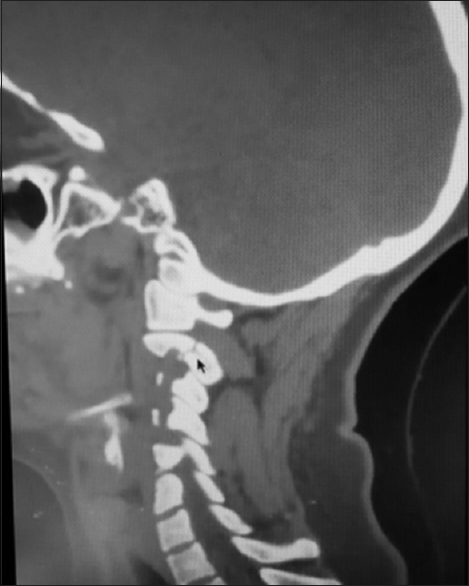
A 40-year-old female, following a motor vehicle accident, presented with severe neck pain and decreased motor function (e.g. 4/5 in the right arm, left arm 2/5 proximally, 1/5 distally, but 5/5 in both lower extremities). Reflexes were diminished in both arms, and she had no sensory deficit. Magnetic resonance imaging (MRI) and computed tomography (MRI) documented a C2 Hangman's fracture type IIa [Figures


Cervical surgery
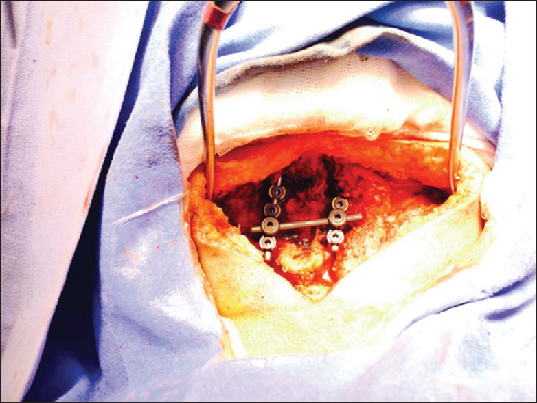
The patient underwent a posterior instrumented fusion performed under fluoroscopy. First, C3 and C4 facet screws were placed followed by bilateral C2 transpedicular screw application. Prior to the placement of 2 titanium bars, the C2-3 subluxation was reduced with active manipulation of the craniocervical joint under direct fluoroscopy. Next, 2 titanium bars/six locks, cross link, and 10 ml of bone matrix were applied [Figures


Postoperatively, the patient regained motor function as follows: right arm 4/5, left arm 3/5 proximal and distal, and both legs remained at 5/5 (Daniels scale). Reflexes in both her arms also improved +/++.
DISCUSSION
Hangman's fractures (bilateral fracture of pars interarticularis) constitue approximately 20% of all C2 injuries, and the optimal management of unstable hangman's fractures remains controversial.[
The optimal management of unstable hangman's fractures (Effendi, Levine, and Edwards types II, IIa and III) remains controversial, and different approaches have been recommended.
Available surgical options include anterior C2-3 discectomy with fusion, posterior C2-3 fusion, r C2 pars pedicle screw fusion alone, and combined anterior and posterior fixation.[
Nonoperative management
Management guidelines in the literature are based on level III evidence. External immobilization is recommended for the initial management of traumatic spondylolisthesis. Most authors suggest nonoperative treatments for stable fracture types.[
Surgical management
Surgical stabilization and fusion are reserved for severe angulation of C2 over C3, disruption of C2-3 disk, and/or inability to achieve/maintain fracture alignment with external immobilization. In 2006, Li et al. reviewed the management of hangman's fractures and concluced that patients with Effendi, Levine, and Edwards IIa and III might be candidates for surgical stabilization and fusion.[
CONCLUSION
Here, the patient presented an unstable C2 Hangman's fracture successfully managed with the placement of bilateral C2 transpedicular screws and bilateral C3-4 transfacet screws.
Declaration of patient consent
The authors certify that they have obtained all appropriate patient consent forms. In the form the patient(s) has/have given his/her/their consent for his/her/their images and other clinical information to be reported in the journal. The patients understand that their names and initials will not be published and due efforts will be made to conceal their identity, but anonymity cannot be guaranteed.
Financial support and sponsorship
Nil.
Conflicts of interest
There are no conflicts of interest.
References
1. Al-Mahfoudh , Beagrie C, Woolley E, Zakaria R, Radon M, Clark S. Management of typical and atypical Hangman's fractures. Global Spine J. 2016. 6: 248-56
2. Effendi B, Roy D, Cornish B, Dussault RG, Laurin CA. Fractures of the ring of the axis: A classification based on the Analysis of 131 cases. J Bone Joint Surg. 1981. 63B: 319-27
3. Garber JN. Abnormalities of the Atlas and Axis Vertebrae—Congenital and Traumatic. J Bone Joint Surg Am. 1964. 46: 1782-91
4. Kovari VZ, Josvai A, Csokay A. Transpedicular direct osteosynthesis of hangman's fracture from a mini-open exposure as a less invasive procedure: A technical note. Trauma Case Rep. 2017. 12: 66-71
5. Levine AM, Edwards CC. The management of traumatic spondylolisthesis of the axis. J Bone Joint Surg Am. 1985. 67: 217-26
6. Li XF, Dai LY, Lu H, Chen XD. A systematic review of the management of hangman's fractures. Eur Spine J. 2006. 15: 257-69
7. Schleicher P, Scholz M, Pingel A, Kandziora F. Traumatic Spondylolisthesis of the Axis vertebra in Adults. Global Spine J. 2015. 5: 346-58
8. Schneider RC, Livingston KE, Cave AJE, Hamilton G. “Hangman's Fracture” of the cervical spine. J Neurosurg. 1965. 22: 141-54
9. Xie N, Khoo LT, Yuan W, Ye XJ, Chen DY, Xiao JR. Combined Anterior C2-C3 Fusion and C2 Pedicle Screw Fixation for the Treatment of Unstable Hangman's Fracture. A contrast to Anterior Approach Only. Spine. 2010. 35: 613-9

