

- Department of Neurosurgery, Medical Center of the University of Regensburg, Regensburg, Germany
- Department of Otorhinolaryngology, Medical Center of the University of Regensburg, Regensburg, Germany
- Department of Institute of Anatomy of the University of Regensburg, Regensburg, Germany
DOI:10.25259/SNI-45-2019
Copyright: © 2019 Surgical Neurology International This is an open-access article distributed under the terms of the Creative Commons Attribution-Non Commercial-Share Alike 4.0 License, which allows others to remix, tweak, and build upon the work non-commercially, as long as the author is credited and the new creations are licensed under the identical terms.How to cite this article: Karl-Michael Schebesch, Alexander Brawanski, Ernst R. Tamm, Thomas S. Kühnel, Julius Höhne. QEVO® - A new digital endoscopic microinspection tool - A cadaveric study and first clinical experiences (case series). 26-Mar-2019;10:46
How to cite this URL: Karl-Michael Schebesch, Alexander Brawanski, Ernst R. Tamm, Thomas S. Kühnel, Julius Höhne. QEVO® - A new digital endoscopic microinspection tool - A cadaveric study and first clinical experiences (case series). 26-Mar-2019;10:46. Available from: https://surgicalneurologyint.com/surgicalint-articles/9238/
Date of Submission
19-Jun-2018
Date of Acceptance
04-Jan-2019
Date of Web Publication
26-Mar-2019
Abstract
Background: The use of endoscopes in neurosurgery is well established, but the integration of a full high definition signaling, 45° angled endoscopic tool into a digital surgical microscope, is new. We report our first experiences in a cadaveric study and a clinical case series using the new microinspection tool QEVO® that serves as a plug-in feature for the recently launched KINEVO 900 digital visualization platform (CARL ZEISS MEDITEC, Oberkochen, Germany). For illustration purposes, we offer video footage.
Methods: The handling, workflow, and visualization patterns of the QEVO® microinspection tool were critically evaluated in cadaver specimens by simulating four standardized neurosurgical approaches: (1) pterional, (2) retrosigmoidal, (3) transsphenoidal, (4) and transcallosal. Similarly, we evaluated the QEVO® tool in corresponding clinical cases of (1) aneurysm clipping, (2) removal of cerebellar cavernoma, (3) and pituitary adenomectomy.
Results: In both the cadaveric study and clinical case series, the QEVO® tool was found to be beneficial in terms of high-quality visualization of fine structures and for displaying hidden anatomical details (“looking around the corner”). The handling was good, and the workflow was easy. However, the use of this tool was restricted by the lack of an external fixation and a working channel, the shortness of the tool, and the impossibility to switch to a 0° or 30° optic.
Conclusion: Despite some restrictions, the QEVO® microinspection tool is an innovative, handheld, endoscopic tool that allows excellent additional visualization of the surgical field. In our opinion, this tool effectively enhances the modern neurosurgical armamentarium.
Keywords: Endoscopy, KINEVO, microinspection tool, QEVO, surgical microscope
BACKGROUND
In modern neurosurgery, endoscopes are often used as an adjunct to surgical microscopes for endoscopic-assisted approaches and combined procedures in cranial, spinal, and peripheral nerve surgery.[
However, there is a growing demand for the visualization of deeply located and tiny hidden structures and areas, particularly in skull base surgery.[
Thus, the combination of a high-end surgical microscope with a plugged-in, hand-held, full high definition (HD) endoscope would significantly enhance the neurosurgical armamentarium. Here, we present our first experiences with the microinspection tool QEVO® as part of the recently introduced surgical microscope KINEVO (Carl Zeiss Meditec, Oberkochen, Germany) in a cadaveric study and first clinical setting. For illustration purposes, video footage is offered.
METHODS
The selected (non-consecutive) clinical cases were collected retrospectively, and the cadaveric study was designed in a prospective fashion. All procedures were performed in the University of Regensburg, Germany.
Description of devices
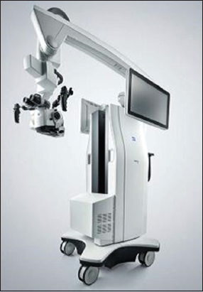
The KINEVO 900 surgical microscope is used for illuminating and magnifying the surgical area to support visualization in surgical procedures[
The QEVO® tool weighs 250 g, measures 12 cm in length, and has a shaft diameter of 3.6 mm and 45 angled view. This device is a completely resterilizable visualization tool for displaying anatomical details that are not accessible by microscope [
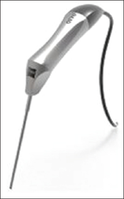
The official instructions for use were recommended using the automatic switch-off mode, in which light and data transmission is turned off after 40 s of permanent use. If the automatic switch-off mode is manually deactivated, the tool should not be used for at least 17 min after 8 min of permanent use so that the hand grip does not get overheated.
The aim of the cadaveric and clinical evaluation was assessing the usability of the QEVO® tool in terms of the quality of visualization, operability, and problems and pitfalls.
Description of the technical environment and description of the cadavers
At the Institute of Anatomy of the University of Regensburg in Germany, logistics and spatial relations were optimal for the surgical and technical evaluation. All surgical instruments were available (e.g., electric Trepan, osseous saw, instruments, and expendable items) to realistically simulate the surgical approaches. The interventions were conducted on an anatomical section table; illumination was provided by the light of the KINEVO 900 [
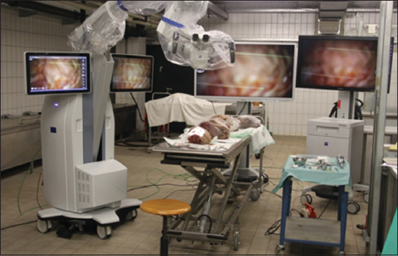
The procedures were captured in video recordings, either through the integrated camera of the KINEVO 900 or through handheld cameras. All cadavers examined were deep-frozen human heads. Although some icy patches were found during preparation, the bony structures and cerebral membranes were intact. Cranial nerves were in a very good state and comparable to in vivo findings. Despite the absence of blood, preparation of the vascular structures was authentic. The parenchyma of the brain of all specimens appeared clearly atrophied and partially liquefied. However, the state of preservation varied widely.
In the anatomical laboratory, four neurosurgical standard approaches were used: (1) the pterional approach to the parasellar region and the anterior skull base, (2) the retrosigmoid approach to the cerebellopontine angle (CPA), (3) the transcallosal approach to the third ventricle, and (4) the transnasal/transsphenoidal approach to the pituitary gland.
Clinical evaluation and case descriptions
For 3 weeks in June 2017, a fully equipped KINEVO 900 was used to conduct cranial neurosurgical procedures at our department. The QEVO® microinspection tool was used for almost every procedure so that its efficacy and usefulness could be assessed by the authors.
According to the approaches employed in the cadaveric study, we demonstrated the use of the microinspection tool in (1) a pterional approach to surgical clipping of an aneurysm of the middle cerebral artery (MCA), (2) a retrosigmoid approach to the CPA to remove a hemorrhagic cerebellar cavernoma, and (3) a transnasal/transsphenoid approach to remove a pituitary adenoma.
RESULTS
Pterional approach – a cadaveric specimen [ Video 1a ]
The skin was incised behind the hairline from the midline to just the front of the ear, the jaw muscle was then mobilized from directly behind and next to the eye, and the bone flap was removed. After opening the dura, the frontal and temporal lobes were exposed, and the Sylvian fissure was opened microsurgically. The frontal lobe was carefully elevated from the anterior skull base and held up by self-retaining retractors. The QEVO® tool was inserted, showing the sphenoid wing. The optic nerve was identified. The internal carotid artery (ICA) was lateral to the optic nerve and optic chiasm. The midline of the anterior skull base (crista galli and falx) was shown with the microinspection tool. Beneath the two frontal lobes, the midline (interhemispheric fissure), the contralateral olfactory nerve, and the contralateral olfactory groove were identified.
Pterional approach – the clinical case of a 61-year-old man with an innocent aneurysm of the left MCA [ Video 1b ]
After skin incision, a small frontotemporal bone flap was created, the dura incised, and the Sylvian fissure dissected. After insertion of the QEVO® tool, we inspected the ICA, the optic nerve, the Sylvian vessels, and the anterior skull base. The left MCA aneurysm was exposed in the bifurcation under microscopic vision. During temporary clipping of the M1 branch, the aneurysm was completely occluded with two slightly curved Yasargil clips. Before and after the clipping procedure, complete occlusion of the aneurysm was confirmed by micro-Doppler sonography and by intraoperative angiography with fluorescein sodium and indocyanine green. Again, the QEVO® tool was inserted to confirm the position of the tips of the clip branches. Then, the situs was closed.
Retrosigmoid approach in a cadaveric specimen [ Video 2a ]
The skin was incised for approximately 8 cm in almost linear fashion directly above the mastoid groove behind the ear. Craniotomy measuring approximately 2–3 cm in width was conducted below the junction of the transverse sinus and sigmoid sinus. The dura was opened under microscopic view showing the lateral part of the cerebellar hemisphere. The cerebellum was gently pushed in medial direction with a handheld retractor to gain access to the CP. The QEVO® tool was inserted to access the cranial nerves N. V, N. VI, N. VII, N. IIX, N. IX, N. X, and N. XI. Consecutively, the internal auditory canal, the jugular foramen, and the underpart of the tentorium were inspected endoscopically.
Retrosigmoid approach – the clinical case of a 37-year-old woman with a hemorrhagic cavernoma in the left lateral cerebellar hemisphere [ Video 2b ]
The patient was in prone position with elevated upper body, and the head was turned to the left and fixed in the head clamp. After skin incision behind the left ear and dissection of the neck muscles, a burr hole was placed directly on the asterion followed by elevation of a bone flap measuring 2 cm. The dura was incised, a self-retracting spatula inserted, and the CPA exposed. The endoscopic view of the QEVO® tool showed the N.V, N.VII, N.IIX, jugular foramen, and internal auditory canal. The hemorrhagic area on the cerebellar surface was identified with the QEVO® tool. The cavernoma was removed microsurgically, and the dissection cavity was inspected for any remnants with the QEVO® tool.
Transnasal/transsphenoidal approach in a cadaveric specimen [ Video 3a ]
The right nostril was entered first with the QEVO® tool, and the natural ostium of the sphenoid bone cave was exposed medially to the middle turbinate. The ostium was then widened with a punch. Then, with the QEVO® tool in the left nostril, the mucosa and parts of the bony septum were removed from the right side with a punch. The sphenoid sinus was entered and the sellar floor was identified. Finally, the sellar floor was opened and partially removed to expose the pituitary gland.
Transnasal/transsphenoidal approach – the clinical case of a 52-year-old woman with a macroadenoma of the pituitary gland [ Video 3b ]
According to our interdisciplinary standard procedure, most single procedures of this surgical intervention were conducted with a conventional 0° angled endoscope. The QEVO® was inserted repeatedly to assess its practicability in this approach.
First, the right ostium to the sphenoid bone cavity was entered to inspect the sphenoid sinus and sellar floor. After opening of the sellar floor, sterile saline was injected into the adenoma capsule to show the excellent visual field under water.
Transcallosal approach in a cadaveric specimen [ Video 4 ]
The right-sided craniotomy was situated around the coronal suture. The dura was opened in a trapdoor fashion based on the sagittal sinus, allowing a microscope view parallel to the falx cerebri. The ipsilateral hemisphere was gently retracted. The corpus callosum was readily identified by its glistening white pallor. After blunt opening, the right-sided lateral ventricle was entered. The QEVO® tool enabled the visualization and illumination of the convoluted lateral ventricle anatomy and the generous exposure of all surgically relevant structures. The small shaft diameter of the QEVO® tool enabled easy and atraumatic passing through the foramen of Monro. Visualization of the very narrow third ventricle looking at the mammillary bodies and at the tip of the basilar artery was uncomplicated.
DISCUSSION
During neurosurgical procedures, a look “around the corner” would often offer valuable additional information. Especially, in skull base surgery, the microvasculature of a tumor is difficult to expose, or tiny remnants are overseen or accidentally left behind, for example, below the sphenoid wing or in the contralateral olfactory groove.[
The above scenarios are just a few situations in which a closer look or a better depiction of hidden structures would offer the surgeon more anatomical details, thus increasing the safety and success of the procedure. The hand-held 45° angled microinspection tool QEVO® was evaluated under ex vivo and in vivo conditions using various cranial approaches and procedures, including aneurysm clipping in the anterior circulation, retrosigmoid approaches with identification of the tentorial bridging veins, cranial nerves, and foramen, transsphenoidal/pituitary surgery, and intraventricular visualization. Furthermore, the resection cavity was checked for remnants after removal of a hemorrhagic cerebellar cavernoma. Basically, our aim was to evaluate the handling and safety of this tool, the quality of the visualization patterns, as well as appraisal of its potential benefit and usability as an endoscopic adjunct.
Both in the cadaveric study and clinical cases, the QEVO® tool was always used by the author or under his supervision. The handling was excellent because the tool is shaped and angled in such a way that it is comparable to a conventional surgical aspirator. Thus, surgeons can adapt to the device very quickly. The visual quality is bright and detailed and hence excellent. We did not encounter any loss of information because the light intensity of the integrated camera can be easily changed on the touch screen of the KINEVO microscope. The tool is a real plug-in device and can be resterilized; thus, the scrub does not need to cover the QEVO® tool with a drape. However, the most important characteristic is the 45° angled view with such excellent visual resolution that even fine nervous branches of the accessory nerve can be identified.
In pterional approaches, the QEVO® tool enabled the view between both optic nerves and below the optic chiasm. This view is pleasantly surprising for the microscope-trained eye and all the more important because remaining tumor tissue arising from the sphenoid plane or the pituitary is often located below the chiasm and concealed from direct view. Furthermore, the often cumbersome task of gaining a perspective of the frontal curvature along the temporal skull base was straightforward in all procedures due to the 45° optics.
In transsphenoidal approaches to the sellar region, the handling of the QEVO® tool was good. For this approach, 30° optics are traditionally used. Nevertheless, the changeover and readjustment to 45° optics were intuitive and possible very quickly. The exposure of all relevant structures within the sphenoid sinus was easy. Working within the sella was feasible, despite or perhaps because of the 45° optics.
Endoscopy in neurosurgery is not new and has been applied, evaluated, and improved during the past decades.[
However, the QEVO® microinspection tool also has some flaws that will hopefully be addressed by the developers in future: (1) at present, no external holder is offered that allows the fixation of the tool to the ring so that both hands can be used, (2) endoscopic visualization of longer duration is not possible due to the integrated automatic shutdown (max. 8 min), (3) the relatively short length of 12 cm of the tool makes handling difficult in a deep surgical situs, (4) no working channel is included which almost disables its use in ventriculostomy, (5) no eligibility of a 0°/30° optic, (6) the use of the tool is only possible in combination with the KINEVO surgical microscope; this restriction may be logistically problematic in procedures in which microscopes are not usually employed (such as full-endoscopic pituitary surgery), and (7) only white light is emitted so that fluorescence cannot be visualized through the QEVO® tool.
Furthermore, since right-handed neurosurgeons are used to holding the aspirator in the left hand and also to using it as a tool for dissection, it might be worthwhile to consider equipping the QEVO® microinspection tool with an additional suction device.
In this cadaveric and clinical case series, we present our first experiences illustrated with video footage. In our opinion, the QEVO® microinspection tool is a reliable, innovative, and beneficial adjunct that increases the safety and success of complex neurosurgical interventions, in both vascular and oncologic procedures. Despite some restrictions, this tool enhances the modern neurosurgical armamentarium.
CONCLUSION
The microinspection tool QEVO® has good haptic properties, can be easily held, and offers a high-quality view of fine morphological structures. The 45° angled view enables the depiction of hidden anatomical details that cannot be displayed by the outside view of the surgical microscope.
Ethical approval
All procedures performed in studies involving human participants were in accordance with the ethical standards of the institutional and/or national research committee (name of institute/committee) and with the 1964 Helsinki Declaration and its later amendments or comparable ethical standards.
Informed consent: informed consent was obtained from all individual participants included in the study.
Financial support and sponsorship
Nil.
Conflicts of interest
K.-M.S, A.B. and J.H. have received honoraria and travel funds from Carl Zeiss Meditec, Germany.
Videos available on: www.surgicalneurologyint.com
References
1. Apuzzo ML, Heifetz MD, Weiss MH, Kurze T. Neurosurgical endoscopy using the side-viewing telescope. J Neurosurg. 1977. 46: 398-400
2. Barone DG, Lawrie TA, Hart MG. Image guided surgery for the resection of brain tumours. Cochrane Database Syst Rev. 2014. 1: CD009685-
3. Cohen AR, Perneczky A, Rodziewicz GS, Gingold SI. Endoscope-assisted craniotomy: Approach to the rostral brain stem. Neurosurgery. 1995. 36: 1128-9
4. Demerdash A, Rocque BG, Johnston J, Rozzelle CJ, Yalcin B, Oskouian R. Endoscopic third ventriculostomy: A historical review. Br J Neurosurg. 2017. 31: 28-32
5. Limbrick DD, Baird LC, Klimo P, Riva-Cambrin J, Flannery AM. Pediatric hydrocephalus: Systematic literature review and evidence-based guidelines. Part 4: Cerebrospinal fluid shunt or endoscopic third ventriculostomy for the treatment of hydrocephalus in children. J Neurosurg Pediatr. 2014. p.
6. Meditec CZLast accessed on 2018 Apr 14. Available from: https://www.zeiss.de/meditec/produkte/neurochirurgie/operationsmikroskope/kinevo-900.html .
7. Nguyen-Huynh A, Blevins NH, Jackler RK. The challenges of revision skull base surgery. Otolaryngol Clin North Am. 2006. 39: 783-99

