Tools

Nancy E. Epstein
  1. Professor of Clinical Neurosurgery, School of Medicine, State University of New York at Stony Brook, New York, and Chief of Neurosurgical Spine and Education, NYU Winthrop Hospital, NYU Winthrop NeuroScience/Neurosurgery, Mineola, New York 11501, United States.

DOI:10.25259/SNI_394_2019

Copyright: © 2019 Surgical Neurology International This is an open-access article distributed under the terms of the Creative Commons Attribution-Non Commercial-Share Alike 4.0 License, which allows others to remix, tweak, and build upon the work non-commercially, as long as the author is credited and the new creations are licensed under the identical terms.

How to cite this article: Nancy E. Epstein. Review of Treatment Options for Smaller Benign Cranial Meningiomas: Observation, Stereotactic Radiosurgery, and Rarely, Open Surgery. 23-Aug-2019;10:167

How to cite this URL: Nancy E. Epstein. Review of Treatment Options for Smaller Benign Cranial Meningiomas: Observation, Stereotactic Radiosurgery, and Rarely, Open Surgery. 23-Aug-2019;10:167. Available from: http://surgicalneurologyint.com/surgicalint-articles/9590/

Date of Submission
06-Jul-2019

Date of Acceptance
11-Jul-2019

Date of Web Publication
23-Aug-2019

Abstract

Background: MR/CT documented smaller cranial meningiomas in asymptomatic patients are often followed for years without requiring any intervention. Only a subset of patients who become symptomatic attributed to significant tumor growth, edema and/or mass effect may require stereotactic radiosurgery (SRS), and rarely, open surgery. Clearly, the decision for choosing any treatment modality must be made on a case by case basis and include an analysis of risks vs. benefits to the individual patient.

Methods: Patients with smaller benign asymptomatic meningiomas are followed with sequential MR studies that typically document lack of tumor progression, edema, or mass effect. Those who become symptomatic with the typical triad (i.e. headaches, seizures, or visual loss) and other focal neurological deficits may warrant SRS, and only occasionally, open surgery. Surgery may indeed be warranted in the presence of certain mitigating factors, (e.g. young age, lesions located adjacent to by not yet invading critical structures etc.).

Results: This review focused largely on smaller benign asymptomatic meningiomas. The non-operative/ conservative management vs. use of SRS vs. open surgery in select cases are discussed, along with a review of the morbidity/mortality of the respective interventions.

Conclusion: There are multiple treatment options for patients with smaller asymptomatic cranial meningiomas. SRS may be warranted for those who exhibit tumor growth, increasing edema, and/or mass effect. Only rarely is open operative intervention necessary; this must include consideration of other factors that may warrant early surgery. Notably, the 5-year survival rates for SRS ranged from 95.2% - 97%, while the 10-year survival rates varied from 88.6% - 94%.

Keywords: Cranial Meningiomas, Conservative Observation, Stereotactic Radiosurgery, Open Surgery, Symptoms, Headaches, Seizures, Visual Changes

INTRODUCTION

Benign cranial meningiomas are often followed for years in asymptomatic patients. Only a subset of patient who become symptomatic from documented tumor growth, onset of significant progressive edema, and/or mass effect, or with other unique clinical features placing patients at increased risk (e.g. young age, critical tumor location - near optic chiasm/skull base, other) may warrant stereotactic radiosurgery (SRS), or rarely, open surgery [ Tables 1 , 2 ].


Table 1:

Treatment of Meningiomas 1995-2011.


Table 2:

Treatment of Meningiomas 2012-2019.


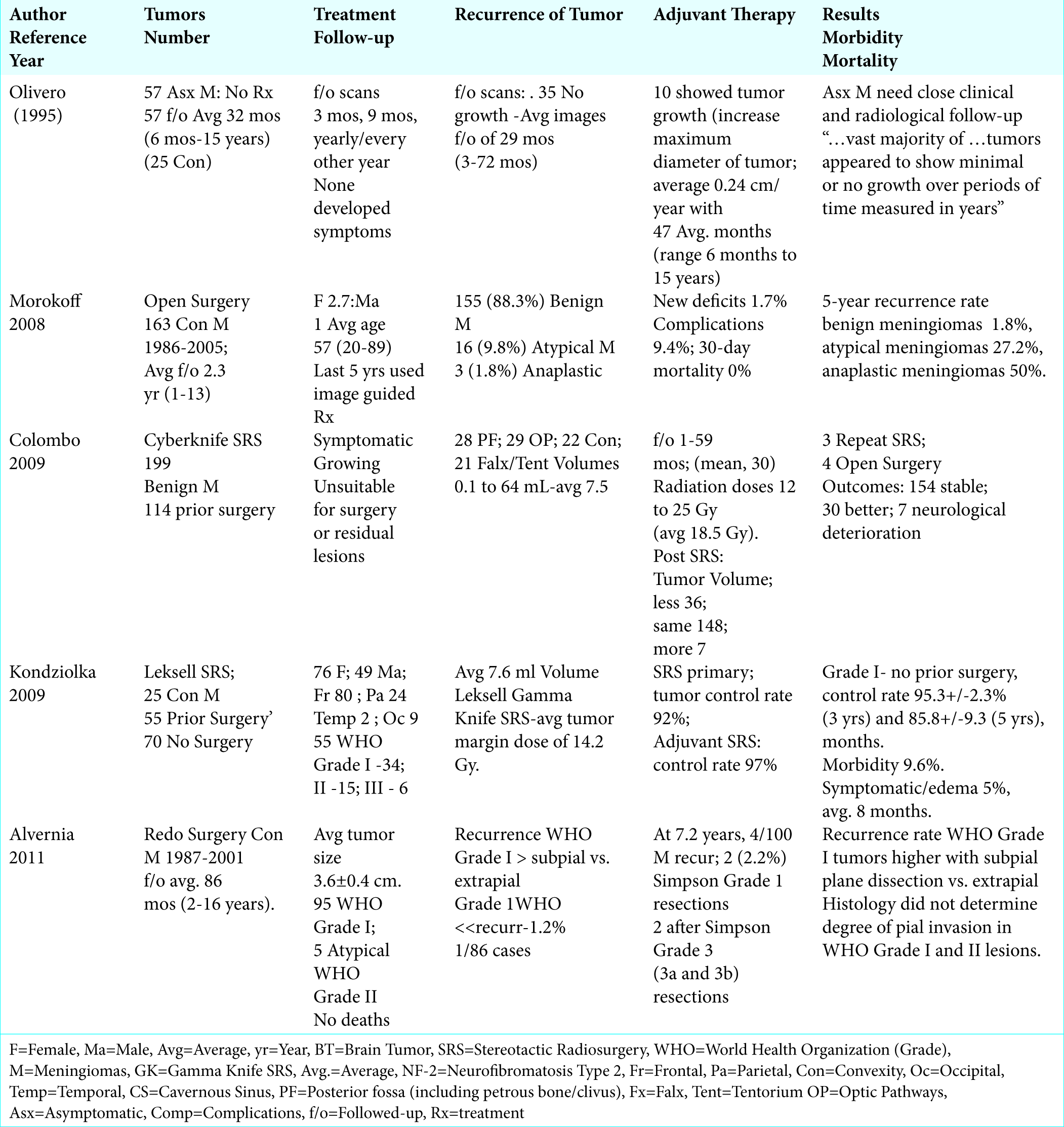
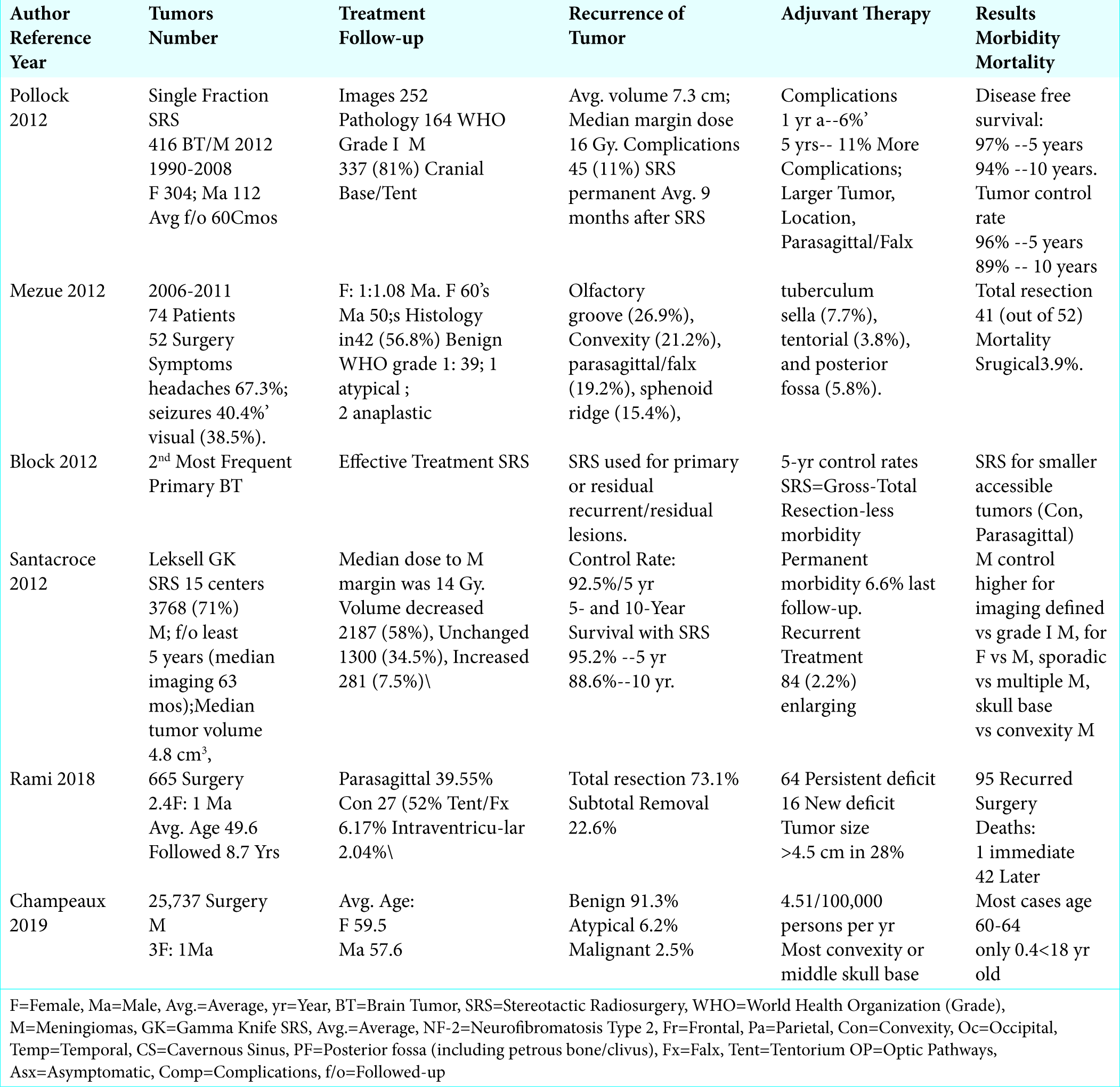
Symptoms of Cranial Meningiomas and Karnofsky Scales Before and After Surgery for Recurrent Lesions

In Mezue’s (2012) series of 74 patients with meningiomas, symptoms included; headaches (67.3%), seizures (40.4%), and visual impairment (38.5%), along with increased motor/sensory deficits [ Table 2 ].[ 6 ] For the 100 recurrent meningiomas from Alvernia et al. 2011 series, the authors reviewed of the Karnofsky Performance Scale scores before and after secondary surgery; 92.6 ± 4.6 and 97.9 ± 2.2 [ Table 1 ].[ 1 ]

Demographics of Cranial Meningiomas

More females than males develop cranial meningiomas, with ratios varying from 1.08:1 to 2.4:1 to 3:1 [ Table 2 ].[ 3 , 6 , 10 ] The average age for all patients undergoing cranial surgery for meningiomas ranged from 49.6 – 64 years of age; males were typically in their fifties (average age 57.6), while females were usually in their late fifties/early sixties (average age 59.5) [ Table 2 ].[ 3 , 6 , 10 ]

Location for Meningiomas

Location of Cranial Meningiomas in General

Cranial meningiomas occur in multiple locations [ Tables 1 , 2].[ 3 , 4 , 6 , 9 , 10 ] Colombo et al. (2009) performed

Cyberknife SRS on 199 lesions; cavernous sinus (99); posterior fossa (28), petrous bone/clivus/anterior optic pathways (29), convexity (22), and falx/tentorium (21) [ Table 1 ].[ 4 ] The 74 meningiomas in Mezue et al. (2012) were located in the; olfactory groove (26.9%), convexity (21.2%), parasagittal/ falx (19.2%), tentorial ridge/sphenoid ridge (15.4%), tuberculum sellae (7.7%), tentorium (3.8%), and posterior fossa (5.8%) [ Table 2 ].[ 6 ] Out of Pollock et al. 2012 series of 416 cranial meningiomas, 337 (81%) were located at the cranial base/tentorium [ Table 2 ].[ 9 ] Rami et al, (2018), found that 665 meningiomas (36% out of 2047 with cranial lesions) originated in the; parasagittal (39.55%), convexity (27.52%), tentorial/falx (6.17%), and intraventricular (2.04%) locations [ Table 2 ].[ 10 ] When Champeaux et al. (2019) evaluated the French database (Programme de Medicalization des Systèmes d’Information (PMSI)) that included 25,737 surgically managed meningiomas (2008-2016: equivalent to 4.5/100, 000 persons/year), the majority were located on the convexity or in the middle fossa/skull base [ Table 2 ].[ 3 ]

Frequency and Locations of Convexity Meningiomas

In two respective series (2012 and 2018), 21.2%-27.5% of 739 meningiomas were convexity lesions [ Table 2 ].[ 6 , 10 ] In Kondziolka et al. (2009), convexity meningiomas were located in the; frontal (80 patients), parietal (24 patients), temporal (12 patients), and occipital (9 patients) regions [ Table 1 ].[ 5 ]

Average Initial and Recurrent Meningioma Size

The average initial size of unoperated cranial meningiomas in Rami et al. (2018) series was > 4.5 cm in 28.07% of 665 cranial meningiomas [ Table 2 ].[ 10 ] The average recurrent size of cranial meningiomas was 3.6 ± 0.4 cm in Alvernia et al. 2011 study, an average of 86 mos. (2-16 years) following the index resection of 100 convexity meningiomas (1987-2001) [ Table 1 ].[ 1 ]

Conservative Follow-Up of Asymptomatic Benign Meningiomas

Many benign cranial meningiomas may be followed conservatively without the necessity for SRS or open surgery. In 1995, Olivero et al. evaluated the growth rate for asymptomatic cranial meningiomas in 57 patients; notably, 25 of 57 tumors were convexity lesions [ Table 1 ].[ 8 ] None of the patients became symptomatic from an enlarging tumor during the follow-up period averaging 32 mos. (range 6 mos.-15 years). The protocol for re-scanning documented meningiomas was originally every 3 months, then every 9 months, then once/year, and finally every other year. Thirty-five patients showed no growth in their tumor size over an average follow-up duration of 29 months (range 3-72 months). Ten patients showed tumor growth, including an average increased maximum tumor diameter of 0.24 cm per year, over the average follow-up of 47 months (range 6 months to 15 years). The authors concluded that although patients with asymptomatic meningiomas need close clinical/radiological follow-up, the “…vast majority of these tumors appeared to show minimal or no growth over periods of time measured in years.”

Stereotactic Radiosurgery (SRS) for Benign Meningiomas

The Cyberknife or Gamma Knife (Leksell) SRS systems have gained increased favor over open surgery for the treatment of smaller primary, residual, and/or recurrent benign meningiomas. SRS is typically utilized when tumors are; located in eloquent areas, show little primary tumor growth, demonstrate mild/moderate non-progressive peritumoral edema, and/or where patients are neurologically stable or show only mild progression of neurological symptoms/signs. In 2009, Colombo et al. used Cyberknife SRS (CyberKnife; Accuray, Inc., Sunnyvale, CA) to treat 199 benign cranial meningiomas over an average period of 30 mos. [ Table 1 ].[ 4 ] Lesions were symptomatic or had grown, and were considered “unsuitable for open surgery” due to their critical location, or represented post-surgical residual or recurrent tumors. Clearly, the choice to utilize SRS must be made on a case by case basis, and the risks vs. benefits must be carefully considered when compared to open surgery especially for patients with unique additional mitigating factors (e.g. young age, lesions near but not yet invading critical structures, etc.).

Patients Undergoing SRS With/Without Prior Surgery

Three series utilized SRS to treat a total of 615 benign cranial meningiomas; 424 never had any surgery, while 191 had prior surgery [ Tables 1 , 2 ].[ 5 , 6 , 9 ] In Kondziolka et al. (2009), 125 patients with convexity meningiomas were treated with SRS over 18 years; 55 had prior surgery, while 70 had no previous operation (SRS or open) [ Table 1 ].[ 5 ] In Pollock et al., 416 patients (published 2012; series 1990-2008) underwent single fraction radiosurgery (SRS), and were followed an average of 60 mos.; 114 had prior surgery [ Table 2 ].[ 9 ] The 74 patients in Mezue et al. series (published 2012; series 2006 – 2011) underwent SRS; 52 (70.3%) had prior open surgery [ Table 2 ].[ 6 ]

Average Volumes of Cranial Meningiomas Treated with SRS

In four series, a total of 4508 cranial meningiomas ranging in size from 0.1 to 7.6 cm3 were treated with SRS [ Tables 1 , 2 ].[ 4 , 5 , 9 , 11 ] Colombo et al. (2009) used Cyberknife SRS to treat 199 tumors ranging from 0.1 to 64 mL (mean, 7.5 mL) [ Table 1 ].[ 4 ] Pollock in 2012 used single-fraction SRS to treat 416 cranial meningiomas averaging 7.3 cm3 in size [ Table 2 ].[ 9 ] Kondziolka et al. (2009) evaluated 125 patients using Gamma Knife (Leksell) SRS with a mean tumor volume of 7.6 ml [ Table 1 ].[ 5 ] Out of 3768 meningiomas treated with Gamma Knife SRS only, Santacroce et al. 2012 (NSGY) documented a median tumor volume or 4.8 cm3 [ Table 2 ].[ 11 ] Again, the decision to choose SRS vs. open surgery for cranial meningiomas must be determined on a case by case basis carefully considering the various risks vs. benefits of each modality, and the unique short and long-term sequelae for each treatment option.

SRS Radiation Doses

In several studies, SRS was typically administered for cranial meningiomas in single radiation doses averaging 14.0--18.5 Gy (range of from 12-25 Gy) [ Tables 1 , 2 ].[ 4 , 5 , 9 , 11 ] Kondziolka et al. (2009) performed SRS for 125 meningiomas using the Leksell Gamma Knife with a mean tumor margin dose of 14.2 Gy [ Table 1 ].[ 5 ] Colombo et al. (2009) employed Cyberknife SRS for 199 meningiomas using doses ranging from 12 to 25 Gy (mean, 18.5 Gy), with treatment isodoses varying from 70 to 90% [ Table 1 ].[ 4 ] For 150 patients with lesions greater than 8 mL in size and/or those located close to critical structures, the dose was delivered in 2 to 5 daily fractions. In 2012, Santacroce et al. evaluated the Gamma Knife SRS treatment of 3768 cranial meningiomas (71% of all brain tumors in series); the median dose to the tumor margin was 14 Gy [ Table 2 ].[ 11 ] Also in 2012, Pollock et al. treated 416 lesions with a single SRS dose averaging 16 Gy [ Table 2 ].[ 9 ]

Residual Volumes and 5 and 10-Year Survival Rates of Meningiomas After SRS

Several studies documented that SRS effectively treated primary, residual (e.g. postoperative), or recurrent cranial meningiomas; the vast majority demonstrated control or decrease in tumor size along with favorable 3, 5, and 10-year outcomes [ Tables 1 , 2 ].[ 2 , 4 , 5 , 9 , 11 ] Colombo et al. (2009) evaluated the results of Cyberknife SRS for 199 benign meningiomas over 1 to 59 months (mean/median 30 months) [ Table 1 ].[ 4 ] Tumor volume decreased in 36 patients, was unchanged in 148 patients, and increased in 7 patients; 3 required repeated radiosurgery, and 4 warranted open operations. Clinical results showed 154 remained clinically stable, 30 showed significant improvement, while 7 patients demonstrated neurological deterioration. When Kondziolka et al. (2009) evaluated 125 patients with meningiomas, 70 underwent primary radiosurgery, while 55 had prior open surgery; of these, 115 had serial imaging (92%) [ Table 1 ].[ 5 ] Tumor control rates at 3 and 5 years were 86.1+/-3.8% and 71.6+/-8.6%, respectively. For Grade I tumors, utilizing primary radiosurgery, the tumor control rate was 92%; with additional adjuvant radiosurgery, the control rate increased to 97%. For benign tumors (Grade I) without prior surgery, the actuarial tumor control rate increased to was 95.3%+/-2.3% at 3 yrs., and 85.8%+/-9.3 at 5 yrs. Delayed resection after radiosurgery was performed in 9 patients (7%) an average of 35 months after SRS. Interestingly, no patient developed a subsequent radiation- induced tumor. Santacroce et al. in 2012 looked at the results from 15 centers involving 3768 meningiomas (71% of the of the total brain tumors) followed at least for 5 years after treatment with Gamma Knife SRS: of these, 2187 lesions (58%) decreased in size, 1300 remained unchanged (34.5%), and 281 lesions (7.5%) increased in size yielding an overall control rate of 92.5% [ Table 2 ].[ 11 ] Further, the control rate/ overall survival was 92.5% at 5 postoperative years, while it was 88.6% at 10 years. In Pollock et al. 2012 study involving 416 patients treated with SRS, the disease-free survival rates were 97% at 5 years, and 94% at 10 years, while the 5- and 10-year local tumor control rates were 96% and 89% respectively [ Table 2 ].[ 9 ] Of interest, the smaller volume, non- operated cranial base or tentorial meningiomas had the best outcomes after single-fraction SRS. Further, Bloch et al. (2012) observed that 5-year control rates with SRS were comparable to initial gross-total resection, but with less morbidity; they recommended initially utilizing SRS to treat smaller readily accessible tumors (e.g. located superficially, at the convexity, or in parasagittal locations) [ Table 2 ].[ 2 ].

SRS Complications/Morbidity

The complication/morbidity rates of SRS for treating benign cranial meningiomas varied from 6.6% to 11% [ Tables 1 , 2 ].[ 5 , 9 , 11 ] In Kondziolka et al. (2009), SRS was utilized in 125 patients, with an overall morbidity rate of 9.6% [ Table 1 ].[ 5 ] Symptomatic peritumoral imaging changes compatible with edema or adverse radiation effects developed in 5%, at a mean of 8 post SRS treatment months. Delayed resection after SRS was warranted in 9 patients (7%) an average of 35 months post-treatment; none developed a subsequent radiation-induced tumor. Out of Santacroce et al. (2012) 3768 tumors, only 84 (2.2%) enlarging tumors required further treatment; the permanent morbidity rate was 6.6% at last follow-up [ Table 2 ].[ 11 ] For Pollock et al. (2012) 416 cranial meningiomas undergoing SRS, 45 patients (11%) developed permanent radiation-related complications a median of 9 months after SRS, and the 1- and 5-year radiation-related complication rate was 6% and 11%, respectively [ Table 2 ].[ 9 ] Risk factors for permanent radiation-related complications included; increasing tumor volume, parasagittal/falx/convexity lesion locations, male sex, and previous surgery. Hence, the morbidity of SRS must be carefully weighed against the risks of open surgery in every case. This is particularly true for younger patients with potential long-term survivals who may develop not only recurrent lesions (e.g. in the absece of initial resection), but also long-term radiation-induced sequelae complications.

Open Surgery for Meningiomas

Demographics for Surgically Managed Meningiomas

For Morokoff et al. (published 2008; series1986-2005) series of 163 patients undergoing open surgery for cranial meningiomas, including 22% convexity lesions, and followed an average of 2.3 years (range 1-13 years), patients averaged 57 years of age (range 20-89 years old), and the female to male ratio was 2.7:1 [ Table 1 ].[ 7 ]

Majority of Cranial Meningiomas Pathologically Documented as Benign (88.3%-91.3%)

The vast majority of cranial meningiomas are pathologically confirmed to be benign [ Tables 1 , 2 ].[ 3 , 7 , 9 ] Following open surgical resection of 163 cranial meningiomas. Morokoff et al. (2008) pathologically confirmed that 144 (88.3%) lesions were benign, 16 (9.8%) were atypical, while only 3 (1.8%) were anaplastic/malignant [ Table 1 ].[ 7 ] In 2012, Pollock et al. had pathological confirmation that 164 cranial meningiomas were benign World Health Organization grade I meningiomas vs. 252 diagnosed as benign on images alone [ Table 2 ].[ 9 ] When Champeaux et al. (2019) evaluated 25,737 surgically confirmed cranial meningiomas predominantly involving the convexity or middle skull base, 91.3% were benign lesions, 6.2% were atypical or of uncertain pathology, while 2.5% were malignant [ Table 2 ].[ 3 ]

Extent of Surgical Resection

The extent of primary surgical resection of benign cranial meningiomas varies [ Table 2 ].[ 6 , 10 ] Rami et al. (2018) noted gross total resections were achieved in 73.1% of 665 meningiomas, while subtotal removal was performed in 22.6% of patients [ Table 2 ].[ 10 ] In Mezue series (2018), gross total resection was achieved in 41out of 52 patients [ Table 2 ].[ 6 ]

Meningioma Recurrence Following Open Surgery

There was an overall 14.2% incidence of recurrent cranial meningiomas following index open surgery for resection of tumors from all pathological groups [ Tables 1 , 2 ]. Specifically, the frequency of recurrent tumors ranged from 1.2% - 1.8% for benign lesions, to 27.2% for atypical, and 50% for anaplastic tumors 50% [ Tables 1 , 2 ].[ 1 , 7 , 10 ] The 5-year recurrence rate following open surgery for benign meningiomas in Morokoff et al. series (2008) was 1.8%, for atypical meningiomas was 27.2%, and anaplastic meningiomas was 50% [ Table 1 ].[ 7 ] When Alvernia et al. 2011 looked at recurrence rates for 100 surgically resected convexity meningiomas (1987-2001) after a mean follow-up of 7.2 years, 4 recurred; 2 (2.2%) were originally Simpson Grade 1, and 2 were initially Simpson Grade 3 (3a and 3b) (2.2%) [ Table 1 ].[ 1 ] Simpson Grade 1/WHO tumors had lower recurrence rates (1.2%; 1 of 86 cases). Notably, the recurrence rate of WHO Grade I tumors was higher in the subpial group vs. the extrapial group (p = 0.025), yet the histological type did not determine the degree of pial invasion for WHO Grade I and II lesions. For Rami et al. (2018), 95 of 665 lesions recurred over an average 8.7 years duration [ Table 2 ].[ 10 ]

Pathology of Recurrent Meningiomas with History of Prior Surgery

Although most recurrent meningiomas were initially benign Grade I tumors, those originally presenting with atypical and anaplastic changes were the most likely to recur [ Tables 1 , 2 ].[ 1 , 5 , 6 ] In 2009, Kondziolka et al. evaluated 55 patients with prior surgery who had recurrent lesions; Grade I WHO tumors recurred in 34 patients (54.5%), WHO Grade II tumors recurred in 15 (27.2%) patients, and WHO Grade III tumors recurred in 6 patients (10.9%) [ Table 1 ].[ 5 ] When Alvernia et al. (2011) evaluated 100 recurrent lesions following prior open surgical resections, 95 were originally benign (WHO Grade I) tumors, while 5 were now atypical lesions (WHO Grade II) [ Table 1 ].[ 1 ] In Mezue et al. series (2012), 52 (70.3%) of 74 patients with intracranial meningiomas treated from 2006–2011 had undergone prior surgery [ Table 2 ].[ 6 ] Histology was available in 42 (56.8%) patients; 39 (92.8%) had benign WHO Grade 1 lesions, while 1 (2.4%) had an atypical, and 2 (4.8%) had anaplastic lesions.

Morbidity for Surgically Treated Meningiomas

Out of 828 patients from two series, following open resection of cranial meningiomas, new postoperative deficits were incurred in from 1.7-2.4% of patients, residual deficits were seen in 9.6%, and overall complications were noted in 9.4% of patients [ Tables 1 , 2 ].[ 7 , 10 ] Specifically, in Morokoff et al. study (2008), the incidence of new postoperative neurological deficits in 163 patients was 1.7%, while the overall complication rate was 9.4% [ Table 1 ].[ 7 ] Out of Rami et al. (2018) series of 665 cranial meningiomas, 64 (9.6%) had persistent postoperative neurological deficits, and 16 (2.4%) patients exhibited new neurological dysfunction [ Table 2 ].[ 10 ] Notably, the relative low morbidity rate for open surgical resection of meningiomas must be taken into account when considering operative vs. SRS treatment options, particularly in younger patients with critically located lesions. Short-term morbidity of open surgery may in some instances prove less risky than potential long-term sequelae of SRS.

Mortality for Surgically Treated Meningiomas

Mortality rates following open surgical resection of cranial meningiomas ranged from 0-1.7% in the short- term (e.g. 30 days), and up to 6.3% over an average of 8.7 postoperative years [ Tables 1 , 2 ].[ 1 , 6 , 7 , 10 ] The 30-day mortality rate for Morokoff et al. (2008) 163 patients undergoing open surgery for cranial meningiomas was 0% [ Table 1 ].[ 7 ] In Alvernia et al. 2011 study of recurrence rates for100 initially surgically resected meningiomas, there were no surgical deaths [ Table 1 ].[ 1 ] The overall mortality rate in Mezue et al (2018) surgical series was 3.9% [ Table 2 ].[ 6 ] Out of Rami et al (2018) 665 operated meningiomas, there were 11 (1.7%) deaths immediately postoperatively, which increased to 42 (6.3%) cases over the average 8.7-year follow-up [ Table 2 ].[ 10 ]

CONCLUSION

Smaller benign cranial meningiomas in asymptomatic patients are often followed for years without requiring any treatment. However, patients who become symptomatic with MR/CT-documented significant tumor growth, peri-tumoral edema, mass effect, new neurological deficits or with other significant confounding factors (i.e. young age, lesions in/near eloquent locations) may warrant stereotactic radiosurgery (SRS), and only rarely, open surgery. Notably, the 5 year and 10-year survival rates following SRS are excellent; 5-year survivals ranged from 95.2% - 97%, and 10-year survivals from 88.6% - 94%. However, for SRS vs. open surgery, the SRS-related long-term sequelae must be carfully weighed against the short-term risks of direct open tumor resection.

References

1. Alvernia JE, Dang ND, Sindou MP. Convexity meningiomas: study of recurrence factors with special emphasis on the cleavage plane in a series of 100 consecutive patients. J Neurosurg. 2011. 115: 491-8

2. Bloch O, Kaur G, Jian BJ, Parsa AT, Barani IJ. Stereotactic radiosurgery for benign meningiomas. J Neurooncol. 2012. 107: 13-20

3. Champeaux C, Weller J, Katsahian S. Epidemiology of meningiomas. A nationwide study of surgically treated tumors on French medico-administrative data. Cancer Epidemiol. 2019. 58: 63-70

4. Colombo F, Casentini L, Cavedon C, Scalchi P, Cora S, Francescon P. Cyberknife radiosurgery for benign meningiomas: short-term results in 199 patients. Neurosurgery. 2009. 64: A7-13

5. Kondziolka D, Madhok R, Lunsford LD, Mathieu D, Martin JJ, Niranjan A. J Stereotactic radiosurgery for convexity meningiomas. Neurosurg. 2009. 111: 458-63

6. Mezue WC, Ohaegbulam SC, Ndubuisi CA, Chikani MC, Achebe DS. Management of Intracranial Meningiomas in Enugu, Nigeria. Surg Neurol Int. 2012. 3: 110-

7. Morokoff AP, Zauberman J, Black PM. Surgery for convexity meningiomas. Neurosurgery. 2008. 63: 427-33

8. Olivero WC, Lister JR, Elwood W. The natural history and growth rate of asymptomatic meningiomas: a review of 60 patients. J Neurosurg. 1995. 83: 222-4

9. Pollock BE, Stafford SL, Link MJ, Brown PD, Garces YI, Foote RL. Single-fraction radiosurgery of benign intracranial meningiomas. Neurosurgery. 2012. 71: 604-12

10. Rami A, Suzan E, Rafeed A, Nooruddeen AM, Lamees A, Duaa A. The Sovereignty of Primary Cranial Tumors-Meningiomas: Vetting the Cardinal Epidemiological Features. Med Arch. 2018. 72: 434-438

11. Santacroce A, Walier M, Régis J, Liščák R, Motti E, Lindquist C. Long-term tumor control of benign intracranial meningiomas after radiosurgery in a series of 4565 patients. Neurosurgery. 2012. 70: 32-9

Leave a Reply

Your email address will not be published. Required fields are marked *