

- Department of Neurosurgery, Grant Medical Center, Columbus, Ohio, USA
- Department of Orthopedics, Grant Medical Center, Columbus, Ohio, USA
Correspondence Address:
Stephen T. Susa
Department of Orthopedics, Grant Medical Center, Columbus, Ohio, USA
DOI:10.4103/sni.sni_347_17
Copyright: © 2018 Surgical Neurology International This is an open access article distributed under the terms of the Creative Commons Attribution-NonCommercial-ShareAlike 3.0 License, which allows others to remix, tweak, and build upon the work non-commercially, as long as the author is credited and the new creations are licensed under the identical terms.How to cite this article: Stephen T. Susa, Chris S. Karas, Nathaniel K. Long. Treatment of glenohumeral arthritis pain utilizing spinal cord stimulation. 01-Mar-2018;9:54
How to cite this URL: Stephen T. Susa, Chris S. Karas, Nathaniel K. Long. Treatment of glenohumeral arthritis pain utilizing spinal cord stimulation. 01-Mar-2018;9:54. Available from: http://surgicalneurologyint.com/surgicalint-articles/treatment-of-glenohumeral-arthritis-pain-utilizing-spinal-cord-stimulation/
Date of Submission
15-Sep-2017
Date of Acceptance
02-Jan-2018
Date of Web Publication
01-Mar-2018
Abstract
Background:Dorsal column stimulation may be utilized to treat non-neuropathic pain attributed to glenohumeral arthritis.
Case Description:An 84-year-old female presented with right shoulder pain for 3 years. She was diagnosed with glenohumeral arthritis and a complete loss of the joint space. She was treated with a dorsal column stimulator, requiring the electrodes to be placed from the inferior aspect of C3 to the superior aspect of T1. Six weeks postoperatively, she reported >90% coverage of her shoulder pain, demonstrated increased right arm function, and a reduction in her use of narcotics.
Conclusion:Dorsal column stimulation of C3–T1 proved to be an effective alternative treatment for drug-resistant glenohumeral arthritis in an 84-year-old female with a complete loss of the joint space.
Keywords: Arthritis, dorsal column stimulation, glenohumeral arthritis, pain management, spinal cord stimulation
INTRODUCTION
Glenohumeral arthritis impacts approximately 32% of individuals over the age of 60.[
CASE REPORT
Clinical parameters
An 84-year-old female presented with pain and stiffness in her right shoulder of 3 years duration. She was diagnosed with glenohumeral joint arthritis, a complete loss of the joint space, the inability to abduct her arm more than 15 degrees, the loss of active or passive motion of her right arm (i.e. frozen shoulder), and severe pain. Prior conservative management had been unsuccessful including several cortisone injections and narcotics. Ultimately, she was offered the placement of a cervical C3–T1 spinal cord stimulator.
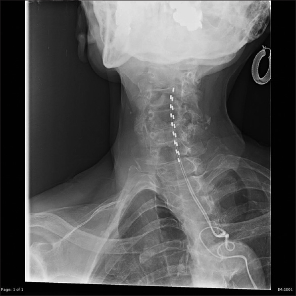
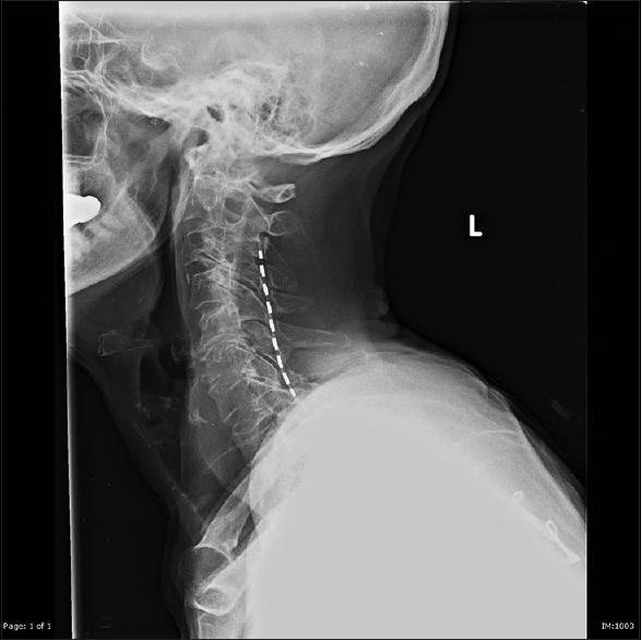
Spinal cord stimulator
A dual-lead, right-sided 16-electrode spinal cord stimulator was placed between the C3–T1 levels (i.e. to cover the C4–C8 roots) utilizing intraoperative X-ray [Figures
DISCUSSION
Glenohumeral arthritis results from the reduction of the joint space between the humerus and the glenoid that leads to grinding friction/inflammation, causing stiffness and pain.[
Rather, a right-sided C3-T1 spinal cord stimulator (SCS) (i.e., dorsal column stimulation of the C4–C8 roots), typically used for treating chronic neuropathic pain, was implanted.[
Financial support and sponsorship
Nil.
Conflicts of interest
There are no conflicts of interest.
References
1. .editors. American Academy of Orthopaedic Surgeons: The Treatment of Glenohumeral Arthritis: Guideline and Evidence Report. Rosemont, IL: American Academy of Orthopaedic Surgeons; 2009. p.
2. . American Academy of Orthopaedic Surgeons: Arthritis of the Shoulder. Rosemont, IL: American Academy of Orthopaedic Surgeons; 2013. p.
3. Aszmann OC, Dellon AL, Birely BT, Mcfarland EG. Innervation of the Human Shoulder Joint and Its Implications for Surgery. Clin Orthop Relat Res. 1996. 330: 202-7
4. Chillemi C, Franceschini V. Shoulder Osteoarthritis. Arthritis. 2013. 2013: 370231-
5. Jeon YH. Spinal Cord Stimulation in Pain Management: A Review. Korean J Pain. 2012. 25: 143-50

