

- Department of Neurosurgery, IRCCS Galeazzi Hospital, Milan, Italy
Correspondence Address:
Edvin Zekaj
Department of Neurosurgery, IRCCS Galeazzi Hospital, Milan, Italy
DOI:10.4103/2152-7806.185779
Copyright: © 2016 Surgical Neurology International This is an open access article distributed under the terms of the Creative Commons Attribution-NonCommercial-ShareAlike 3.0 License, which allows others to remix, tweak, and build upon the work non-commercially, as long as the author is credited and the new creations are licensed under the identical terms.How to cite this article: Zekaj E, Saleh C, Servello D. Intramedullary cyst formation after removal of multiple intradural spinal arachnoid cysts: A case report. Surg Neurol Int 07-Jul-2016;7:
How to cite this URL: Zekaj E, Saleh C, Servello D. Intramedullary cyst formation after removal of multiple intradural spinal arachnoid cysts: A case report. Surg Neurol Int 07-Jul-2016;7:. Available from: http://surgicalneurologyint.com/surgicalint_articles/intramedullary-cyst-formation-removal-multiple-intradural-spinal-arachnoid-cysts-case-report/
Abstract
Background:A rare cause of spinal cord compression is spinal arachnoid cysts. Symptoms are caused by spinal cord compression, however, asymptomatic patients have been also reported. Treatment options depend upon symptom severity and clinical course.
Case Description:We report the case of a 47-year-old patient who developed an intramedullary arachnoid cyst after removal of an intradural extramedullary cyst.
Conclusion:Surgery should be considered early in a symptomatic disease course. Longstanding medullary compression may reduce the possibility of neurological recovery as well as secondary complications such as intramedullary cyst formation.
Keywords: Complication, spinal arachnoid cyst, spinal cord compression
INTRODUCTION
Spinal arachnoid cysts are a rare cause of spinal cord compression.[
CASE REPORT
We report a 47-year-old female patient with multiple intradural extramedullary spinal arachnoid cysts. The patient's symptoms started 3 years prior to the surgery with right dorsal radicular pain (D5–D6). She was admitted in a neurology department and was treated for herpes zoster with poor improvement of the pain. Two years after the first clinical symptoms, the patient presented with a progressive weakness of the left lower limb. The patient underwent a whole spine MRI where dorsal MRI showed multiple intradural arachnoid cysts which caused an important spinal cord compression [Figure
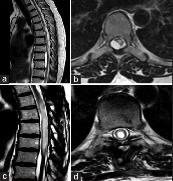
Figure 1
(a) Sagittal T2 dorsal magnetic resonance imaging (MRI) sequences showing multiple arachnoid cyst causing an important compression on spinal cord. (b) Axial T2 dorsal MRI sequences showing a cyst that compresses the spinal cord. (c) A sagittal T2 dorsal MRI showing cyst removal and intramedullary dilatation. (d) Axial T2 dorsal MRI showing intramedullary central canal dilatation

Figure 2
Intrasurgical photos obtained using operative microscope (Opmi Pentero). (a) Pre-removal arachnoid cyst. A diffuse thickening of arachnoid with calcification. (b) After removal of the arachnoid cyst, when the spinal cord expanded, we left only a part that was tightly attached to the spinal cord
DISCUSSION
To the best of our knowledge, this is the first report of intramedullary arachnoid cyst formation after removal of an intradural extramedullary cyst. To date, the patient is without symptoms; we have decided to follow her up conservatively every 6 months using a spine MRI. Only in case of neurological worsening, we will adopt the surgical option. We think that the intramedullary cyst developed due to scar adhesions in the central medullary canal secondary to the long duration of the disease (more than 3 years).
The aim of our report is to bring attention to this rarely known disorder and to highlight that surgery should be proposed in patients with neurological deficits as soon as possible, considering longstanding medullary compression may on one side reduce the possibility of neurological recover and on the other because of a secondary complication such as intramedullary cyst formation. In this context, we believe that radiological follow-up with MRI has to be done at least once during the first year.
Financial support and sponsorship
Nil.
Conflicts of interest
There are no conflicts of interest.
References
1. Kazan S, Ozdemir O, Akyuz M, Tuncer R. Spinal intradural arachnoid cysts located anterior to the cervical spinal cord. Report of two cases and review of the literature. J Neurosurg. 1999. 91: 211-5
2. Kumar S, Desai A, Bhatia L, Garg A. Multiple Spinal Arachnoid Cysts in a Child. Pediatr Neurol. 2016. 55: 76-7
3. Lee HJ, Cho DY. Symptomatic spinal intradural arachnoid cysts in the pediatric age group: Description of three new cases and review of the literature. Pediatr Neurosurg. 2001. 35: 181-7
4. Medved F, Seiz M, Baur MO, Neumaier-Probst E, Tuettenberg J. Thoracic intramedullary arachnoid cyst in an infant. J Neurosurg Pediatr. 2009. 3: 132-6
5. Novegno F, Umana G, Di Muro L, Fraioli B, Fraioli MF. Spinal intramedullary arachnoid cyst: Case report and literature review. Spine J. 2014. 14: e9-15


Donna Faye ledger
Posted February 4, 2019, 2:09 pm
I am a 75 year old women who was diagnosed with arachnoid cysts in 2014. I have had three surgeries since 2014. I am in a failing state and need expert option on where to go. Have all records, Mri and reports. Barely able to walk and things are getting worse