

- Department of Neurosurgery, Kinki University, Faculty of Medicine, Osaka, Japan
Correspondence Address:
Hisashi Kubota
Department of Neurosurgery, Kinki University, Faculty of Medicine, Osaka, Japan
DOI:10.4103/2152-7806.158206
Copyright: © 2015 Kubota H. This is an open-access article distributed under the terms of the Creative Commons Attribution License, which permits unrestricted use, distribution, and reproduction in any medium, provided the original author and source are credited.How to cite this article: Kubota H, Sanada Y, Nagatsuka K, Kato A. A case of angiographically occult, distal small anterior inferior cerebellar artery aneurysm. Surg Neurol Int 04-Jun-2015;6:97
How to cite this URL: Kubota H, Sanada Y, Nagatsuka K, Kato A. A case of angiographically occult, distal small anterior inferior cerebellar artery aneurysm. Surg Neurol Int 04-Jun-2015;6:97. Available from: http://surgicalneurologyint.com/surgicalint_articles/case-angiographically-occult-distal-small-anterior-inferior/
Abstract
Background:A small aneurysm at an unusual location, such as a distal anterior inferior cerebellar artery (AICA) aneurysm, may conceal as a computed tomography angiography (CTA) and digital subtraction angiography (DSA)-occult aneurysm.
Case Description:We herein present the case of a patient suffering from a subarachnoid hemorrhage (SAH) with two aneurysms in which the AICA aneurysm was negative by CTA and DSA. CTA demonstrated a right anterior choroidal artery aneurysm, which was revealed to be an unruptured aneurysm after surgical exploration. A small distal AICA aneurysm was detected by 3D rotational angiography (3DRA). The patient fully recovered except for left-side hearing loss four months after the second operation.
Conclusion:We recommend a meticulous diagnosis by 3DRA in patients with SAH in which the distribution is not coincident with a typical aneurysmal location.
Keywords: Distal anterior inferior cerebellar aneurysm, diagnosis, subarachnoid hemorrhage, 3D rotational angiography, 3D computed tomography angiography
INTRODUCTION
A distal anterior inferior cerebellar artery (AICA) aneurysm is relatively rare, and is estimated to comprise approximately 1–2% of all intracranial aneurysms.[
CASE REPORT
A 71-year-old male was transferred to our hospital with severe headache. A computed tomography (CT) scan showed subarachnoid hemorrhage (SAH), the distribution of which was dominant in the left side and relatively localized in the posterior fossa [
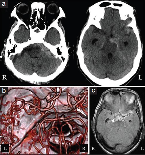
Figure 1
(a) A CT scan taken on admission showed a subarachnoid hemorrhage (SAH) in the left cerebellopontine cistern, ambient cistern and sylvian fissure. (b) The 3D computed tomographic angiography depicted the right anterior choroidal artery (AChA) aneurysm (arrow). (c) Magnetic resonance fluid attenuated inversion recovery images demonstrated that the SAH was continuing from the AChA aneurysm (arrow head); L: Left, R: Right
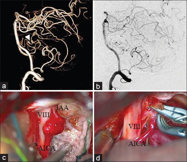
Figure 2
(a) The 3D rotational angiography showed a small aneurysm (arrow head) on the left anterior inferior cerebellar artery (AICA). (b) The lateral view of the 2D digital subtraction angiogram failed to depict any aneurysms. (c) An intraoperative photograph. (d) Neck clipping of the aneurysm; VIII: Acoustic nerve, IAA: Internal auditory artery
DISCUSSION
Although the incidence of distal AICA aneurysms varies,[
The diagnosis for a small peripheral aneurysm commonly requires careful investigation during radiological examinations. Especially if the aneurysm is small (<3 mm), the aneurysm may be defined as an angiographic occult aneurysm, or as a CTA-occult aneurysm.[
CONCLUSION
3DRA is desirable to detect a small peripheral aneurysm when the distribution of SAH is not in agreement with the aneurysmal location. A distal AICA aneurysm is a relatively rare aneurysm, but such a possibility has to be taken into consideration when making a differential diagnosis, especially if the SAH is observed in the posterior fossa.
References
1. Bambakidis NC, Manjila S, Dashti S, Dashti S, Tarr R, Megerian CA. Management of anterior inferior cerebellar artery aneurysms: An illustrative case and review of literature. Neurosurg Focus. 2009. 26: E6-
2. Gonzalez LF, Alexander MJ, McDougall CG, Spetzler RF. Anteriorinferior cerebellar artery aneurysms: Surgical approaches and outcomes-a review of 34 cases. Neurosurgery. 2004. 55: 1025-35
3. Iwanaga S, Shrier DA, Okawara S, Numaguchi Y. Value of CT angiography in the evaluation of a peripheral anterior inferior cerebellar artery aneurysm: Case report. Clin Imaging. 1999. 23: 77-80
4. Li X, Zhang D, Zhao J. Anterior inferior cerebellar artery aneurysms: Six cases and a review of the literature. Neurosurg Rev. 2012. 35: 111-9
5. Mahmoud M, El Serwi A, Alaa Habib M, Abou Gamrah S. Endovascular treatment of AICA flow dependent aneurysms: A report of three cases and review of the literature. Interv Neuroradiol. 2012. 18: 449-57
6. McKinney AM, Palmer CS, Truwit CL, Karagulle A, Teksam M. Detection of aneurysm by 64-section multidetector CT angiography in patients acutely suspected of having an intracranial aneurysm and comparison with digital subtraction and 3D rotational angiography. AJNR Am J Neuroradiol. 2008. 29: 594-602
7. Rodríguez-Hernández A, Zador Z, Rodríguez-Mena R, Lawton MT. Distal aneurysms of intracranial arteries: Application of numerical nomenclature, predilection for cerebellar arteries, and results of surgical management. World Neurosurg. 2013. 80: 103-12
8. Schwartz HG. Arterial aneurysm of the posterior fossa. J Neurosurg. 1948. 5: 312-6
9. van Rooij WJ, Peluso J, Sluzewski M, Beute GN. Additional value of 3D rotational angiography in angiographically negative aneurysmal subarachnoid hemorrhage: How negative is negative?. AJNR Am J Neuroradiol. 2008. 29: 962-6
10. Yamakawa H, Hattori T, Tanigawara T, Sahashi Y, Ohkuma A. Intracanalicular aneurysm at the meatal loop of the distal anterior inferior cerebellar artery: A case report and review of the literature. Surg Neurol. 2004. 61: 82-8
11. Zager EL, Shaver EG, Hurst RW, Flamm ES. Distal anterior inferior cerebellar artery aneurysms: Report of four cases. J Neurosurg. 2002. 97: 692-6

