

- Department of Neurosurgery, Avicenne Military Hospital, Marrakech,
- Mohammed V University, Rabat,
- School of Science and Engineering, Al Akhawayn University, Ifrane, Morocco.
Correspondence Address:
Ali Akhaddar
School of Science and Engineering, Al Akhawayn University, Ifrane, Morocco.
DOI:10.25259/SNI_554_2019
Copyright: © 2019 Surgical Neurology International This is an open-access article distributed under the terms of the Creative Commons Attribution-Non Commercial-Share Alike 4.0 License, which allows others to remix, tweak, and build upon the work non-commercially, as long as the author is credited and the new creations are licensed under the identical terms.How to cite this article: Akhaddar A, Akhaddar H. A new learning approach for identifying cortical brain areas around the central sulcus using the name of Allah. Surg Neurol Int 13-Dec-2019;10:244
How to cite this URL: Akhaddar A, Akhaddar H. A new learning approach for identifying cortical brain areas around the central sulcus using the name of Allah. Surg Neurol Int 13-Dec-2019;10:244. Available from: https://surgicalneurologyint.com/surgicalint-articles/9803/
Date of Submission
08-Nov-2019
Date of Acceptance
14-Nov-2019
Date of Web Publication
13-Dec-2019
To the Editor,
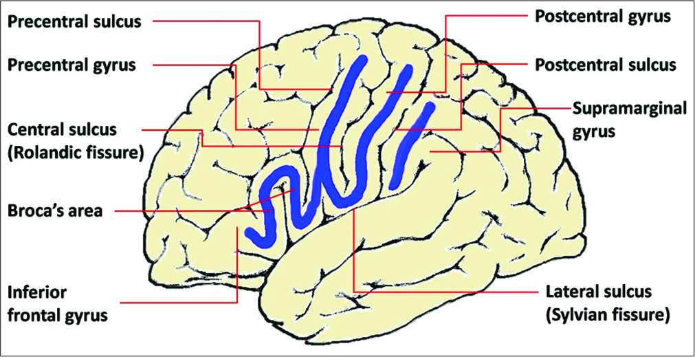
Normal brain cortex is very convoluted due to the development of a large cerebral surface area confined within the intracranial space. This cerebral curved surface consists of grooves named sulci and raised ridges between the grooves called gyri or convolutions.[
Nevertheless, teaching neuroanatomy to students is particularly challenging. This is in part attributed to the complexity of the brain and to the students’ inability to apply their knowledge of basic sciences to clinical situations (neurophobia).[
Here, we describe a new way to facilitate identification and memorization of some of the most important/critical cortical brain areas, for example, around the central sulcus.
If we contemplate the shape of the convolutions on the external surface of the left cerebral hemisphere (the dominant hemisphere in the right-hand man), we find that they form the Arabic name of “Allah” (الله) around the central sulcus (Rolandic fissure) area.
As shown in
This new learning approach was successfully implemented in a small group of young medical students; the feedback was positive. For future work, we plan to apply this new simple method for a larger number of medical students studying neuroanatomy.
Opinions among readers of this paper will surely vary, but we hope that this modest contribution will at least stimulate further scholarly and scientific supports to this fascinating topic.
References
1. Arantes M, Arantes J, Ferreira MA. Tools and resources for neuroanatomy education: A systematic review. BMC Med Educ. 2018. 18: 94-
2. Dornald I, Newman WA.editorsDorland’s Illustrated Medical Dictionary. Philadelphia, PA: Saunders, Elsevier; 2012. p.
3. Jozefowicz RF. Neurophobia: The fear of neurology among medical students. Arch Neurol. 1994. 51: 328-9
4. Lazić D, Marinković S, Tomić I, Mitrović D, Starčević A, Milić I. Brain and art: Illustrations of the cerebral convolutions. A review. Folia Morphol (Warsz). 2014. 73: 247-58
5. Moxham B, McHanwell S, Plaisant O, Pais D. A core syllabus for the teaching of neuroanatomy to medical students. Clin Anat. 2015. 28: 706-16
6. Shin DS, Kim DH, Park JS, Jang HG, Chung MS. Evaluation of anatomy comic strips for further production and applications. Anat Cell Biol. 2013. 46: 210-6


Dr. Mohamed E. El-Fiki
Posted December 16, 2019, 2:16 pm
The article is really interesting and I think it is worse spreading.
I believe this a very good article that deserves verification from both Muslim and muslim neurosurgery, neurology a d neuroscience academics and practitioners
Pr Akhaddar
Posted July 10, 2020, 10:34 pm
Thank you very much Professor El Fiki.
I salute your support and commitment to young scientists in developing countries.
All the Best.
Warm Regards.
Naveed Ashraf
Posted July 27, 2020, 7:27 pm
its an excellent abstract way of memorising even for those who are very familiar with the anatomy of the region. Another feature could be to ascribe various functions of these regions to the letters of the word Allah. The upper and lower stems of ا and the two ل and the curve joining them and finally the convolutions of ہ۔