- Division of Neurosurgery, Riverside University Health System, Moreno Valley, Fontana, CA, USA
- Department of Neurosurgery, Fontana, CA, USA
- Department of Pathology, Fontana, CA, USA
- Department of Head and Neck Surgery, Fontana, CA, USA
Correspondence Address:
Christopher J. Elia
Department of Neurosurgery, Fontana, CA, USA
DOI:10.4103/sni.sni_54_18
Copyright: © 2018 Surgical Neurology International This is an open access journal, and articles are distributed under the terms of the Creative Commons Attribution-NonCommercial-ShareAlike 4.0 License, which allows others to remix, tweak, and build upon the work non-commercially, as long as appropriate credit is given and the new creations are licensed under the identical terms.How to cite this article: Christopher J. Elia, Marc Cabanne, Zhe Piao, Andrew Lee, Todd Goldenberg, Vaninder Chhabra. A rare case of intracranial teratocarcinosarcoma: Case report and review of literature. 14-Aug-2018;9:167
How to cite this URL: Christopher J. Elia, Marc Cabanne, Zhe Piao, Andrew Lee, Todd Goldenberg, Vaninder Chhabra. A rare case of intracranial teratocarcinosarcoma: Case report and review of literature. 14-Aug-2018;9:167. Available from: http://surgicalneurologyint.com/surgicalint-articles/a-rare-case-of-intracranial-teratocarcinosarcoma-case-report-and-review-of-literature/
Abstract
Background:Teratocarcinosarcoma (TCS) is a rare malignant neoplasm with epithelial and mesenchymal components such as fibroblasts, cartilage, bone and smooth muscle. With less than 100 total reported cases, this malignant neoplasm is rarely encountered by neurosurgeons because it primarily involves the nasal cavity and paranasal sinuses.
Case Description:A 55-year-old male with chronic frontal headaches was found to have a frontal mass with involvement of nasal sinus and right ethmoid sinus. The patient underwent preoperative embolization of tumor followed by bilateral frontal craniotomy for near total resection of the tumor. Patient did well postoperatively without new neurological deficits.
Conclusion:Although cases with intracranial involvement are scarce, treatment with surgical resection with or without adjuvant treatments of chemotherapy and radiation therapy is the most widely accepted with goal for gross total resection. In our case, we achieved near total resection and the patient continues to do well without any gross neurological deficits.
Keywords: Brain tumor, neuro-oncology, neurosurgery, oncology, teratocarcinosarcoma
INTRODUCTION
Teratocarcinosarcoma (TCS) is a rare malignant neoplasm with epithelial and mesenchymal components, such as fibroblasts, cartilage, bone, and smooth muscle, with less than 100 cases described in the literature.[
Nearly all reported cases of TCS arise within the nasal cavity or paranasal sinuses, with a few reports having described intracranial invasion into the dura mater or the frontal lobe at presentation or progression, as in our case.[
CASE REPORT
History and presentation
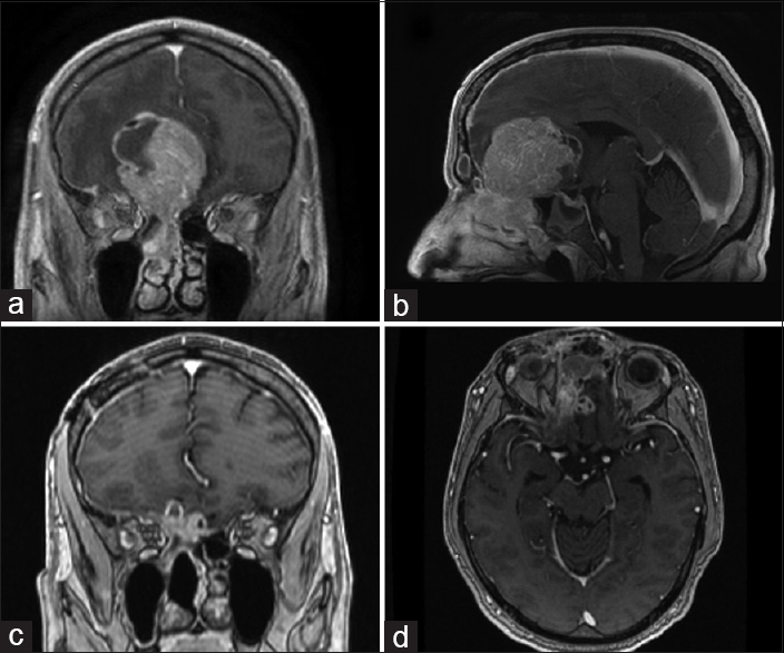
A 55-year-old male presented with frontal headaches 3 times a week with progressive memory problems, word-finding difficulties, and new-onset seizures. Magnetic resonance imaging (MRI) of the brain revealed a 7.0 × 6.2 × 4.4 cm enhancing mass extending across the cribriform plate and into the right anterior cranial fossa and right nasal cavity [
Operative intervention
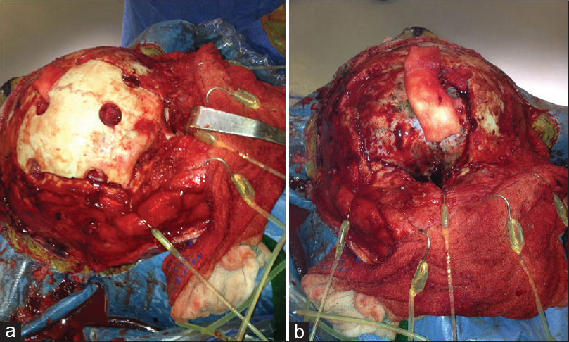
Given the preoperative diagnosis via transnasal biopsy and presumed hypervascularity of the neoplasm, the patient underwent preoperative embolization followed by bilateral frontal craniotomy via a bicoronal incision with pericranial flap preservation. ENT assisted with exposure of the tumor using the transglabellar subcranial approach. Tumor was found to be pale and hypervascular with a dural breach in the low frontal plane with involvement of the superior ethmoid sinus [
Histological staining
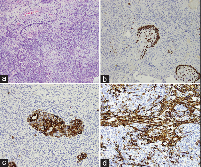
Staining was positive for CK-PanAE1-3CAM52, vimentin, EMA, S100, CK 5/6, P63, synaptophysin, chromogranin, and GFAP. Staining negative for CD99 and neurofilament [
Postoperative course
The postoperative course was significant for pulmonary embolism and was otherwise unremarkable. Twenty-four months postoperatively the patient is and doing well without any seizure-like activity with return to baseline neurological status. MRI showed no evidence of recurrence with expected postoperative changes [
DISCUSSION
TCS is a rare malignant neoplasm with epithelial and mesenchymal components. The most common presenting signs and symptoms are nasal obstruction and epistaxis, found in ~75% to 90% of documented cases.[
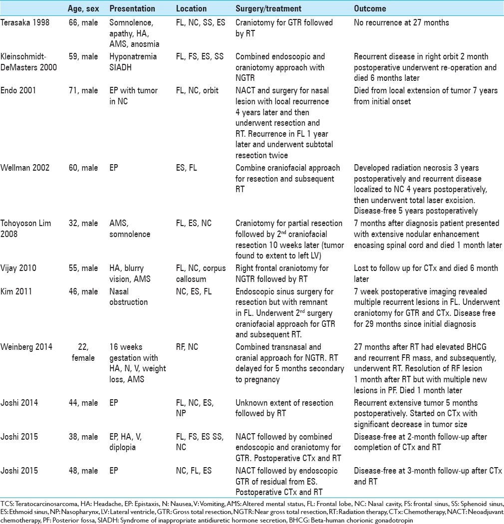
A review of literature revealed 11 cases of TCS with intracranial invasion [
Given the multitude of cell types in this neoplasm, biopsy alone is not of high yield. TCS requires adequate sampling for proper diagnosis. Possible erroneous diagnosis include olfactory neuroblastoma, squamous cell carcinoma, undifferentiated carcinoma, adenocarcinoma, malignant craniopharyngioma, malignant mixed tumor of salivary gland type, mucoepidermoid carcinoma, adenosquamous carcinoma, and transitional carcinoma of Schneiderian type.[
The combined transglabellar/subcranial approach gives simultaneous exposure of the upper and lower limits of the tumor in the anterior fossa, ethmoid sinus, and sphenoid sinus for improved completeness of resection.[
CONCLUSIONS
TCS is a rare neoplasm rarely encountered by the neurosurgeon given its predilection for sinonasal involvement with majority of cases presenting with sinonasal obstruction and/or epistaxis. Neurological signs and symptoms are very rare with this neoplasm. To our knowledge, this is the first reported case with seizure as a presenting sign. Diagnosis requires ample tissue sample. Once diagnosis is established, the surgeon can plan for the optimal surgical approach to facilitate gross total resection. Goal of gross total surgical resection is standard with or without chemotherapy and/or radiation therapy. Combined ENT and neurosurgical approaches can limit the number of surgical procedures required for gross total resection. Metastasis is uncommon but has been reported in cases with postoperative residual mass
Financial support and sponsorship
Nil.
Conflicts of interest
There are no conflicts of interest.
References
1. Agrawal N, Chintagumpala M, Hicks J, Eldin K, Paulino AC. Sinonasal teratocarcinosarcoma in an adolescent male. J Pediatr Hematol Oncol. 2012. 34: e304-7
2. Cardesa A, Luna MA, Barnes L, Evenson JW, Reichart P, Sidransky D.editors. Germ cell tumors. Pathology and Genetics of Head and Neck Tumors. Lyon: IARC; 2005. 9: 76-7
3. Endo H, Hirose T, Kuwamura KI, Sano T. Case report: Sinonasal teratocarcinosarcoma. Pathol Int. 2001. 51: 107-12
4. Fatima SS, Minhas K, Din NU, Fatima S, Ahmed A, Ahmad Z. Sinonasal teratocarcinosarcoma: A clinicopathologic and immunohistochemical study of 6 cases. Ann Diagn Pathol. 2013. 17: 313-8
5. Heffner DK, Hyams VJ. Teratocarcinosarcoma (malignant teratoma?) of the nasal cavity and paranasal sinuses A clinicopathologic study of 20 cases. Cancer. 1984. 53: 2140-54
6. Joshi A, Dhumal SB, Manickam DR, Noronha V, Bal M, Patil VM. Recurrent sinonasal teratocarcinosarcoma with intracranial extension: Case report. Indian J Cancer. 2014. 51: 398-400
7. Joshi A, Noronha V, Sharma M, Dhumal S, Juvekar S, Patil VM. Neoadjuvant chemotherapy in advanced sinonasal teratocarcinosarcoma with intracranial extension: Report of two cases with literature review. J Cancer Res Ther. 2015. 11: 1003-5
8. Kim JH, Maeng YH, Lee JS, Jung S, Lim SC, Lee MC. Sinonasal teratocarcinosarcoma with rhabdoid features. Pathol Int. 2011. 61: 762-7
9. Kleinschmidt-DeMasters BK, Pflaumer SM, Mulgrew TD, Lillehei KO. Sinonasal teratocarcinosarcoma (“mixed olfactory neuroblastoma-craniopharyngioma”) presenting with syndrome of inappropriate secretion of antidiuretic hormone. Clin Neuropathol. 2000. 19: 63-9
10. Misra P, Husain Q, Svider PF, Sanghvi S, Liu JK, Eloy JA. Management of sinonasal teratocarcinosarcoma: A systematic review. Am J Otolaryngol. 2014. 35: 5-11
11. Raveh J, Vuillemin T, Sutter F. Subcranial management of 395 combined frontobasal-midface fractures. Arch Otolaryngol Head Neck Surg. 1988. 114: 1114-22
12. Shanmugaratnam K, Kunaratnam N, Chia KB, Chiang GS, Sinniah R. Teratocarcinosarcoma of the paranasal sinuses. Pathology. 1983. 15: 413-9
13. Shorter C, Nourbakhsh A, Dean M, Thomas-Ogunniyi J, Lian TS, Guthikonda B. Intracerebral metastasis of a sinonasal teratocarcinosarcoma: A case report. Skull Base. 2010. 20: 393-6
14. Smith SL, Hessel AC, Luna MA, Malpica A, Rosenthal DI, El-Naggar AK. Sinonasal teratocarcinosarcoma of the head and neck: A report of 10 patients treated at a single institution and comparison with reported series. Arch Otolaryngol Head Neck Surg. 2008. 134: 592-5
15. Sweety Vijay S, Kumar TD, Srikant B, Vithal SH, Vijay KS, Gurunath P. Intracranial presentation of teratocarcinosarcoma. J Clin Neurosci. 2010. 17: 1347-9
16. Takasaki K, Sakihama N, Takahashi H. A case with sinonasal teratocarcinosarcoma in the nasal cavity and ethmoid sinus. Eur Arch Otorhinolaryngol. 2006. 263: 586-91
17. Tchoyoson Lim CC, Thiagarajan A, Sim CS, Khoo ML, Shakespeare TP, Ng I. Craniospinal dissemination in teratocarcinosarcoma. J Neurosurg. 2008. 109: 321-4
18. Terasaka S, Medary MB, Whiting DM, Fukushima T, Espejo EJ, Nathan G. Prolonged survival in a patient with sinonasal teratocarcinosarcoma with cranial extension. Case report. J Neurosurg. 1998. 88: 753-6
19. Thomas J, Adegboyega P, Iloabachie K, Mooring JW, Lian T. Sinonasal teratocarcinosarcoma with yolk sac elements: A neoplasm of somatic or germ cell origin?. Ann Diagn Pathol. 2011. 15: 135-9
20. Weinberg BD, Newell KL, Wang F. A case of a beta-human chorionic gonadotropin secreting sinonasal teratocarcinosarcoma. J Neurol Surg Rep. 2014. 75: e103-7
21. Wellman M, Kerr PD, Battistuzzi S, Cristante L. Paranasal sinus teratocarcinosarcoma with intradural extension. J Otolaryngol. 2002. 31: 173-6


