- Department of Neurosurgery, Helsinki University Central Hospital, Helsinki, Finland
Correspondence Address:
Fransua Sharafeddin
Department of Neurosurgery, Helsinki University Central Hospital, Helsinki, Finland
DOI:10.4103/2152-7806.199559
Copyright: © 2017 Surgical Neurology International This is an open access article distributed under the terms of the Creative Commons Attribution-NonCommercial-ShareAlike 3.0 License, which allows others to remix, tweak, and build upon the work non-commercially, as long as the author is credited and the new creations are licensed under the identical terms.How to cite this article: Fransua Sharafeddin, Ahmad Hafez, Martin Lehecka, Rahul Raj, Roberto Colasanti, Ahmadreza Rafiei, Joham Choque, Behnam R. Jahromi, Mika Niemelä, Juha Hernesniemi. A5 segment aneurysm of the anterior cerebral artery, imbedded into the body of the corpus callosum: A case report. 06-Feb-2017;8:18
How to cite this URL: Fransua Sharafeddin, Ahmad Hafez, Martin Lehecka, Rahul Raj, Roberto Colasanti, Ahmadreza Rafiei, Joham Choque, Behnam R. Jahromi, Mika Niemelä, Juha Hernesniemi. A5 segment aneurysm of the anterior cerebral artery, imbedded into the body of the corpus callosum: A case report. 06-Feb-2017;8:18. Available from: http://surgicalneurologyint.com/surgicalint_articles/a5-segment-aneurysm-of-the-anterior-cerebral-artery-imbedded-into-the-body-of-the-corpus-callosum-a-case-report/
Abstract
Background:The A5 segment aneurysms of the anterior cerebral artery are rare, approximately 0.5% of all intracranial aneurysms. They are small with a wide base located in the midline, with the domes mostly projecting upward or backward.
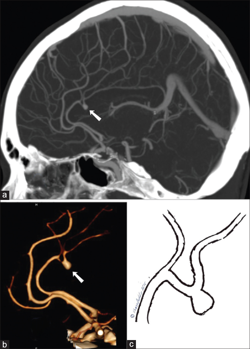
Case Description:The authors describe a unique case of A5 segment aneurysm, with the dome embedded into the body of the corpus callosum. This 41-year-old female was admitted to the neurology department for possible multiple sclerosis investigation. Computed tomography angiogram (CTA) revealed a 4-mm right-sided pericallosal artery aneurysm, with rare configuration, which was caudally projected, embedded into the body of the corpus callosum. Considering the family history, patient underwent a prophylactic ligation surgery. The postoperative CT and CTA showed no complication and successful occlusion of the aneurysm with no ischemia or hemorrhage in the corpus callosum.
Conclusion:To the best of our knowledge, this is the first case of an aneurysm with this configuration. Our rare case of A5 segment aneurysm demonstrates the importance of planning of the surgery, choosing the appropriate approach, and knowing the detailed anatomy of the region, as well as the necessity of microsurgical clipping of small unruptured AdistAs.
Keywords: Aneurysm, anterior cerebral artery, clipping, callosomarginal artery, corpus callosum, pericallosal artery
INTRODUCTION
The A5 segment aneurysms of anterior cerebral artery (ACA) are rare, approximately 0.5% of all intracranial aneurysms (IA).[
CLINICAL PRESENTATION
A 41-year-old healthy non-smoking Caucasian female was admitted to the neurology department to be investigated for possible multiple sclerosis, vertigo, coordination problems, tingling, and numbness in her left arm. A neurological examination showed no focal abnormalities.
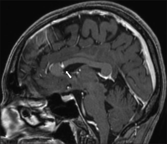
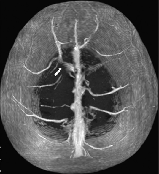
A computed tomography angiogram (CTA) was performed, which showed a 4-mm right-sided pericallosal artery aneurysm, with a rare configuration [
Considering the family history (mother and uncle from mother's side died from aneurysmal SAH in their forties), the patient was motivated for prophylactic ligation surgery.
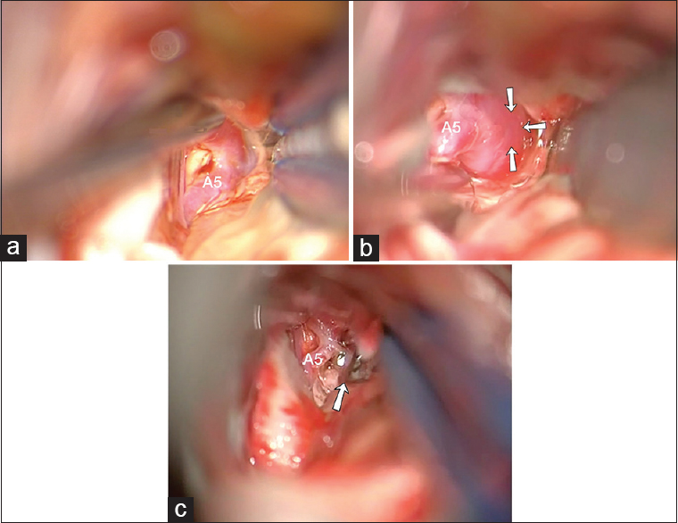
The patient was placed in a supine position, with the head fixed in a head frame. The head was elevated 20° above the heart level in neutral position with the nose pointing upward and somewhat flexed. Because of the presence of a bridging vein at the shortest trajectory projection of the aneurysm to the scull convex that was visualized at the preoperative CTA and magnetic resonance angiogram (MRA), the approach was planned frontally to the vein [
After transferring to the intensive care unit, the patient was oriented, without any signs of neurological deficit. At first postoperative day, CT and CTA was performed, that showed no complication and successful occlusion of the aneurysm with no ischemia or hemorrhage in the corpus callosum. The same day the patient was transferred to the ward.
DISCUSSION
According to Fischer, the ACA can be divided into 5 segments, namely, A1 to A5.[
CONCLUSION
To the best of our knowledge, this is the first case of an aneurysm with this configuration. Our rare case of A5 segment aneurysm demonstrates the importance of planning of the surgery, choosing the appropriate approach, and knowing the detailed anatomy of the region, as well as the necessity of microsurgical clipping of small unruptured AdistAs.
Financial support and sponsorship
Nil.
Conflicts of interest
There are no conflicts of interest.
References
1. de Sousa AA, Dantas FL, de Cardoso GT, Costa BS. Distal anterior cerebral artery aneurysms. Surg Neurol,. 1999. 52: 128-35
2. Fischer E. Die Lageabweichungen der vorderen Hirnarterie im Gefassbild. Zentralbl Neurochir. 1938. 3: 300-12
3. Gomes FB, Dujovny M, Umansky F, Berman SK, Diaz FG, Ausman JI. Microanatomy of the anterior cerebral artery. Surg Neurol. 1986. 26: 129-41
4. Hernesniemi J, Tapaninaho A, Vapalahti M, Niskanen M, Kari A, Luukkonen M. Saccular aneurysms of the distal anterior cerebral artery and its branches. Neurosurgery. 1992. 31: 994-8
5. Inci S, Erbengi A, Ozgen T. Aneurysms of the distal anterior cerebral artery: Report of 14 cases and a review of the literature. Surg Neurol. 1998. 50: 130-9
6. Kakou M, Destrieux C, Velut S. Microanatomy of the pericallosal arterial complex. J Neurosurg. 2000. 93: 667-75
7. Kawashima M, Matsushima T, Sasaki T. Surgical strategy for distal anterior cerebral artery aneurysms: Microsurgical anatomy. J Neurosurg. 2003. 99: 517-25
8. Laitinen L, Snellman A. Aneurysms of the pericallosal artery: A study of 14 cases verified angiographically and treated mainly by direct surgical attack. J Neurosurg. 1960. 17: 447-58
9. Lehecka M, Dashti R, Hernesniemi J, Niemelä M, Koivisto T, Ronkainen A. Microneurosurgical management of aneurysms at A3 segment of anterior cerebral artery. Surg Neurol. 2008. 70: 135-51
10. Lehecka M, Dashti R, Hernesniemi J, Niemelä M, Koivisto T, Ronkainen A. Microneurosurgical management of aneurysms at A4 and A5 segments and distal cortical branches of anterior cerebral artery. Surg Neurol. 2008. 70: 352-67
11. Lehecka M, Dashti R, Hernesniemi J, Niemelä M, Koivisto T, Ronkainen A. Microneurosurgical management of aneurysms at the A2 segment of anterior cerebral artery (proximal pericallosal artery) and its frontobasal branches. Surg Neurol. 2008. 70: 232-46
12. Lehecka M, Lehto H, Niemelä M, Juvela S, Dashti R, Koivisto T. Distal anterior cerebral artery aneurysms: Treatment and outcome analysis of 501 patients. Neurosurgery. 2008. 62: 590-601
13. Mann KS, Yue CP, Wong G. Aneurysms of the pericallosal-callosomarginal junction. Surg Neurol. 1984. 21: 261-6
14. Ohno K, Monma S, Suzuki R, Masaoka H, Matsushima Y, Hirakawa K. Saccular aneurysms of the distal anterior cerebral artery. Neurosurgery. 1990. 27: 907-12
15. Perlmutter D, Rhoton AL. Microsurgical anatomy of the distal anterior cerebral artery. J Neurosurg. 1978. 49: 204-28
16. Proust F, Toussaint P, Hannequin D, Rabenenoïna C, Le Gars D, Fréger P. Outcome in 43 patients with distal anterior cerebral artery aneurysms. Stroke. 1997. 28: 2405-9
17. Sindou M, Pelissou-Guyotat I, Mertens P, Keravel Y, Athayde AA. Pericallosal aneurysms. Surg Neurol. 1988. 30: 434-40
18. Snyckers FD, Drake CG. Aneurysms of the distal anterior cerebral artery. A report on 24 verified cases. S Afr Med J. 1973. 47: 1787-91
19. Steven DA, Lownie SP, Ferguson GG. Aneurysms of the distal anterior cerebral artery: Results in 59 consecutively managed patients. Neurosurgery. 2007. 60: 227-33
20. Ugur HC, Kahilogullari G, Esmer AF, Comert A, Odabasi AB, Tekdemir I. A neurosurgical view of anatomical variations of the distal anterior cerebral artery: An anatomical study. J Neurosurg. 2006. 104: 278-84
21. Wisoff JH, Flamm ES. Aneurysms of the distal anterior cerebral artery and associated vascular anomalies. Neurosurgery. 1987. 20: 735-41
22. Yasargil MG, Carter LP. Saccular aneurysms of the distal anterior cerebral artery. J Neurosurg. 1974. 40: 218-23
23. Yoshimoto T, Uchida K, Suzuki J. Surgical treatment of distal anterior cerebral artery aneurysms. J Neurosurg. 1979. 50: 40-4


