- Department of Neurological Surgery, Juntendo University Urayasu Hospital, Urayasu, Japan.
- Department of Pathology, Juntendo University Urayasu Hospital, Urayasu, Japan.
Correspondence Address:
Dr. Satoshi Tsutsumi, Department of Neurological Surgery, Juntendo University Urayasu Hospital, Urayasu, Japan.
DOI:10.25259/SNI_520_2022
Copyright: © 2022 Surgical Neurology International This is an open-access article distributed under the terms of the Creative Commons Attribution-Non Commercial-Share Alike 4.0 License, which allows others to remix, transform, and build upon the work non-commercially, as long as the author is credited and the new creations are licensed under the identical terms.How to cite this article: Hiroki Sugiyama1, Satoshi Tsutsumi1, Akane Hashizume2, Kiyotaka Kuroda1, Natsuki Sugiyama1, Hideaki Ueno1, Hisato Ishii1. Calvarial angiomatous meningioma developed in the diploe. 29-Jul-2022;13:326
How to cite this URL: Hiroki Sugiyama1, Satoshi Tsutsumi1, Akane Hashizume2, Kiyotaka Kuroda1, Natsuki Sugiyama1, Hideaki Ueno1, Hisato Ishii1. Calvarial angiomatous meningioma developed in the diploe. 29-Jul-2022;13:326. Available from: https://surgicalneurologyint.com/surgicalint-articles/11752/
Abstract
Background: Angiomatous meningioma is a rare subtype of meningiomas. To the best of our knowledge, there have been no reports of intradiploic angiomatous meningioma.
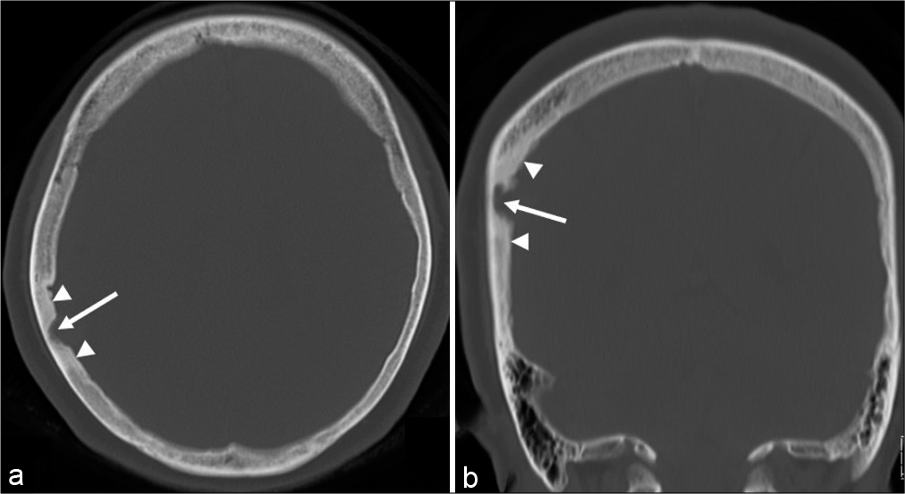
Case Description: A 53-year-old previously healthy woman was diagnosed with a calvarial lesion during a brain checkup. Cerebral magnetic resonance imaging showed an intradiploic tumor, 11 × 14 × 12 mm, in the right parietal bone. It was an enhancing, lobular tumor presenting as isointensity on T1- and hyperintensity on T2-weighted sequences, with an intense enhancement of the adjacent dura mater. Computed tomography revealed bone erosion at the tumor site, extending predominantly into the inner side, and sclerotic changes in the surrounding bone. Total resection was performed. Microscopically, the tumor tissue comprised cells with low-grade meningioma and intervening prominent vasculatures, consistent with angiomatous meningioma.
Conclusion: Angiomatous meningioma should be considered as a differential diagnosis when an intradiploic tumor shows a lobular structure, intense enhancement of the adjacent dura mater, and sclerotic changes in the surrounding skull. These findings can support prompt tumor resection.
Keywords: Angiomatous meningioma, Diploe, Lobular structure, Osteosclerosis
INTRODUCTION
Meningiomas are the most common primary brain tumors in adulthood. They commonly grow in subdural sites, whereas approximately 1% of them are estimated to arise in extradural sites involving the calvarium or diploe.[
Here, we present a unique case of intradiploic angiomatous meningioma with distinctive radiological and intraoperative findings.
CASE PRESENTATION
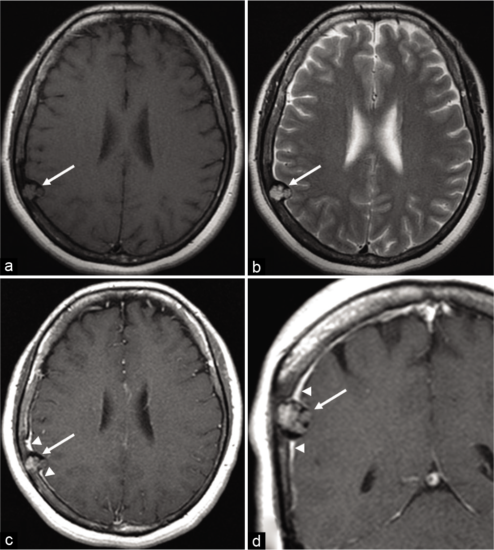
A 53-year-old previously healthy woman was diagnosed with a calvarial lesion on a brain checkup and was referred to the hospital. She had not undergone cranial radiotherapy in her life. Cerebral magnetic resonance imaging (MRI) revealed an intradiploic tumor in the right parietal bone protruding into the cranial cavity. It was a lobular mass 11 × 14 × 12 mm, presented isointensity on T1- and hyperintensity on T2-weighted sequences, and was inhomogeneously enhanced with intense enhancement of the surrounding dura mater. The inner table and dura underlying the tumor appeared intact. There was no identifiable peritumoral brain edema [
Figure 1:
Axial T1- (a), T2- (b), and postcontrast axial (c) and coronal (d) T1-weighted magnetic resonance images show the intradiploic mass in the right parietal bone, protruding into the cranial cavity (arrow). It involves a lobular mass 11 × 14 × 12 mm in diameter, presenting isointensity on T1- and hyperintensity on T2-weighted sequences, respectively, and is inhomogeneously enhanced with an intense enhancement of the surrounding dura mater (c and d, arrowheads). The inner table and dura underlying the tumor appear intact. There is no identifiable peritumoral brain edema (b).
Figure 4:
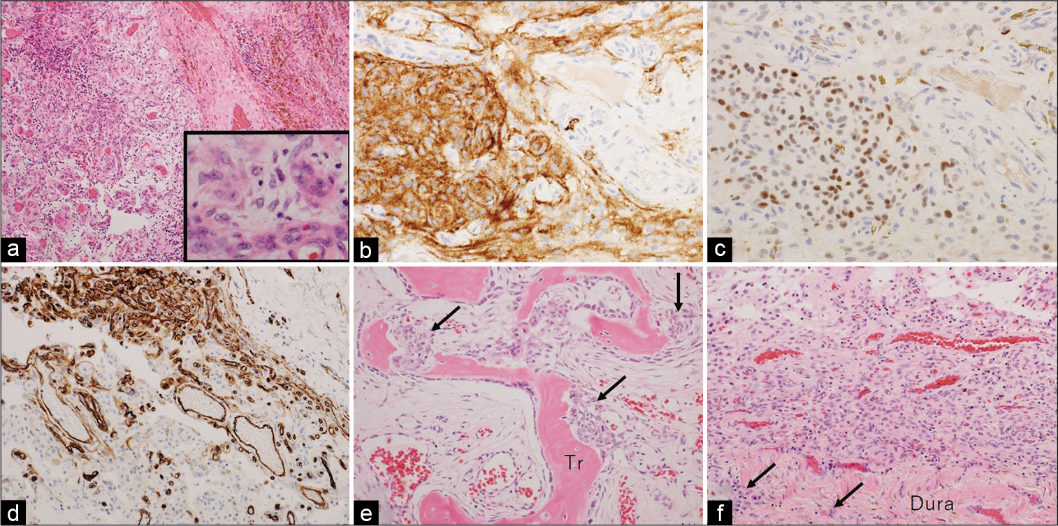
Photomicrographs of the resected specimens showing tumor tissue comprising cells with oval-shaped nuclei and intervening vasculatures with varying sizes (a and its inset, magnified view). There are few mitotic figures. Immunohistochemical examination shows positive staining for epithelial membrane antigen (b) and progesterone receptor (c), while negative for CD34 (d). Tumor invasions into the adjacent bone (e, arrows) and dura mater (f, arrows) are noted. (a) Hematoxylin and eosin stain, ×40; (b) epithelial membrane antigen, ×200; (c) progesterone receptor, ×200; (d) CD34, ×100; and (e and f) hematoxylin and eosin stain, ×100. Tr: trabecula.
DISCUSSION
Although the diploe can be affected by varying pathologies, intradiploic meningioma has been rarely reported.[
Whereas CT revealed sclerotic changes in the skull surrounding the tumor. Furthermore, the histological appearance of the resected tumor was consistent with that of an angiomatous meningioma. Therefore, we assumed that the findings identified on the MRI and CT might represent the characteristic appearance of intradiploic angiomatous meningioma.
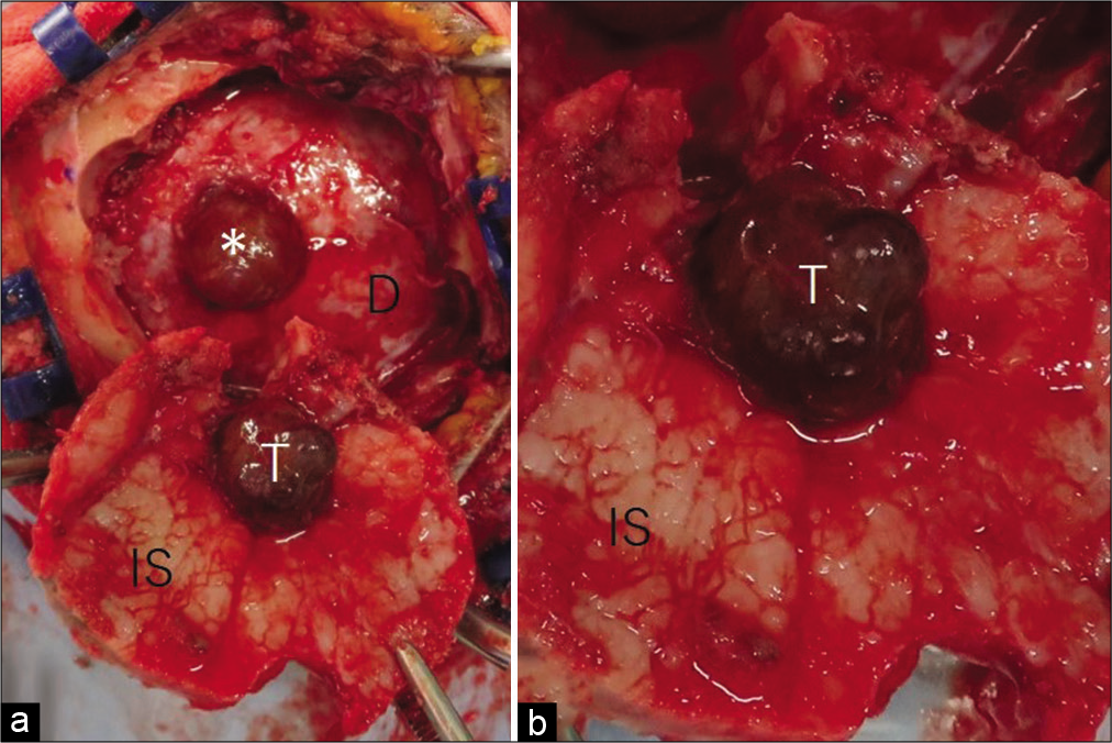
The present case involved a small intradiploic tumor incidentally detected during a brain checkup. However, intraoperative observation revealed a defective inner table due to compression by the intradiploic tumor. Furthermore, histological examination showed overt tumor invasion into the surrounding bone and underlying dura mater. Therefore, prompt tumor resection is recommended if an asymptomatic intradiploic tumor is assumed to be an angiomatous meningioma.
In addition, the present intradiploic tumor showed unevenly inward extension into the cranial cavity, with the outer table intact. This may be because the inner table of the patient was less resistant to compression by the tumor than the outer table. Further case studies would clarify the pros and cons of these speculations.
CONCLUSION
Angiomatous meningioma should be considered as a differential diagnosis when an intradiploic tumor shows a lobular structure, intense enhancement of the adjacent dura mater, and sclerotic changes in the surrounding skull. These findings can support prompt tumor resection.
Declaration of patient consent
The authors certify that they have obtained all appropriate patient consent.
Financial support and sponsorship
Nil.
Conflicts of interest
There are no conflicts of interest.
References
1. Asil K, Aksoy YE, Yaldiz C, Kahyaglu ZK. Primary intraosseous meningioma mimicking osteosarcoma: Case report. Turk Neurosurg. 2015. 25: 174-6
2. Borkar SA, Tripathi AK, Satyarthee GD, Rishi A, Kale SS, Sharma BS. Fronto-orbital intradiploic transitional meningioma. Neurol India. 2008. 56: 205-6
3. Chang JH, Chang JW, Park YG, Kim TS, Kim JA, Chung SS. Simple bone cyst occurring in calvarium. Acta Neurochir (Wien). 2003. 145: 927-8
4. Chatterjee S, Foy P, Diengdoh V. Intra-diploic meningioma in a child. Br J Neurosurg. 1993. 7: 315-7
5. Cirak B, Guven MB, Ugras S, Kutluhan A, Unal O. Frontoorbitonasal intradiploic meningioma in a child. Pediatr Neurosurg. 2000. 32: 48-51
6. Crea A, Grimod G, Scalia G, Verlotta M, Mazzeo L, Rossi G. Fronto-orbito-ethmoidal intradiploic meningiomas: A case study with systematic review. Surg Neurol Int. 2021. 12: 485
7. Desai KI, Nadkarni TD, Bhayani RD, Goel A. Intradiploic meningioma of the orbit: A case report. Neurol India. 2004. 52: 380-2
8. Halpin SF, Britton J, Wilkins P, Uttley D. Intradiploic meningiomas. A radiological study of two cases confirmed histologically. Neuroradiology. 1991. 33: 247-50
9. Hua L, Luan S, Li H, Zhu H, Tang H, Liu H. Angiomatous meningiomas have a very benign outcome despite frequent peritumoral edema at onset. World Neurosurg. 2017. 108: 465-73
10. Iannelli A, Pieracci N, Bianchi MC, Becherini F, Castagna M. Primary intra-diploic meningioma in a child. Childs Nerv Syst. 2008. 24: 7-11
11. Jeong DJ, Lee B, Yang K. Intradiploic encephalocele at the parietal bone: A case report and literature review. Brain Tumor Res Treat. 2022. 10: 38-42
12. Kumar M, Joshi A, Meena RK, Nalin S. Atypical intradiploic meningioma: A case report and review of the literature. Surg Neurol Int. 2022. 13: 46
13. Liu Y, Wang H, Shao H, Wang C. Primary extradural meningiomas in head: A report of 19 cases and review of literature. Int J Clin Pathol. 2015. 8: 5624-32
14. Nagase S, Ogura K, Ashizawa K, Sakaguchi A, Hotchi S, Hishii M. Intraosseous lipoma of the calvaria in the early stage resembling normal fatty marrow. J Neurol Surg Rep. 2022. 83: e29-32
15. Nasi D, Di Somma L, Iacoangeli M, Liverotti V, Zizzi A, Dobran M. Calvarial bone cavernous hemangioma with intradural invasion: An unusual aggressive course-Case report and literature review. Int Surg Case Rep. 2016. 22: 79-82
16. Pompili A, Caroli F, Cattani F, Iachetti M. Intradiploic meningioma of the orbital roof. Neurosurgery. 1983. 12: 565-8
17. Reale F, Delfini R, Cintorino M. An intradiploic meningioma of the orbital roof: Case report. Ophthalmologica. 1978. 177: 82-7
18. Sambasivan M, Sanal KP, Mahesh S. Primary intradiploic meningioma in the pediatric age-group. J Pediatr Neurosci. 2010. 5: 76-8
19. Smirniotopoulos JG, Chiechi MV. Teratomas, dermoids, and epidermoids of the head and neck. Radiographics. 1995. 15: 1437-55
20. Spinnato S, Cristofori L, Iuzzolino P, Pinna G, Bricolo A. Intradiploic meningioma of the skull. Case report and review of literature. J Neurosurg Sci. 1999. 43: 149-52
21. Vital RE, Hamamoto Filho PT, Lapate RL, Martins VZ, de Oliveira Lima F, Romero FR. Calvarial ectopic meningothelial meningioma. Int J Surg Case Rep. 2015. 10: 69-72
22. West MS, Russell EJ, Breit R, Sze G, Kim KS. Calvarial and skull base metastases: Comparison of no enhanced and GdDTPA-enhanced MR images. Radiology. 1990. 174: 85-91
23. Yamaguchi R, Yoshida T, Nakazato Y, Yoshimoto Y. Solitary intraosseous neurofibroma of the frontal bone. Case report. Neurol Med Chir (Tokyo). 2010. 50: 683-6
24. Yamaguchi S, Hirohata T, Sumida M, Arita K, Kurisu K. Intradiploic arachnoid cyst identified by diffusion-weighted magnetic resonance imaging–Case report. Neurol Med Chir (Tokyo). 2002. 42: 137-9
25. Yang L, Ren G, Tang J. Intracranial angiomatous meningioma: A clinicopathological study of 23 cases. Int J Gen Med. 2020. 13: 1653-9
26. Yener U, Bayrakli F, Vardereli E, Sav A, Peker S. Intradiploic meningioma mimicking calvarial metastasis: Case report. Turk Neurosurg. 2009. 19: 297-301


