

- Unit of Neurosurgery, AOUP “Paolo Giaccone”, Postgraduate School in Neurosurgery, Department of Experimental Biomedicine and Clinical Neuroscience, School of Medicine, University of Palermo, Palermo, Italy
Correspondence Address:
Giuseppe Roberto Giammalva
Unit of Neurosurgery, AOUP “Paolo Giaccone”, Postgraduate School in Neurosurgery, Department of Experimental Biomedicine and Clinical Neuroscience, School of Medicine, University of Palermo, Palermo, Italy
DOI:10.4103/sni.sni_223_18
Copyright: © 2018 Surgical Neurology International This is an open access journal, and articles are distributed under the terms of the Creative Commons Attribution-NonCommercial-ShareAlike 4.0 License, which allows others to remix, tweak, and build upon the work non-commercially, as long as appropriate credit is given and the new creations are licensed under the identical terms.How to cite this article: Giuseppe Roberto Giammalva, Domenico Gerardo Iacopino, Francesca Graziano, Carlo Gulì, Maria Angela Pino, Rosario Maugeri. Clinical and radiological features of Forestier's disease presenting with dysphagia. 28-Nov-2018;9:236
How to cite this URL: Giuseppe Roberto Giammalva, Domenico Gerardo Iacopino, Francesca Graziano, Carlo Gulì, Maria Angela Pino, Rosario Maugeri. Clinical and radiological features of Forestier's disease presenting with dysphagia. 28-Nov-2018;9:236. Available from: http://surgicalneurologyint.com/surgicalint-articles/9098/
Date of Submission
05-Jul-2018
Date of Acceptance
02-Aug-2018
Date of Web Publication
28-Nov-2018
Abstract
Background:Diffuse idiopathic skeletal hyperostosis (DISH), also known as Forestier's disease, is a rheumatologic condition characterized by ossification of the spinal ligaments and tendons. Large anterior osteophytes are typically present in the lower cervical levels, while upper cervical ossification resulting in dysphagia is very rare.
Methods:Here, we presented a patient with Forestier's disease involving massive ossification of the anterior longitudinal ligament extending from C3 to C4 downward contributing to severe dysphagia.
Results:A 65-year-old male presented with cervical pain and dysphagia. The computed tomography of the cervical spine demonstrated massive anterior longitudinal ligament ossification (DISH) extending from C3 to C7. There was an additional large osteophyte at the C3-C4 level, and also a high-grade intracanalicular C6-C7 cervical stenosis due to ossification of the posterior longitudinal ligament. The patient was offered surgical intervention (e.g., resection of the C3-C7 anterior DISH and anterior cervical discectomy/fusion at the C6-C7 level), but he declined.
Conclusions:When conservative management fails to resolve severe dysphagia for cervical DISH/Forestier's disease, anterior surgical resection is typically performed. In this case, the patient refused surgery and opted for conservative management strategies.
Keywords: Cervical spine, diffuse idiopathic skeletal hyperostosis, Forestier's disease, non-surgical options
INTRODUCTION
Diffuse idiopathic skeletal hyperostosis (DISH), also known as Forestier's disease, is a rheumatologic condition characterized by ossification of the spinal ligaments and tendons. It occurs in up to 30% of the population, but is mostly seen in patients between the ages of 60 and 80.[
Cervical DISH usually appears as large anterior longitudinal ligament osteophytes involving the lower cervical spinal canal; upper cervical DISH resulting in dysphagia is very rare. Here we presented a patient who exhibited C3-C7 anterior cervical DISH with dysphagia, accompanied by C6-C7 intracanalicular cord compression due to ossification of the posterior longitudinal ligament.
METHODS
Clinical history
A 65-year-old male presented with cervical pain and dysphagia (e.g., inability to eat solid food for the past year). The neurological examination revealed no motor or sensory deficits; the patient only exhibited diffuse hyperreflexia to the upper and lower extremities, without Babinski responses.
Radiographic findings
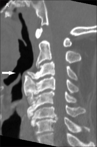
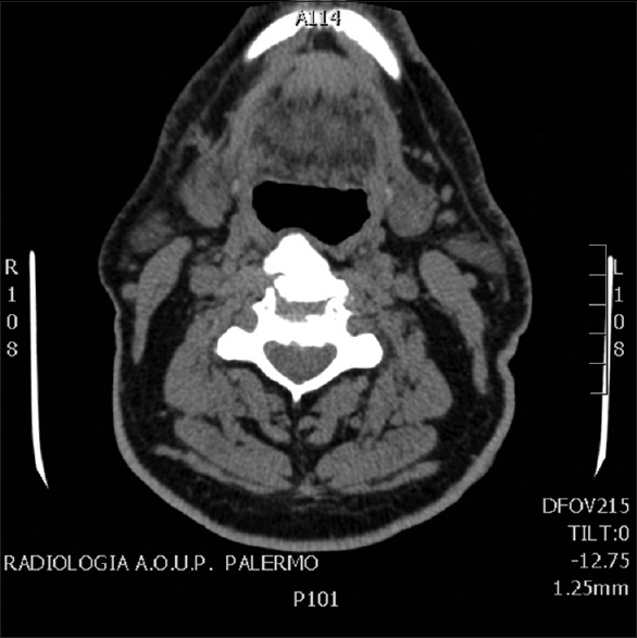
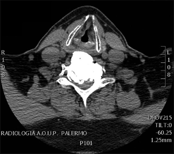
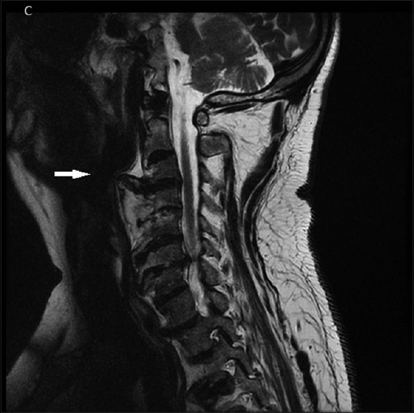
The computed tomography of the cervical spine demonstrated massive anterior longitudinal ligament ossification extending from C3 to C7 (DISH); there was an additional large osteophyte at C3-C4, and high-grade C6-C7 intracanalicular cervical stenosis due to ossification of the posterior longitudinal ligament [Figures

Refusal of surgery
Despite the dysphagia attributed to C3-C7 DISH and C3-C4 osteophyte, along with C6-C7 ossification of the posterior longitudinal ligament (OPLL), the patient refused surgery. Rather, the patient opted for conservative management, and was discharged on steroids and postural therapy.
DISCUSSION
The diagnosis of Forestier's disease or DISH is primarily radiological; its etiology is still unknown.[
Treatment of diffuse idiopathic skeletal hyperostosis
The initial treatment of cervical DISH is typically conservative therapy, for example, the use of anti-inflammatory medication, steroids, muscle relaxants, and postural education to optimize swallowing.[
Financial support and sponsorship
Nil.
Conflicts of interest
There are no conflicts of interest.
References
1. Carlson ML, Archibald DJ, Graner DE, Kasperbauer JL. Surgical management of dysphagia and airway obstruction in patients with prominent ventral cervical osteophytes. Dysphagia. 2011. 26: 34-40
2. Egerter AC, Kim ES, Lee DJ, Liu JJ, Cadena G, Panchal RR. Dysphagia secondary to anterior osteophytes of the cervical spine. Global Spine J. 2015. 5: e78-83
3. Forestier J, Rotes-Querol J. Senile ankylosing hyperostosis of the spine. Ann Rheum Dis. 1950. 9: 321-30
4. Giammalva GR, Iacopino DG, Maugeri R. Natura abhorret a vacuo. Future perspectives of autologous fibrin glue. Is it time for reappraisal?. Surg Neurol Int. 2017. 8: 57-
5. Goh PY, Dobson M, Iseli T, Maartens NF. Forestier's disease presenting with dysphagia and dysphonia. J Clin Neurosci. 2010. 17: 1336-8
6. Graziano F, Certo F, Basile L, Maugeri R, Grasso G, Meccio F. Autologous fibrin sealant (Vivostat(®)) in the neurosurgical practice: Part I: Intracranial surgical procedure. Surg Neurol Int. 2015. 6: 77-
7. Maugeri R, Giammalva GR, Graziano F, Iacopino DG. May autologue fibrin glue alone enhance ossification? An unexpected spinal fusion. World Neurosurg. 2016. 95: 611-2
8. Resnick D, Niwayama G. Radiographic and pathologic features of spinal involvement in diffuse idiopathic skeletal hyperostosis (DISH). Radiology. 1976. 119: 559-68
9. Sebaaly A, Boubez G, Sunna T, Wang Z, Alam E, Christopoulos A. Diffuse idiopathic hyperostosis manifesting as dysphagia and bilateral cord paralysis: A case report and literature review. World Neurosurg. 2018. 111: 79-85
10. Verlaan JJ, Boswijk PF, de Ru JA, Dhert WJ, Oner FC. Diffuse idiopathic skeletal hyperostosis of the cervical spine: An underestimated cause of dysphagia and airway obstruction. Spine J. 2011. 11: 1058-67

