

- Department of Neurosurgery, Maulana Azad Medical College, Lok Nayak Jai Prakash Narayan Hospital, Guru Nanak Eye Centre and G. B. Pant Institute of Postgraduate Medical Education and Research (G.I.P.M.E.R.), New Delhi, India
- Department of Pathology, Maulana Azad Medical College, Lok Nayak Jai Prakash Narayan Hospital, Guru Nanak Eye Centre and G. B. Pant Institute of Postgraduate Medical Education and Research (G.I.P.M.E.R.), New Delhi, India
- Department of Paediatrics, Sri Guru Ram Das Institute of Medical Sciences and Research, Amritsar, Punjab, India
Correspondence Address:
Charandeep S. Gandhoke
Department of Neurosurgery, Maulana Azad Medical College, Lok Nayak Jai Prakash Narayan Hospital, Guru Nanak Eye Centre and G. B. Pant Institute of Postgraduate Medical Education and Research (G.I.P.M.E.R.), New Delhi, India
DOI:10.4103/sni.sni_34_18
Copyright: © 2018 Surgical Neurology International This is an open access journal, and articles are distributed under the terms of the Creative Commons Attribution-NonCommercial-ShareAlike 4.0 License, which allows others to remix, tweak, and build upon the work non-commercially, as long as appropriate credit is given and the new creations are licensed under the identical terms.How to cite this article: Charandeep S. Gandhoke, Simran K. Syal, Hukum Singh, Daljit Singh, Ravindra K. Saran. Dorsal accessory ectopic breast with polythelia – A marker of occult spinal dysraphism. 24-Jul-2018;9:143
How to cite this URL: Charandeep S. Gandhoke, Simran K. Syal, Hukum Singh, Daljit Singh, Ravindra K. Saran. Dorsal accessory ectopic breast with polythelia – A marker of occult spinal dysraphism. 24-Jul-2018;9:143. Available from: http://surgicalneurologyint.com/surgicalint-articles/dorsal-accessory-ectopic-breast-with-polythelia-a-marker-of-occult-spinal-dysraphism/
Date of Submission
11-Feb-2018
Date of Acceptance
05-Jun-2018
Date of Web Publication
24-Jul-2018
Abstract
Background:Accessory breast, also known as supernumerary breasts, polymastia, or mammae erraticae, is a clinical condition of having an additional breast. Accessory breasts are usually seen along the embryonic milk line, with the majority located in the axilla. Polythelia is the presence of an additional nipple. We report a rare case of dorsal accessory ectopic breast with three nipples (two well formed and one rudimentary) occurring along with lipomeningomyelocele and diastematomyelia.
Case Description:We report the case of an 18-year-old female who presented with chief complaints of swelling over the upper back since birth and spastic weakness of bilateral lower limbs with inability to walk since 2 years. Three-dimensional computed tomography scan of the dorsal spine was suggestive of a wide bony defect in the posterior spinal elements from D3 to D9 vertebrae. Diastematomyelia was also seen. Magnetic resonance imaging of the dorsal spine was suggestive of a complex spinal dysraphism with lipomeningomyelocele and diastematomyelia. During surgery, the patient's accessory breast was removed, lipomatous tissue and bony septum were excised, and dural repair was done. Histopathological examination was consistent with accessory ectopic breast with lipomeningomyelocele.
Conclusion:Dorsal accessory breast, although a rare entity, whenever present should alert the clinician regarding the possibility of an underlying occult spinal dysraphism (OSD). Therefore, dorsal accessory breast can also be considered as a marker of OSD.
Keywords: Dorsal, ectopic breast, lipomeningomyelocele, occult spinal dysraphism, polythelia
INTRODUCTION
Accessory breast, also known as supernumerary breasts, polymastia, or mammae erraticae, is a clinical condition of having an additional breast. Accessory breasts are usually seen along the embryonic milk line, with the majority located in the axilla. Polythelia is the presence of an additional nipple. We report a rare case of dorsal accessory ectopic breast with three nipples (two well formed and one rudimentary) occurring along with lipomeningomyelocele and diastematomyelia. The association of dorsal accessory breast with meningomyelocele and diastematomyelia has been reported only once in the world literature, however, the association of dorsal accessory breast with polythelia with lipomeningomyelocele and diastematomyelia has never been reported.[
CASE DESCRIPTION
We report the case of an 18-year-old female who presented with chief complaints of swelling over the upper back since birth and spastic weakness of bilateral lower limbs with an inability to walk since 2 years. On examination, the swelling was soft-to-firm, nontender, midline in location, which started increasing in size at puberty and progressed since then to the present size of 14 cm × 12 cm [
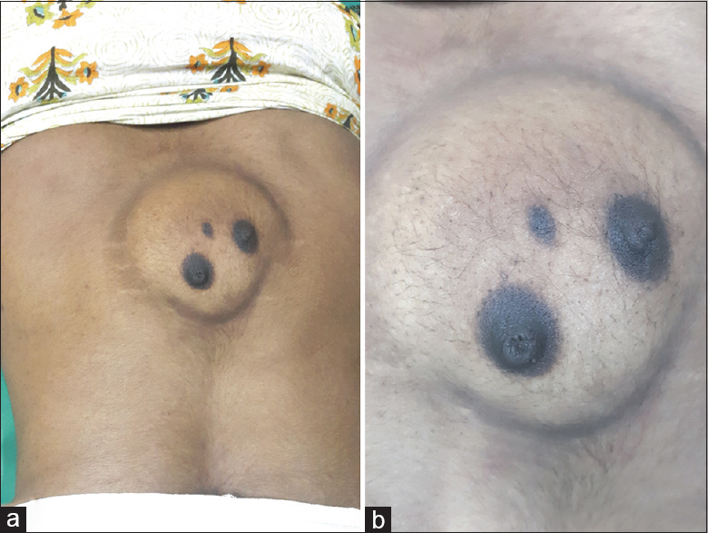
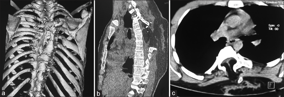
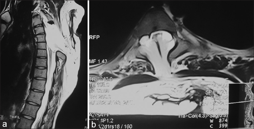
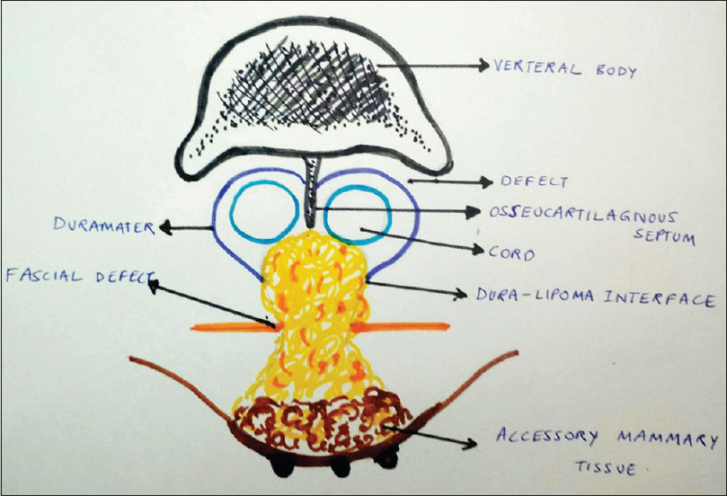
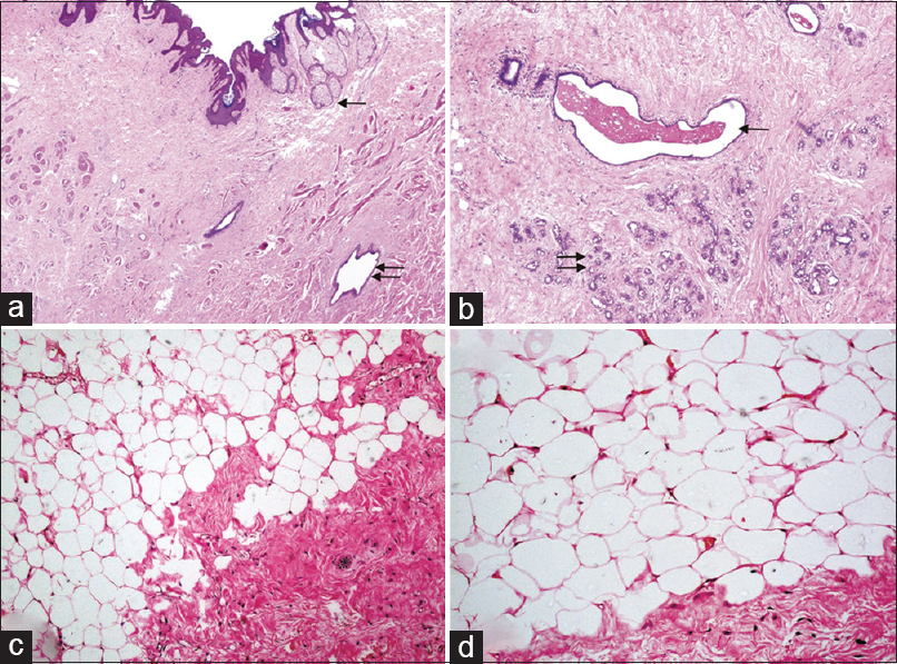
Figure 5
(a) Hematoxylin and eosin stained section (×40) showing normal skin with appendages (single arrow); Subepithelial tissue shows lactiferous duct (double arrow). (b) Hematoxylin and eosin stained section (×80) showing lactiferous duct with secretion inside (single arrow) along with terminal duct lobular units of breast (double arrow). (c and d) Hematoxylin and eosin stained section (×20 and ×40 respectively) showing lipomatous component composed of sheets of mature adipocytes with adjacent dural fibroblasts
The postoperative period was uneventful. Spasticity and power in the lower limbs improved and the patient was able to walk with support at the time of discharge.
DISCUSSION
Multiple theories have been proposed to explain the occurrence of accessory breast. These include Darwin's theory which stated that “traits which have disappeared generations before, can reappear,” Pfeifer's theory of metaplasia of sweat glands or modified sweat glands, Hughes's theory of random migration of primordial breast cells away from the mammary crest, and Schultz's theory of displacement of milk lines, laterally or caudally.[
Studies have shown that an ectopic breast usually increases in size after hormonal stimulation during puberty, pregnancy, or lactation.[
Occult spinal dysraphism (OSD) is characterized by skin-covered lesions without exposed neural tissue. Cutaneous markers of OSD include lipoma, fibroma pendulum, human or faun tail, dermal sinus, atypical dimple (larger than 5 mm and more than 2.5 cm from the anus), hamartoma, aplasia cutis congenita, deviation of the gluteal furrow, port wine stain, hemangioma, hypertrichosis, and acrochordon (skin tag).[
CONCLUSION
Dorsal accessory breast, although a rare entity, whenever present should alert the clinician regarding the possibility of an underlying OSD. Therefore, dorsal accessory breast can also be considered as a marker of OSD.
Declaration of patient consent
The authors certify that they have obtained all appropriate patient consent forms. In the form the patient(s) has/have given his/her/their consent for his/her/their images and other clinical information to be reported in the journal. The patients understand that their names and initials will not be published and due efforts will be made to conceal their identity, but anonymity cannot be guaranteed.
Financial support and sponsorship
Nil.
Conflicts of interest
There are no conflicts of interest.
References
1. Burdick AE, Thomas KA, Welsh E, Powell J, Elgart GW. Axillary polymastia. J Am Acad Dermatol. 2003. 49: 1154-6
2. Darwin C.editors. The descent of man and selection in relation to sex. New York: Appleton and Co; 1892. p. 36-7
3. Guggisberg D, Hadj-Rabia S, Viney C, Bodemer C, Brunelle F, Zerah M. Skin markers of occult spinal dysraphism in children: A review of 54 cases. Arch Dermatol. 2004. 140: 1109-15
4. Gupta VK, Kapoor I, Punia RS, Attri AK. Dorsal ectopic breast in a case of spinal dysraphism: A rare entity. Neurol India. 2015. 63: 392-4
5. Hughes ES. The development of the mammary gland: Arris and Gale Lecture, delivered at the Royal College of Surgeons of England on 25 th October, 1949. Ann R Coll Surg Engl. 1950. 6: 99-119
6. Pfeifer JD, Barr RJ, Wick MR. Ectopic breast tissue and breast-like sweat gland metaplasias: An overlapping spectrum of lesions. J Cutan Pathol. 1999. 26: 190-6
7. Schultz A.editors. Pathologische anatomic der brustdruse In: Handbuch der speziellen Pathologischen Anatomie and Histologie. Berlin: Julius Springer; 1933. p.
8. Shin SJ, Sheikh FS, Allenby PA, Rosen PP. Invasive secretory (juvenile) carcinoma arising in ectopic breast tissue of the axilla. Arch Pathol Lab Med. 2001. 125: 1372-4

