

- Department of Neurosurgery, Nakamura Memorial South Hospital, Sapporo, Hokkaido, Japan.
- Department of Neurosurgery, Nakamura Memorial Hospital, Sapporo, Hokkaido, Japan.
Correspondence Address:
Hideki Endo, Department of Neurosurgery, Nakamura Memorial South Hospital, Sapporo, Hokkaido, Japan.
DOI:10.25259/SNI_864_2021
Copyright: © 2021 Surgical Neurology International This is an open-access article distributed under the terms of the Creative Commons Attribution-Non Commercial-Share Alike 4.0 License, which allows others to remix, tweak, and build upon the work non-commercially, as long as the author is credited and the new creations are licensed under the identical terms.How to cite this article: Megumi Matsuda1, Hideki Endo1, Kohei Ishikawa1, Ryota Nomura1, Tomoaki Ishizuka1,2, Koji Oka1, Hirohiko Nakamura2. Extremely tortuous superior cerebellar artery mimicking an aneurysm. 23-Nov-2021;12:569
How to cite this URL: Megumi Matsuda1, Hideki Endo1, Kohei Ishikawa1, Ryota Nomura1, Tomoaki Ishizuka1,2, Koji Oka1, Hirohiko Nakamura2. Extremely tortuous superior cerebellar artery mimicking an aneurysm. 23-Nov-2021;12:569. Available from: https://surgicalneurologyint.com/surgicalint-articles/11246/
Date of Submission
27-Aug-2021
Date of Acceptance
01-Sep-2021
Date of Web Publication
23-Nov-2021
Abstract
Background: An extremely tortuous superior cerebellar artery is a rare anomaly. We report a case of an extremely tortuous superior cerebellar artery mimicking an aneurysm.
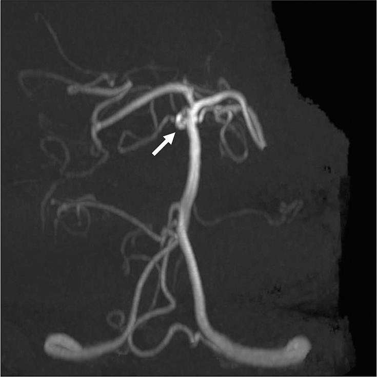
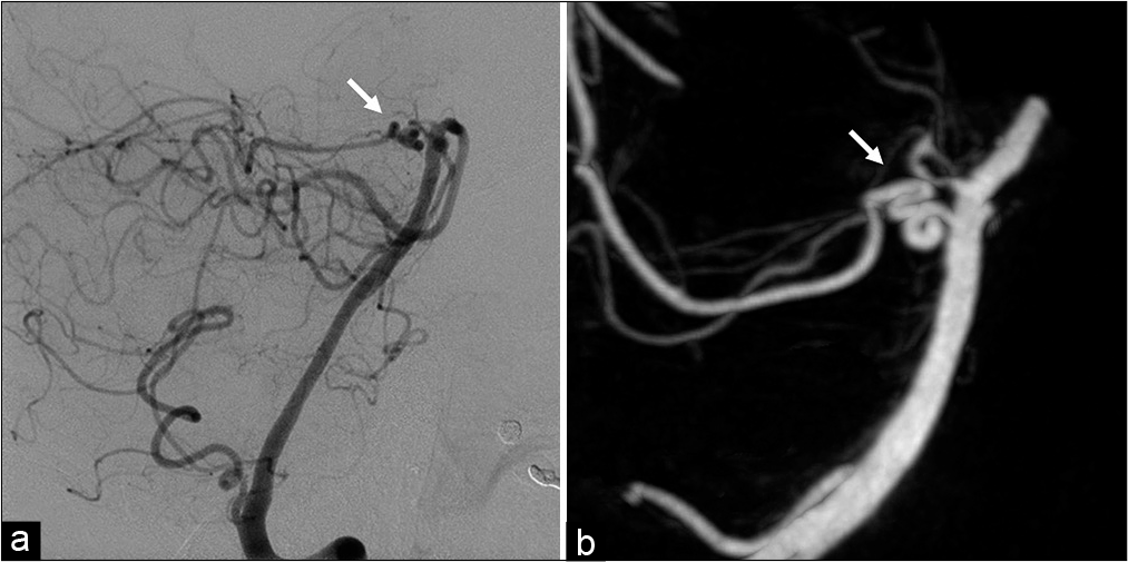
Case Description: A 77-year-old woman was initially diagnosed with unruptured cerebral aneurysm at the right basilar artery-superior cerebellar artery junction by magnetic resonance angiography. Catheter angiogram revealed that there was no apparent aneurysm at the basilar artery-superior cerebellar artery junction and the lesion was actually an extremely tortuous superior cerebellar artery.
Conclusion: Although an extremely tortuous superior cerebellar artery is rare, it should be considered when examining other vascular lesions.
Keywords: Anatomical anomaly, Cerebral aneurysm, Segmental dysgenesis, Superior cerebellar artery, Vascular malformations
CASE REPORT
A 77-year-old woman was initially diagnosed with unruptured cerebral aneurysms by magnetic resonance angiography. She had a family history of subarachnoid hemorrhage. The aneurysms were located at the right middle cerebral artery (MCA) and the right basilar artery-superior cerebellar artery (BA-SCA) junction. Sixteen years later, routine magnetic resonance angiography examination showed no apparent change in the BA-SCA lesion [
DISCUSSION
An extremely tortuous SCA is a rare anomaly. Anatomical variations of the SCA have been described in the past literature, including duplication, triplication, early bifurcation, fenestration, aplasia, originating from the posterior cerebral artery, and originating as a common trunk with the posterior cerebral artery.[
CONCLUSION
Here, we report a 77-year-old woman with an extremely tortuous SCA mimicking an aneurysm. Although this anomaly is rare, it should be considered when examining other vascular lesions.
Declaration of patient consent
The authors certify that they have obtained all appropriate patient consent.
Financial support and sponsorship
Nil.
Conflicts of interest
There are no conflicts of interest.
Acknowledgements
We thank Daishi Yamaguchi, Daisuke Mori, Seiji Ara, Tamio Ito, and Yoshihiro Sumi for their support. We also thank Justin Dean from Edanz (https://jp.edanz.com/ac) for editing a draft of this manuscript.
References
1. Brinjikji W, Cloft HJ, Flemming KD, Comelli S, Lanzino G. Pure arterial malformations. J Neurosurg. 2018. 129: 91-9
2. Krzyżewski RM, Stachura MK, Stachura AM, Rybus J, Tomaszewski KA, Klimek-Piotrowska W. Variations and morphometric analysis of the proximal segment of the superior cerebellar artery. Neurol Neurochir Pol. 2014. 48: 229-35
3. Songur A, Gonul Y, Ozen OA, Kucuker H, Uzun I, Bas O. Variations in the intracranial vertebrobasilar system. Surg Radiol Anat. 2008. 30: 257-64
4. Uchino A, Abe M, Sawada A, Takase Y, Kudo S. Extremely tortuous superior cerebellar artery. Eur Radiol. 2003. 13: L237-8
5. Uchino A, Sawada A, Takase Y, Kudo S. Variations of the superior cerebellar artery: MR angiographic demonstration. Radiat Med. 2003. 21: 235-8

