- School of Medicine, New York Medical College, Valhalla, New York, United States.
- Department of Neurosurgery, University of New Mexico Hospital, Albuquerque, New Mexico, United States.
- Department of Neurology, University of South Carolina School of Medicine, Columbia, South Carolina, United States.
- Department of Pathology, University of New Mexico Hospital, Albuquerque, New Mexico, United States.
Correspondence Address:
Christian A. Bowers, M.D. Vice Chair for Clinical Affairs, Department of Neurosurgery, University of New Mexico Health Sciences Center, 1 University New Mexico, Department of Neurosurgery, MSC10 5615. Albuquerque, NM 81731.
DOI:10.25259/SNI_169_2021
Copyright: © 2021 Surgical Neurology International This is an open-access article distributed under the terms of the Creative Commons Attribution-Non Commercial-Share Alike 4.0 License, which allows others to remix, tweak, and build upon the work non-commercially, as long as the author is credited and the new creations are licensed under the identical terms.How to cite this article: Jonathan V. Ogulnick1, Syed Faraz Kazim2, Andrew P. Carlson2, Smit Shah3, Alis J. Dicpinigaitis1, Karen SantaCruz4, Meic H. Schmidt2, Christian A. Bowers2. Fenestration of intracranial neurenteric cyst: A case report. 14-Jun-2021;12:287
How to cite this URL: Jonathan V. Ogulnick1, Syed Faraz Kazim2, Andrew P. Carlson2, Smit Shah3, Alis J. Dicpinigaitis1, Karen SantaCruz4, Meic H. Schmidt2, Christian A. Bowers2. Fenestration of intracranial neurenteric cyst: A case report. 14-Jun-2021;12:287. Available from: https://surgicalneurologyint.com/?post_type=surgicalint_articles&p=10880
Abstract
Background: Neurenteric cysts are rare congenital lesions of endodermal origin which result from the failure of the neurenteric canal to close during embryogenesis. The majority of neurenteric cysts occur in the spinal cord, though in rare instances can occur intracranially, typically in the posterior fossa anterior to the pontomedullary junction (80%) or in the supratentorial region adjacent to the frontal lobes (20%).
Case Description: We present the case of a 75-year-old woman with an extra-axial cystic lesion centered in the premedullary cistern causing brainstem compression. The lesion was later histopathologically confirmed to be a neurenteric cyst. She presented initially with a 4-month history of worsening headache, dizziness, and unsteady gait. We performed a left retrosigmoid craniotomy for cyst fenestration/biopsy with the aid of operating microscope and stealth neuronavigation. Following the procedure, the patient recovered without complications or residual deficits.
Conclusion: This case illustrates the successful fenestration of an intracranial neurenteric cyst with good clinical outcome. We present the pre- and post-operative imaging findings, a technical video of the procedure, histopathological confirmation, and a brief review of the relevant clinical literature on the topic.
Keywords: Cerebellopontine angle, Fenestration, Intracranial neurenteric cyst, Premedullary cistern, Retrosigmoid craniotomy
INTRODUCTION
Neurenteric (endodermal) cysts are uncommon, benign lesions of the central neuraxis that typically occur intraspinally because of aberrant embryological development of the notochord.[
CASE REPORT
History and examination
A 75-year-old woman presented with a 4-month history of worsening headaches along with progressive weakness, dizziness, and lightheadedness mainly when going from supine or sitting to standing. On neurological examination, the patient was noted to have mild resting tremor in the left hand and horizontal diplopia when attempting to look to the left, along with impaired convergence. The patient was also noted to have gait unsteadiness and poor balance.
Pre-operative neuroimaging
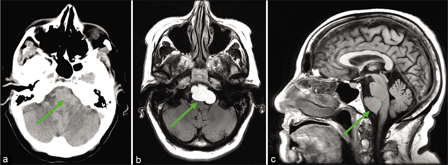
Computed tomography (CT) of the head without contrast showed a mildly hyperdense large extra-axial mass at the upper aspect of the premedullary cistern extending mildly to the inferior left cerebellopontine angle with compression of the brainstem [
Figure 1:
Pre-operative neuroimaging. (a) Axial computed tomography-head image showing mildly hyperdense large extraaxial mass at the upper aspect of the premedullary cistern extending mildly to the inferior left cerebellopontine angle with compression of the brainstem. (b) Axial magnetic resonance imaging (MRI)-brain T2- fluid-attenuated inversion recovery (FLAIR) image showing hyperintense left premedullary cistern and cerebellopontine angle mass. (c) Sagittal MRI-brain T1-FLAIR image showing isointense premedullary cistern mass. Arrow points to the left premedullary cistern mass later histopathologically confirmed to be a neurenteric cyst.
Magnetic resonance imaging (MRI) with/without contrast of the brain showed a T2-fluid-attenuated inversion recovery (FLAIR), well-circumscribed, hyperintense mass in the left cerebellopontine angle and premedullary cistern [
Operative details
A left retrosigmoid craniotomy was performed for neuroenteric cyst fenestration/biopsy with the use of operating microscope and use of Medtronic Stealth neuronavigation. A yellowish cyst was identified which was opened using an arachnoid knife and yellowish fluid immediately returned [
Video 1
Histopathological findings
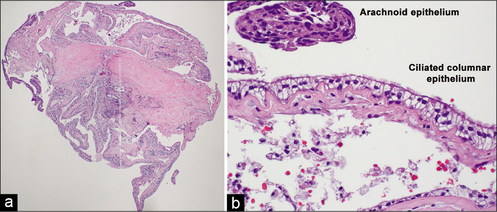
Microscopic examination of the tissue sections demonstrated a benign fibromembranous cystic lesion lined by pseudostratified, ciliated columnar epithelium, and providing a histopathological diagnosis of neurenteric cyst. [
Figure 2:
Microscopic examination of the cyst lining reveals a pseudostratified ciliated columnar epithelium. (a) Low-magnification hematoxylin and eosin (H and E) staining showing fibromembranous tissue lined by columnar epithelium. Imaged at ×4 magnification. (b) High-magnification H and E staining showing pseudostratified ciliated columnar epithelium and adjacent arachnoidal epithelium. Imaged at ×40 magnification.
Postoperative imaging and course
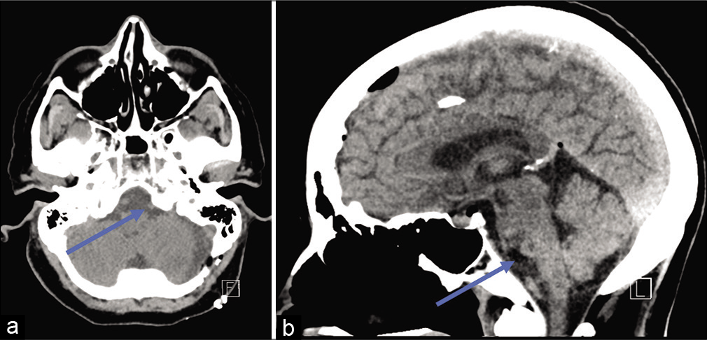
Postoperatively, the patient recovered well, and post-operative CT of the head without contrast showed no residual mass [
DISCUSSION
Clinical characteristics
A neurenteric cyst is a central nervous system lesion of endodermal origin which most frequently occurs in the spine. Intracranial neurenteric cysts have been estimated to represent about 17.9% of occurrences and of these, the vast majority occur in the posterior fossa. Rare cases of neurenteric cyst have been noted to occur in the brainstem, fourth ventricle, and supratentorial region.[
Imaging
The radiological characteristics of neurenteric cysts are heterogeneous. CT of neurenteric cysts typically reveal hypodense lesions, but can also appear isodense or, more rarely, hyperdense. CT-head in our case revealed a mildly hyperdense lesion. In addition, our patient’s cyst did not show internal enhancement, which is consistent with the literature.[
On MRI-brain, a classic neurenteric cyst usually presents as a well-circumscribed cystic mass which is enhancing and slightly iso- or hyperintense on T1-weighted sequences and slightly more hyperintense on T2-weighted-FLAIR sequences with mild restriction diffusion noted on diffusion-weighted imaging secondary to its high proteinaceous content.[
Surgical management
The standard of care for patients with a symptomatic neurenteric cyst is complete surgical excision. However, complete excision can be difficult if the cyst is adherent to important local structures, as was the case with our patient’s cyst which was adherent to the nearby vertebral and basilar artery. Although difficult, it is important to achieve as neurenteric cysts have an 11.9–37% recurrence rate during a span of 4 months to 14 years, thus resulting in the recommended 10-year follow-up period.[
In the present case report, the cyst was fenestrated using a left retrosigmoid approach. This is the primary means employed to gain access to the cerebellopontine angle - an anatomically complex site with many critical structures. Various vascular and neoplastic lesions that are approached in this manner include vestibular schwannomas, meningiomas, and aneurysms of nearby arteries.[
Histopathology
Three major histopathologic sub-types of neurenteric cysts have been noted in literature: Types A, B, and C.[
CONCLUSION
Neurenteric cysts are a rare, benign lesion of the central nervous system, typically occurring in the spine and less commonly intracranially. In the present report, we describe the case of a 75-year-old woman with a symptomatic lesion centered in the premedullary cistern causing brainstem compression. Fenestration of the intracranial cyst through retrosigmoid craniotomy resulted in good post-operative outcome. The associated imaging findings, operative video detailing fenestration of the intracranial neuroenteric cyst, and histopathologic findings are presented, along with relevant literature on the topic.
Declaration of patient consent
Patient’s consent not required as patients identity is not disclosed or compromised.
Financial support and sponsorship
Nil.
Conflicts of interest
There are no conflicts of interest.
Video available on:
www.surgicalneurologyint.com
References
1. Anderson T, Kaufman T, Murtagh R. Intracranial neurenteric cyst: A case report and differential diagnosis of intracranial cystic lesions. Radiol Case Rep. 2020. 15: 2649-54
2. Baek WK, Lachkar S, Iwanaga J, Oskouian RJ, Loukas M, Oakes WJ. Comprehensive review of spinal neurenteric cysts with a focus on histopathological findings. Cureus. 2018. 10: e3379
3. Brooks BS, Duvall ER, el Gammal T, Garcia JH, Gupta KL, Kapila A. Neuroimaging features of neurenteric cysts: Analysis of nine cases and review of the literature. AJNR Am J Neuroradiol. 1993. 14: 735-46
4. Cheng JS, Cusick JF, Ho KC, Ulmer JL. Lateral supratentorial endodermal cyst: Case report and review of literature. Neurosurgery. 2002. 51: 493-9
5. Cikla U, Kujoth GC, Baskaya MK. A stepwise illustration of the retrosigmoid approach for resection of a cerebellopontine meningioma. Neurosurg Focus. 2014. 36: 1
6. Eynon-Lewis NJ, Kitchen N, Scaravilli F, Brookes GB. Neurenteric cyst of the cerebellopontine angle: Case report. Neurosurgery. 1998. 42: 655-8
7. Gauden AJ, Khurana VG, Tsui AE, Kaye AH. Intracranial neuroenteric cysts: A concise review including an illustrative patient. J Clin Neurosci. 2012. 19: 352-9
8. Harris CP, Dias MS, Brockmeyer DL, Townsend JJ, Willis BK, Apfelbaum RI. Neurenteric cysts of the posterior fossa: Recognition, management, and embryogenesis. Neurosurgery. 1991. 29: 893-7
9. Heman-Ackah SE, Cosetti MK, Gupta S, Golfinos JG, Roland JT. Retrosigmoid approach to cerebellopontine angle tumor resection: Surgical modifications. Laryngoscope. 2012. 122: 2519-23
10. Kim CY, Wang KC, Choe G, Kim HJ, Jung HW, Kim IO. Neurenteric cyst: Its various presentations. Childs Nerv Syst. 1999. 15: 333-41
11. Osborn AG, Preece MT. Intracranial cysts: Radiologicpathologic correlation and imaging approach. Radiology. 2006. 239: 650-64
12. Preece MT, Osborn AG, Chin SS, Smirniotopoulos JG. Intracranial neurenteric cysts: Imaging and pathology spectrum. AJNR Am J Neuroradiol. 2006. 27: 1211-6
13. Tan GS, Hortobagyi T, Al-Sarraj S, Connor SE. Intracranial laterally based supratentorial neurenteric cyst. Br J Radiol. 2004. 77: 963-5
14. Wang L, Zhang J, Wu Z, Jia G, Zhang L, Hao S. Diagnosis and management of adult intracranial neurenteric cysts. Neurosurgery. 2011. 68: 44-52
15. Waqas M, Khan I, Khawaja R, Quddusi A, Enam SA. Self-resolving prepontine cyst. Surg Neurol Int. 2017. 8: 215


