- Department of Neurosurgery, G.B. Pant Institute of Post Graduate Medical Education and Research (G.I.P.M.E.R.), New Delhi, India
Correspondence Address:
Ankit S. Shah
Department of Neurosurgery, G.B. Pant Institute of Post Graduate Medical Education and Research (G.I.P.M.E.R.), New Delhi, India
DOI:10.4103/sni.sni_277_18
Copyright: © 2018 Surgical Neurology International This is an open access journal, and articles are distributed under the terms of the Creative Commons Attribution-NonCommercial-ShareAlike 4.0 License, which allows others to remix, tweak, and build upon the work non-commercially, as long as appropriate credit is given and the new creations are licensed under the identical terms.How to cite this article: Ankit S. Shah, Robin Gupta, Ghanshyam Singhal, Anita Jagetia, Daljit Singh. Fourth ventricle meningioma with cervical extension: An unusual entity. 28-Nov-2018;9:237
How to cite this URL: Ankit S. Shah, Robin Gupta, Ghanshyam Singhal, Anita Jagetia, Daljit Singh. Fourth ventricle meningioma with cervical extension: An unusual entity. 28-Nov-2018;9:237. Available from: http://surgicalneurologyint.com/surgicalint-articles/9097/
Keywords: Clear cell meningioma, dural attachment, fourth ventricle
A 35-year-old female patient was admitted with complaints of intermittent headache for 9 months and progressive gait disturbance for 6 months. She also complained of swaying while walking and disabling vertigo. Neurological examination on admission revealed ataxic dysarthria and ataxia of gait along with deranged cerebellar functions. Fundoscopy revealed papilledema.
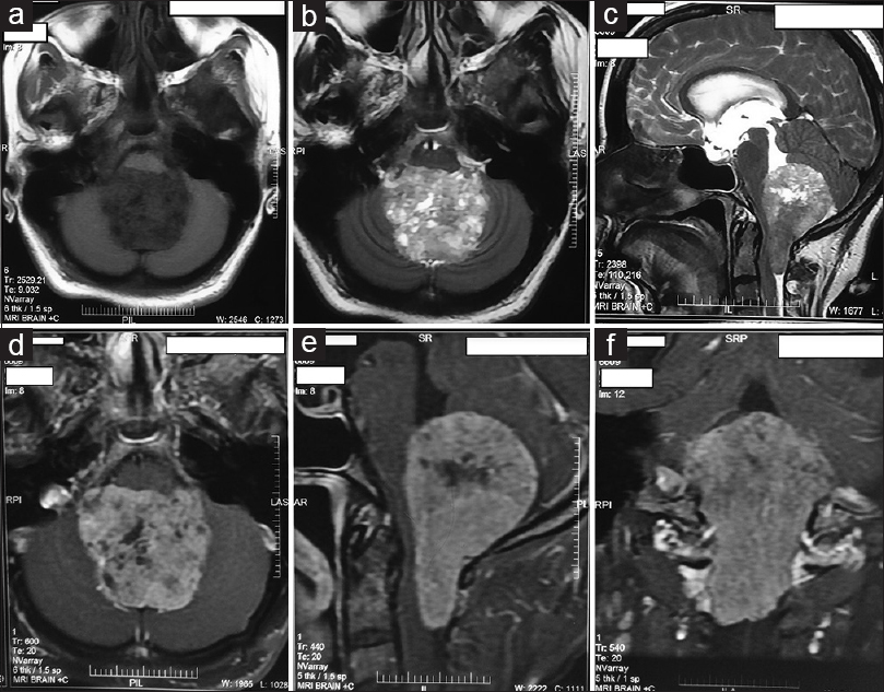
Computed tomography (CT) showed a homogeneous hyperdense lesion occupying the whole fourth ventricle, with proximal enlargement of ventricular horns and heterogeneous enhancement on contrast administration. There were neither intralesional calcifications nor cyst formations. Magnetic resonance imaging [Figure
Figure 1
Contrast-enhanced MRI of brain showing fourth ventricle occupying mass lesion extending into bilateral foramen of Luschka and into cervical cord which is hypointense on T1W axial (a); heterogenous hyperintense on T2W axial (b) and sagittal (c); and heterogeneously enhancing on contrast administration on axial (d), sagittal (e), and coronal (f) images
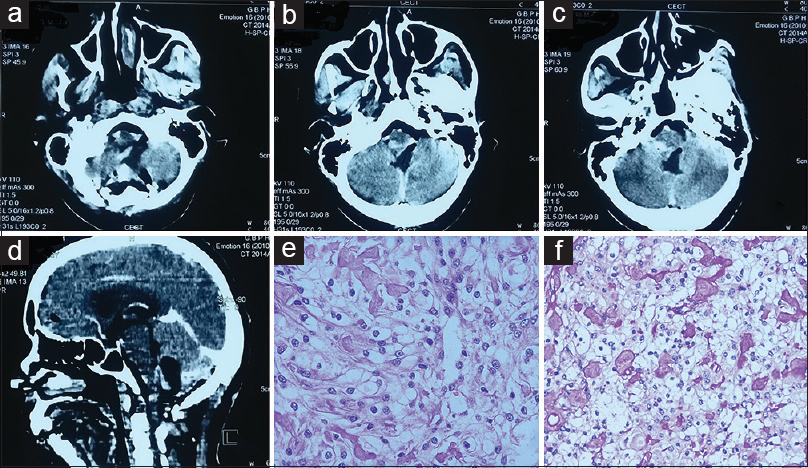
Surgery was done in prone position via suboccipital craniotomy and C1 posterior arch excision. Tumor was encountered on opening dura. It was firm, well-vascularized, grayish, encapsulated, and extending into cervical canal compressing the spinal cord. No attachment to dura or choroid plexus was encountered. Gross total excision was achieved and postoperative contrast-enhanced CT showed no residual [Figure
Figure 2
Postoperative contrast enhanced computed tomography. (CECT) imaging showing complete excision of mass lesion on axial (a–c) and sagittal (d) images. Histopathological photographs of hematoxylin–eosin stain section (20× magnification) (e) showing patternless arrangement of tumor cells with interspersed collagen fibrils and clear cytoplasm. Periodic acid–Schiff–diastase staining (f) showing breakdown of glycogen and clear cytoplasm suggestive of clear-cell meningioma
Histopathological evaluation [Figure
Meningiomas occurring primarily in fourth ventricle are rare entities and are believed to be originating from choroid plexus or tela choroidae.[
Preoperative diagnosis of meningioma is difficult but should be kept in mind as differential as they differ in terms of surgical challenge and clinical outcome as compared to other tumors in this location.
Declaration of patient consent
The authors certify that they have obtained all appropriate patient consent forms. In the form the patient(s) has/have given his/her/their consent for his/her/their images and other clinical information to be reported in the journal. The patients understand that their names and initials will not be published and due efforts will be made to conceal their identity, but anonymity cannot be guaranteed.
Financial support and sponsorship
Nil.
Conflicts of interest
There are no conflicts of interest.
References
1. Alver I, Abuzayed B, Kafadar AM, Muhammedrezai S, Sanus GZ, Akar Z. Primary fourth ventricular meningioma: Case report and review of the literature. Turk Neurosurg. 2011. 21: 249-53
2. Burgan O, Bahl A, Critcher V, Zaki H, McMullan P, Sinha S. Clear cell meningioma of the fourth ventricle in a child: A case report and literature review. Pediatr Neurosurg. 2010. 46: 462-5
3. Shrestha R, Yue-Kang Z, Chao Y. Fourth ventricular meningioma in an adult: Case report and review of the literature. Indian J Neurosurg. 2012. 01: 161-4


