

- Glasgow Neuro Society, Wolfson School of Medicine, University of Glasgow, Scotland, United Kingdom,
- Department of Neurology, Institute of Neurological Sciences, Queen Elizabeth University Hospital, Glasgow, Scotland, United Kingdom,
- Department of Neurosurgery, Institute of Neurological Sciences, Queen Elizabeth University Hospital, Glasgow, Scotland, United Kingdom.
Correspondence Address:
Mohammad Ashraf, President, Glasgow Neuro Society, Wolfson School of Medicine, University of Glasgow, Scotland, United Kingdom.
DOI:10.25259/SNI_268_2022
Copyright: © 2022 Surgical Neurology International This is an open-access article distributed under the terms of the Creative Commons Attribution-Non Commercial-Share Alike 4.0 License, which allows others to remix, transform, and build upon the work non-commercially, as long as the author is credited and the new creations are licensed under the identical terms.How to cite this article: Ashraf M1, Ismahel H1, Lub S1, Middleton E1, Chaudhary A1, Gardee A1, Salloum LA1, Szilagyi-Nagy E1, Wales R1, Omar S1, Baird TA2, O’Kane R3, , Holden J, Abdelsadg M, Burr P, Decruz AB, Badran K, Amato-Watkins A, Alakandy L, Boardman R, Nicely L, Tandon V, Banerjee S, Keenlyside A, Hughes M, Ho JW, Sieradzki J, My Phan EP, James MacDougall NJ, Azad Bashir AB B, Hutton DL, F. Doley AS, Oochit KK, Kontorinis G, Wong YY. Glasgow Neuro Society 2021 Conference Proceedings. Surg Neurol Int 29-Apr-2022;13:173
How to cite this URL: Ashraf M1, Ismahel H1, Lub S1, Middleton E1, Chaudhary A1, Gardee A1, Salloum LA1, Szilagyi-Nagy E1, Wales R1, Omar S1, Baird TA2, O’Kane R3, , Holden J, Abdelsadg M, Burr P, Decruz AB, Badran K, Amato-Watkins A, Alakandy L, Boardman R, Nicely L, Tandon V, Banerjee S, Keenlyside A, Hughes M, Ho JW, Sieradzki J, My Phan EP, James MacDougall NJ, Azad Bashir AB B, Hutton DL, F. Doley AS, Oochit KK, Kontorinis G, Wong YY. Glasgow Neuro Society 2021 Conference Proceedings. Surg Neurol Int 29-Apr-2022;13:173. Available from: https://surgicalneurologyint.com/surgicalint-articles/11570/
Date of Submission
20-Mar-2022
Date of Acceptance
26-Mar-2022
Date of Web Publication
29-Apr-2022
Welcoming Editorial
As Sir Isaac Newton once said, “If I have seen further, it is by standing on the shoulder of giants.” With immense pleasure and privilege accorded to me by the giants before me, I write as President of Glasgow Neuro Society 2021–2022. With humble beginnings in 2010, Glasgow Neuro Society was founded by Mr Allan Hall, a current specialist registrar neurosurgeon at the Institute of Neurological Sciences (INS) Glasgow, who back then, was a medical student at the University of Glasgow. The initial aim was to provide undergraduates and junior doctors interested in neurology, neurosurgery, and clinical neuroscience; an opportunity to improve their knowledge and inspire all toward a field that traditionally was and still is neglected in terms of exposure to the average medical student. But with overwhelming support from world-leading neurosurgeon and founder of the Glasgow Coma Scale, Sir Professor Graham Teasdale, Allan built the society to a nationally reputable position where it commanded support from pioneers in neurology and neurosurgery both nationally and internationally.
Over the years, as our standards and outreach grew, Glasgow Neuro gained support and sponsorship from international companies, including major medical charities, airlines, and corporations, to support the Society’s events, of which, the flagship is our annual conference. Over the past decade, we have been able to bring a range of speakers to Scotland who are authorities from across the globe, including the chief of neurosurgery at John Hopkins, Henry Brem, and the previous director of pediatric neurosurgery George Jallo. May other esteemed speakers like the chief of neurosurgery at Harvard, Robert Martuza, Professor Sir Graham Teasdale, Professor Charlie Teo, Professor James Goodrich, Professor Henry Marsh, and pioneer of deep brain stimulation Professor Alim Louis Benabid have also honored the occasion. We never shied away from controversial topics and brought Professor Sergio Canavero, who revealed his ongoing head-transplant research at our 2016 conference. After Allan, the past presidents and my predecessors, who imparted considerable inspiration to me, and gave me a society already at the forefront of Neuro Societies in the UK, are my giants.
Our annual conference attracts delegate from across the United Kingdom who present their research and attend talks by our national and international invited faculty. It was our pleasure this year to host Professor Gary Steinberg, Founder, and Previous Chair of Neurosurgery at Stanford University, Founder of Stanford Stroke Centre and Moya Moya Centre, Mr Alistair Jenkins, President Society of British Neurological Surgeon, Professor Naveed Ashraf, Founder and Previous Chairman Neurosurgery (Unit 3), Lahore General Hospital, and Founder and Previous Chair of the Department of Neurosurgery Jinnah Hospital Lahore, Pakistan, Mr Patrick Mitchell, Consultant Neurosurgeon and Previous Editor-in-Chief of the British Journal of Neurosurgery, and Dr Paul Bentley, Clinical Director of Imperial College Network Of Excellence in Rehabilitation Technology and Consultant Neurologist at Imperial College London. As always, outstanding feedback demonstrated that our speakers’ talks left delegates and committee as humbled as much inspired. Verily, those at the forefront of their fields left students and junior doctors awestruck, opening their eyes to just how far neurology and neurosurgery have gone and the exciting prospects of rapid life-changing advancement in the years to come.
With support from the neurosurgical faculty at the INS Glasgow, we provided delegates with hands-on workshops. Mr Calan Mathieson engaged with delegates in a Q&A on life as a consultant neurosurgeon in the UK; Mr Michael Cearns provided targeted advice guiding delegates on meeting the requisites of the much-feared national selection for neurosurgery which represents one of the most rigorous and competitive training programs in the UK. Professor Ashraf conducted our flagship, hands-on intracranial pressure monitoring workshop, providing delegates with the closest possible simulation to one of the most common neurosurgical procedures. We are extremely grateful to Mr Tony Poutney, Scottish Territory Manager for Delta Surgical, who provided Raumedic’s cranial access kits and ICP bolts for this educational endeavor. As a personal reflection, it was enlightening for me and delegates to appreciate neurotrauma, a subspecialty of neurosurgery which is the arguably oldest (sub)specialty and makes up the majority of the workload for most neurosurgeons. Despite being so, it has only recently been acknowledged in its own right. Professor Ashraf, while being recognized for his pioneering work on cerebrovascular surgery in Pakistan, whilst especially AVMs, cerebral revascularization, and bypasses nationwide took up the arduous task to advance neurotrauma in Pakistan. He began multimodal monitoring, introducing ICP, brain parenchymal oxygen and temperature monitoring, and cerebral microdialysis to the National Health Service in a composite manger by introducing the concept of neurosurgeon driven Neuro Critical Care in the country, lighting the path for future generations of neurosurgeons to engage in neurotrauma and head injury research.
I want to extend my sincerest gratitude to my committee, each of whom in their own right led aspects of organizing this annual conference and who were burdened by the ever-evolving uncertainty of restrictions and unexpected turn of events due to the pandemic that negatively impacted organization in the run-up to the conference. I am humbled to have worked with such dynamic individuals who allowed Glasgow Neuro to execute another conference that matched the caliber of the previous years through their sheer dedication and commitment. We want to thank our honorary Presidents, Mr Roddy O’Kane, and Dr Tracey Baird from INS Glasgow, who supported and aided in organizing this conference, and chaired the abstract presentation videos. In addition, we owe a debt of gratitude to Mr Likhith Alakandy, Mr Calan Matheison and Ms Emer Campbell, consultant neurosurgeons, and Mr Michael Cearns and Mr Allan Hall, Neurosurgical Registrars, at INS Glasgow, all of whom led workshop stations and chaired sessions at this conference. We want to acknowledge the support of the University of Glasgow’s Medical School and Dr John Paul Leach, Professor of Neurology and Dean of Medicine, who shared our societies passion and devotion to galvanize students toward neuroscience and thank him for his constant guidance and support to Glasgow Neuro.
The following abstracts were presented at Glasgow Neuro’s 9th Annual Conference held at the Royal College of Physicians and Surgeons of Glasgow on November 20, 2021. These are accompanied by their respective video presentations in a panel discussion format on Surgical Neurology’s newest electronic format, E-SNI, which we are honored to be a part of. The concept of a panel discussion was conceived by Professor James Ausman (Founder and Emeritus Editor-in-Chief of SNI) and aimed to make research accessible to everyone across the globe. We are privileged to be one of the first medical school neuro-society within the UK, to have their conference proceedings published in a major neurosurgical journal. We are thrilled to play a small role in Professor Ausman’s pioneering vision on improving the dissemination of research in conference proceedings from a standard written vanilla format to adding a more engaging touch by accompanying a practical discussion. We are grateful to Professor Ausman, Professor Nancy Epstein (SNI Editor-in-Chief) and Mr James Cook (managing editor) for their constant guidance in the production of these videos and publication of these proceedings.
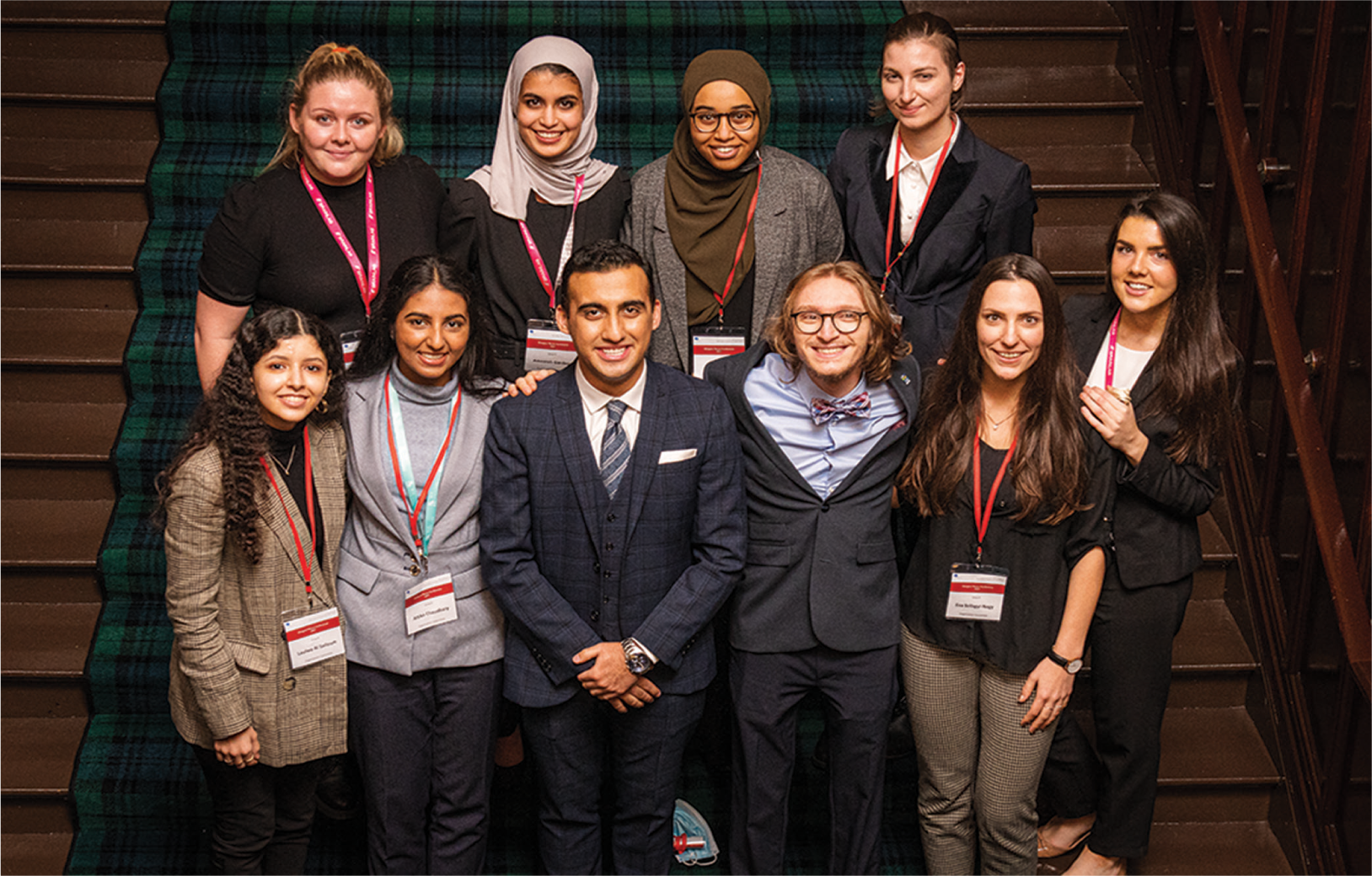
Glasgow Neuro Society Committee 2021–2022. Top Row from left: Eilidh Middleton (Treasurer), Ameerah Gardee (Educational Officer), Samia Omar (Clinical Skills Officer), Sytske Lub (Secretary), and Rachel Wales (Junior Representative). Bottom Row from left: Laulwa Al Salloum (Conference Convenor-Neurosurgery), Attika Chaudhary (Senior Representative), Mohammad Ashraf (President), Hassan Ishmael (Vice-President), and Eva Szilagyi-Nagy (Conference Convenor-Neurology). Photographed at the Royal College of Physicians and Surgeons of Glasgow. Glasgow Neuro Society Committee 2021–2022. Top Row from left: Eilidh Middleton (Treasurer), Ameerah Gardee (Educational Officer), Samia Omar (Clinical Skills Officer), Sytske Lub (Secretary), and Rachel Wales (Junior Representative). Bottom Row from left: Laulwa Al Salloum (Conference Convenor-Neurosurgery), Attika Chaudhary (Senior Representative), Mohammad Ashraf (President), Hassan Ishmael (Vice-President), and Eva Szilagyi-Nagy (Conference Convenor-Neurology). Photographed at the Royal College of Physicians and Surgeons of Glasgow.
ABSTRACTS
ABSTRACT 1
(Accompanying video can be seen here:
In silico stratification of extracellular phosphoproteome reveals five secreted exCK substrates as prognostic biomarkers of glioblastoma
Lynden Guy Nicely, Vasudha Tandon, Sourav Banerjee
Department of Cellular Medicine, School of Medicine, University of Dundee, Scotland, United Kingdom.
Background: Stage IV glioma (glioblastoma) is one of the most aggressive malignancies in humans. To date, there are no prognostic biomarkers for glioma. Overexpression of the atypical protein kinase exCK has been shown to correlate with poor survival outcomes in glioblastoma. Recent discoveries show that exCK phosphorylates a large number of substrates in the extracellular space and secretory pathway. Here, we tested the hypothesis that substrates and affiliated proteins of exCK contribute toward the oncogenic affiliations of exCK.
Methods: Using Genome Expression Profiling Interactive Analysis 2 (GEPIA2) and The Chinese Glioma Genome Atlas (CGGA) data, we individually stratified substrates and related family members of exCK through various rounds of shortlisting. Initial shortlisting of all reported exCK substrates and associated proteins was carried out using the GEPIA2 to identify overexpression in low-grade glioma and glioblastoma. We then used CGGA data to identify proteins with a consistent oncogenic profile.
Results: Our results show that 53 exCK-related proteins are overexpressed in glioma. We reveal five substrates – MMP14, SPP1, IL-6, MMP7, and PDIA1 – that are promising biomarkers in glioma with consistent oncogenic profiles of an effective biomarker in glioblastoma with the literature supporting their roles in gliomagenesis and progression.
Conclusion: The five shortlisted substrates of exCK are likely to play a role in the oncogenic affiliations of exCK with glioma. The identification of these five proteins as plausible prognostic or stratification biomarkers in the early detection and risk stratification of glioma provides an initial step for the development of novel therapeutics and interventions for brain cancer.
ABSTRACT 2
(Accompanying video can be seen here:
Effect of total body weight and body mass index on lymphocyte counts in Scottish multiple sclerosis patients treated with cladribine
Elizabeth Phuong My Phan1,2, Niall John James MacDougall1,2,3
1Wolfson School of Medicine, University of Glasgow, Scotland, United Kingdom, 2Department of Neurology, University Hospital Hairmyres, East Kilbride, South Lanarkshire, 3Department of Neurology, Institute of Neurological Sciences, Queen Elizabeth University Hospital, Glasgow, Scotland, United Kingdom.
Background: Scotland and Lanarkshire have some of the highest global incidences of multiple sclerosis (MS) and higher obesity rates than much of Europe. Cladribine for treating MS is dosed based on total body weight (TBW). Having observed higher rates of lymphopenia after pivotal cladribine clinical trials, we hypothesized that obesity may have some effect on lymphopenia rates in our cohort. We investigated correlations between TBW and body mass index (BMI) and severe lymphopenia in cladribine-treated people with MS (PwMS).
Methods: Data on general demographics, weight, height, cladribine doses, the previous disease-modifying therapies, and lowest lymphocyte counts (in years 1 and 2 of taking cladribine) were collected from 68 PwMS. Data were analyzed using Pearson Correlation Tests and regression analyzes. If P < 0.05, correlation was considered significant.
Results: About 41.7% and 44.4% of patients in Year 1 and 2, respectively, were obese (BMI > 30 kg/m2). Mean BMI was 30.6 in Year 1 and 32.0 in Year 2. About 11.0% and 42.2% of patients in this group had Grade 3 lymphopenia in Years 1 and 2, respectively, compared to only 4.0% and 11.3% in the literature. No significant correlation was found between TBW and lowest lymphocyte count or total cladribine dose and lowest lymphocyte count. There was a weak but significant negative linear correlation between BMI and lowest lymphocyte count in Year 2 of taking cladribine.
Conclusion: Higher BMI appeared to correlate with the lower lymphocyte count in cladribine-treated patients in Year 2. Cladribine doses may be overestimated in obese patients. Our small sample size reduces the certainty of these results; hence, similar studies with larger cohorts would clarify this relationship. Alternative dosing scalars more accurate than TBW could be explored for cladribine dosing.
ABSTRACT 3
(Accompanying video can be seen here:
Quality of epilepsy care in a rural and deprived Scottish population
John Holden
BBT2 Trainee, NHS Ayrshire and Arran, Crosshouse, Scotland, United Kingdom.
Background: Epilepsy is among the most common neurological disorders globally. Appropriate prescription and good adherence to anticonvulsants can achieve seizure freedom rates of 70%. Scotland is an affluent nation with free at point-of-access healthcare but there remain significant health-care inequalities, particularly associated with deprivation. Anecdotally, people with epilepsy in rural Ayrshire rarely engage with health-care services. We describe the prevalence and management of epilepsy in a deprived and rural Scottish population.
Methods: Electronic records were used to obtain the following for patients with coded diagnoses of “Epilepsy” or “Seizures” within a general practice list of 3500 patients: demographics, diagnosis, seizure types, date and level (primary and secondary) of last review, last seizure date, anticonvulsant prescription, and adherence as well as any clinic discharge due to nonattendance.
Results: Ninety-two patients were coded as above. Fifty-six had a current diagnosis of epilepsy (prev 16.1/100,000). About 69% had good adherence. About 56% had good seizure control, with adherence associated with control. Of the 68% managed by primary care, 33% were uncontrolled and 13% had an epilepsy review in the previous year. About 45% of patients referred to secondary care were discharged for nonattendance.
Discussion: We demonstrate a high prevalence of epilepsy, low anticonvulsant adherence, and sub-optimal rates of seizure freedom. These may be linked to poor attendance at specialist clinics. Management in primary care is challenging as evidenced by low review rates and high rates of ongoing seizures. We propose that the synergistic factors of uncontrolled epilepsy, deprivation, and rurality make it difficult to attend clinics, with resultant health inequalities.
ABSTRACT 4
(Accompanying video can be seen here:
The digital Cullen chart: A red color perimetry aid for neuro-opthalmological examination
Andrew Keenlyside1, Mark Hughes2, Jen Wae Ho3, Jake Sieradzki4
1School of Medicine, University of Dundee, Dundee, Scotland, United Kingdom,
2Department of Clinical Neurosciences, BioQuarter, Edinburgh,
3Department of Neurosurgery, North Bristol Trust, Bristol,
4School of Informatics, University of Edinburgh, Edinburgh, United Kingdom.
Background: Thirty years ago, a paper chart was developed by Cullen et al. and validated as a swift, supplementary method for perimetric evaluation of the central 25 degrees of the visual field. We have redeveloped this concept in digital form (as a smartphone application) and sought to assess its sensitivity and specificity in detecting field loss, through comparison with formal machine-based perimetry.
Methods: Following refinement of app usability, a case series pilot study was developed to test app-based Cullen chart efficacy in generating indicative data by concordance of app and formal machine-based perimetry data on visual field loss. Eighteen nonsecretory and secretory pituitary adenoma, craniopharyngioma, and parasellar meningioma patients at Western General Hospital, Edinburgh were involved in this study. Patients underwent formal visual field perimetry as part of standard care. They also underwent assessment using the smartphone-based Cullen chart as part of routine outpatient assessment. Thirty-seven eye episodes were assessed, incorporating pre- and post-treatment assessment for a range of potentially compressive pathologies.
Results: The digital Cullen chart had a sensitivity of 75% and specificity of 98% compared with machine-based perimeters. The positive predictive value was 93% and negative predictive value was 92%.
Conclusion: In the context of visual field assessment for patients with sellar/ parasellar tumors, this smartphone-based chart shows good concordance with machine-based perimeters. It is, therefore, an accessible visual field screening and monitoring tool for clinicians. With further study, there is also potential for approval as a remote patient-led visual field monitoring method.
ABSTRACT 5
(Accompanying video can be seen here:
Speech-induced oromandibular dystonia: A case report
Aneesah Bashir Binti Azad Bashir
Woflson School of Medicine, University of Glasgow, Scotland, United Kingdom.
Background: Oromandibular dystonia (OMD) is a focal neurological disorder characterized by repetitive involuntary movements of the face, jaw, and/or tongue. It could either be idiopathic (primary) or acquired (secondary) where it is attributed to brain damage, neurodegenerative disorders, neuroleptic medications, or infections.
Case Presentation: A 49-year-old man presented with a 1 month history of involuntary jaw opening and tongue protrusion when speaking. He had no medical or family history of any neurological conditions. An interesting clinical feature was that the patient experienced a relief of symptoms and saw an improvement in speech whenever he had chewing gum or toothpick in his mouth and when he applied slight pressure on his jaw or wore a mask (sensory tricks). On conducting a detailed neurological examination, the patient’s sensory, motor, coordination, and reflexes were found to be normal. Laboratory and imaging tests were carried out to rule out possible secondary causes of OMD. The patient’s full blood count, serum electrolytes, renal function tests, liver function tests, creatine phosphokinase, and thyroid profile were unremarkable. An MRI of the brain showed no abnormalities. He was prescribed Trihexyphenidyl (2 mg) twice daily which was changed to thrice daily during his follow-up visit after 1 month. Baclofen (10 mg) was also prescribed to be taken once at night. Oral medications showed significant improvement in jaw movement but not in tongue protrusion. He then received electromyography-guided botulinum toxin injected to the tongue muscles through a superficial approach. The patient is still undergoing treatment and is showing good prognosis with significant speech improvement.
Conclusion: OMD is a challenging diagnosis which should not be missed as appropriate management can provide good long-term results. It is important to take an accurate history and to not miss out details such as the presence of sensory tricks which could aid in making an accurate diagnosis.
ABSTRACT 6
(Accompanying video can be seen here:
IgG4-related disease involving the temporal bone: A systematic review
Krishna K. Oochit1, Georgios Kontorinis1,2, Yun Yan Wong1
1Wolfson School of Medicine, University of Glasgow, Scotland, United Kingdom, 2Department of Otorhinolaryngology-Head, Neck and Skull Base Surgery, Queen Elizabeth University Hospital, Glasgow, Scotland, United Kingdom.
Background: IgG4-related disease (IgG4-RD) is an emerging fibroinflammatory condition that can involve any anatomic site. Hamano et al. first described the disease in 2001 involving the pancreas. IgG4-RD involving the temporal bone is an uncommon and underrecognized anatomical location of the disease and is often mistaken for malignancy, infection, or other immune-related diseases. This systematic review is the first that aims to analyze IgG4-RD of the temporal bone and review the presentation, management, and prognosis of the disease.
Methods: A detailed literature search was performed from four different databases: Ovid Medline, Embase, Cochrane library, and Google scholar using a combination of keywords. The bibliographies of these articles were also searched for articles that our database search missed. Seventeen studies with 22 cases were included after a thorough risk of bias assessment.
Results: The most common presenting symptoms were hearing loss, otalgia, and headache. The mastoid and petrous portions of the bone were most commonly involved. Both computed tomography and magnetic resonance were used to assess the extent of bony involvement. Biopsies showed the characteristic lymphoplasmacytic infiltrate in all patients, 10/22 cases reported the storiform fibrous pattern while only 3/22 reported obliterative phlebitis. About 63.6% reported ratio IgG4+: total IgG+ > 40%. Most patients were treated with corticosteroids ± surgery or combination of other immunosuppressants. About 95.5% of patients demonstrated positive symptomatic response with their initial treatment regimen. Seven patients relapsed after initial symptomatic response. All patients were symptom free at their past follow-up. (Mean follow-up time 14.5 months).
Conclusion: This review is the most comprehensive analysis of the disease to date. IgG4-RD of the temporal bone can present as mass occupying lesions causing cranial nerve palsies to locally invasive lesions involving the middle/ inner ear. Diagnosis involves a thorough workup (clinical examination, imaging, and biopsy). Treatment involves corticosteroids and/or surgery depending on extent of disease involvement.
ABSTRACT 7
(Accompanying video can be seen here:
Can endoscopic third ventriculostomy replace ventriculoperitoneal shunts in the treatment of pediatric hydrocephalus: A critical review
Rosie Boardman
School of Medicine, University of St Andrews, St Andrews, Scotland, United Kingdom.
Background: The pediatric population has the highest incidence of hydrocephalus, with approximately 1 case/1000 children. The standard treatment is currently ventriculoperitoneal shunting (VPS); however, VPS is associated with high complication rates, and surgeons are now beginning to investigate the use of endoscopic third ventriculostomy (ETV) as an alternative. The primary objective was to conduct a review of the present literature, to determine whether ETV is a viable alternative and to identify the most effective treatment option available.
Methods: A search was carried out across five databases, using key words to identify studies comparing any aspect of the use of ETV and VPS in pediatric hydrocephalus. Studies were then processed using Endnote, and inclusion and exclusion criteria were applied. Studies identified as suitable by these criteria were then further screened for validity and bias using relevant checklists, before being summarized in evidentiary tables. Thirty-four full-text articles were assessed for eligibility, 20 of which were excluded, leaving 14 studies to be included in the review.
Results: Clinical effectiveness, rate of complications, effect on development and quality of life, and effectiveness in infant cohorts, as well as economic efficiency were identified as key areas for comparison. ETV was largely found to be more effective for long term control; however, this was age and etiology dependent, with infants benefiting the least. Likewise, ETV was found to carry fewer complications. VPS appeared to offer more rapid control, and so improved quality of life and cognitive development, especially in younger cohorts, but the data were not statistically significant. The data for economic efficiency were also not statistically significant due to varying follow-up periods and the tendency for late failure/complications in VPS.
Conclusion: While ETV may not yet be appropriate in completely replacing VPS to treat pediatric hydrocephalus, it can continue to provide a promising alternative, with hope that the future research will guide evidence-based guidelines and enable its widespread use in this population.
ABSTRACT 8
(Accompanying video can be seen here:
Investigating the association between Type 2 diabetes and Parkinson’s disease using electronic medical records in the GoDARTS bioresource
Dana L. Hutton, Alexander S. F. Doley
Division of Population Health and Genomics, Ninewells Hospital and Medical School, University of Dundee, Dundee, Scotland, United Kingdom
Background: Pre-clinical and clinical data have linked Type 2 diabetes (T2D) and Parkinson’s disease (PD), suggesting pathological mechanisms associated with insulin resistance may influence the development and progression of PD. This prospective case–control study utilized the GoDARTS bioresource to investigate the impact of T2D on PD incidence. GoDARTS represents a large case–control study of T2D with longitudinal follow-up in electronic medical records (EMR).
Methods: The GoDARTS bioresource was used to determine the risk of T2D status for incident PD that accounted for competing risk of death. PD patients were passively identified in the EMR, using a combination of General Practice Read-Codes, hospital admission data, hospital death data, and prescribing data. Cause-specific and sub-distributional hazards were used to compare hazards of PD. Age and sex were included in all models. Individuals were followed up from 45 years of age, until the earliest PD event recorded in the EMR. Censoring was the earliest date of non-PD death or the end of available EMR data. Individuals with evidence of PD before entry date were excluded from the analysis. It may be that other genetic and lifestyle factors are implicated in both T2D and PD. As the leading cause of death in T2D patients is cardiovascular disease (CVD), we investigated the impact of various cardiovascular risk factors. We used the T2D cohort to examine differences in characteristics between subjects with or without a PD event. We also considered a genome-wide polygenic risk score for CVD (CVDprs) to see whether genetic factors influencing CVD outcomes were important in PD.
Results: T2D status was associated with a significant risk of PD (cause-specific hazard ratio = 1.40; 95% CI = 1.14–1.73). However, this association was weakened when accounting for competing risk of death (subdistribution HR = 1.18; 95% CI = 0.96–1.45). Subjects who developed PD were significantly older, of higher SIMD5 index, lower BMI, and had lower diastolic blood pressure, total cholesterol, and triglyceride levels. No significant difference was seen with systolic blood pressure, alcohol status, physical activity, or CVDprs. Smoking was a protective factor.
Conclusion: Our study indicates that T2D is associated with increased risk of PD. The lack of association of CVD risk factors among T2D patients suggests that the link between T2D and PD may be explained by insulin resistance. T2D and PD remain incurable and continue to increase in incidence. However, further investigating their association may enable implementation of preventative lifestyle and clinical interventions.
ABSTRACT 9
(Accompanying video can be seen here:
Retrospective study looking at suspected CSF shunt(s) malfunction patients in out of hours setting, aiming to identify factors affecting duration of hospital stay
Mohamed Abdelsadg1, Pierce Burr1, Antony Ben Decruz2, Khaled Badran1, Anthony Amato-Watkins1,2,3, Likhith Alakandy1
1Department of Neurosurgery, Institute of Neurological Sciences, Queen Elizabeth University Hospital, Glasgow, Scotland, United Kingdom,
2Wolfson School of Medicine, University of Glasgow, Scotland, United Kingdom, 3Department of Pediatric Neurosurgery, The Royal Hospital For Children, Glasgow, Scotland, United Kingdom.
Background: Papilledema represents an important sign of CSF shunt failure and symptoms of shunt malfunction may warrant an ophthalmology referral. The objectives of this study are to identify the number of patients admitted with suspected CSF shunt malfunction to the emergency neurosurgical service in a tertiary neurosurgical center within an 18-month period. The next objective is to identify demographics and presenting complaints for patients admitted out of hours with suspected CSF shunt(s) malfunction. Finally, this study aims to identify any factors affecting the duration of stay in hospital for this cohort.
Methods: The retrospective study examining the prospectively populated-emergency referral database in a tertiary neurosurgical unit, to identify and subsequently analyze out of hours admissions with suspected CSF shunt(s) malfunction. Patients with suspected CSF shunt(s) malfunction referred out of hours (17:00–08:00) to the on call neurosurgical registrar in the institute of neurosciences, at QEUH-Glasgow between January 1, 2019, and June 30, 2020. Once the cohort of patients identified from the on call referral database during the specified period, the electronic patients record and the operative database was examined to note the admission and the operative course when applicable.
Results: During the study period, 96 admissions were made for suspected shunt malfunction among 43 patients. Apart from a peak of admissions during the first decade of life, they were generally spread out across the age groups. Seventeen patients were admitted once while 26 had recurrent admissions (defined as two or more admissions). The mean number of admissions per patient was 2.23 while the median number of admissions was 3. The greatest number of admissions per patient within 2 years was six admissions. It is noted that there is a general decline in the number of patients having recurrent admissions after the first decade; moreover, there is a positive relationship between the number of admissions and the mean days of hospital stay. The three most common presenting complaints were headache (31 patients) and nausea and vomiting (24 patients) followed by a suspected shunt infection (15 patients). Among the admitted patients, 13 patients had nil interventions done. The remaining admissions had a total of 111 interventions and the mean number of interventions per admission was 1.23. Except for 0 interventions, there is generally an inverse relationship between the number of interventions and the number of admissions. Conversely, the more the number of interventions, the higher the mean days of stay in hospital. Among the patients admitted, 25% received an ophthalmology referral. The mean number of days of stay for those referred to ophthalmology was 9.38 while the median number of days was 5. About 23% of the referrals which required no intervention received an ophthalmology referral but mean days of stay were 2 days.
Conclusion: The study reviewed patients admitted with suspected CSF shunt malfunction to the neurosurgical department. It highlighted the relationship between recurrent admissions and different age groups also identified an interesting pattern of an inverse relationship between the number of interventions and the number of admissions at least during the study period. The study also highlighted the duration of hospital stay for patients awaiting formal ophthalmology opinion in CSF shunt(s) patients, a solution of which would be to improve out of hours access to such specialized test(s).

