- Department of Neurosurgery, Kwong Wah Hospital, Hong Kong,
- Division of Neurosurgery, Department of Surgery, Prince of Wales Hospital, Hong Kong,
- Division of Neurosurgery, Department of Surgery, Queen Mary Hospital, Hong Kong,
- Department of Neurosurgery, National Neuroscience Institute, Singapore,
- Department of Neurosurgery, Albert Einstein Hospital, Sao Paulo, Brazil.
Correspondence Address:
Peter Y. M. Woo, Department of Neurosurgery, Kwong Wah Hospital, Hong Kong.
DOI:10.25259/SNI_70_2022
Copyright: © 2022 Surgical Neurology International This is an open-access article distributed under the terms of the Creative Commons Attribution-Non Commercial-Share Alike 4.0 License, which allows others to remix, transform, and build upon the work non-commercially, as long as the author is credited and the new creations are licensed under the identical terms.How to cite this article: Peter Y. M. Woo1, Danise Au1, Natalie M. W. Ko1, Oscar Wu1, Emily K. Y. Chan2, Kevin K. F. Cheng3, Alain K. S. Wong1, Ramez Wadie Kirollos4, Guilherme Ribas5, Kwong-Yau Chan1. Gods and monsters: Greek mythology and Christian references in the neurosurgical lexicon. 25-Feb-2022;13:67
How to cite this URL: Peter Y. M. Woo1, Danise Au1, Natalie M. W. Ko1, Oscar Wu1, Emily K. Y. Chan2, Kevin K. F. Cheng3, Alain K. S. Wong1, Ramez Wadie Kirollos4, Guilherme Ribas5, Kwong-Yau Chan1. Gods and monsters: Greek mythology and Christian references in the neurosurgical lexicon. 25-Feb-2022;13:67. Available from: https://surgicalneurologyint.com/surgicalint-articles/11414/
Abstract
Background: Myths and religion are belief systems centered around supernatural entities that attempt to explain the observed world and are of high importance to certain communities. The former is a collection of stories that belong to a cultural tradition and the latter are organized faiths that determine codes of ethics, rituals and philosophy. Deities or monstrous creatures in particular act as archetypes instructing an individual’s conduct. References to them in Greek mythology and Christianity are frequently manifested in the modern neurosurgical vernacular.
Methods: A review of the medical literature was performed using the PubMed and MEDLINE bibliographic databases. Publications from 1875 to 2021 related to neurosurgery or neuroanatomy with the medical subject headings (MeSH) terms mythology, religion, Christianity and Catholicism were reviewed. References pertaining to supernatural beings were classified to either a deity or a monstrosity according to their conventional cultural context.
Results: Twelve narratives associated with neurosurgery were identified, nine relating to Greek mythology and three associated with the Christian-Catholic faith. Eight accounts concerned deities and the remaining with monstrous creatures.
Conclusion: This article explores the etymology of commonly utilized terms in daily neurosurgical practice in the context of mythology and religion. They reveal the ingenuity and creativity of early pioneers who strived to understand the brain.
Keywords: Christianity, Greek mythology, Neuroanatomy, Neurosurgery, Religion
INTRODUCTION
The word mythology is derived from the combination of the Greek words “study” (logos) and “myth” (mûthos), a symbolic narrative concerning the early history of a people or a natural phenomenon associated with religious belief. As a means to rationalize the inexplicable nature of observed events, myths have played important functions in providing continuity to a culture and creating archetypes for how one should behave. In particular, the tales of supernatural gods and monsters served as cautionary allegories for exemplary or reprehensible conduct. Greek mythology is considered one of the oldest and richest of all mythologies.[
MATERIALS AND METHODS
A review of the medical literature was performed using the PubMed and MEDLINE bibliographic databases. Using the Boolean operator “AND,” publications related to neurosurgery or neuroanatomy with the medical subject headings terms mythology, religion, Christianity, and Catholicism were reviewed. The following search limits were: publication date from January 1875 to December 2021; reports concerning studies of humans; and reports in English, French or German. The citations from the included reports were also manually searched to find other studies that were not identified initially. The final search was performed on January 3, 2022.
RESULTS
The Gods
Poseidon’s steed and the horns of Ammon: The hippocampus
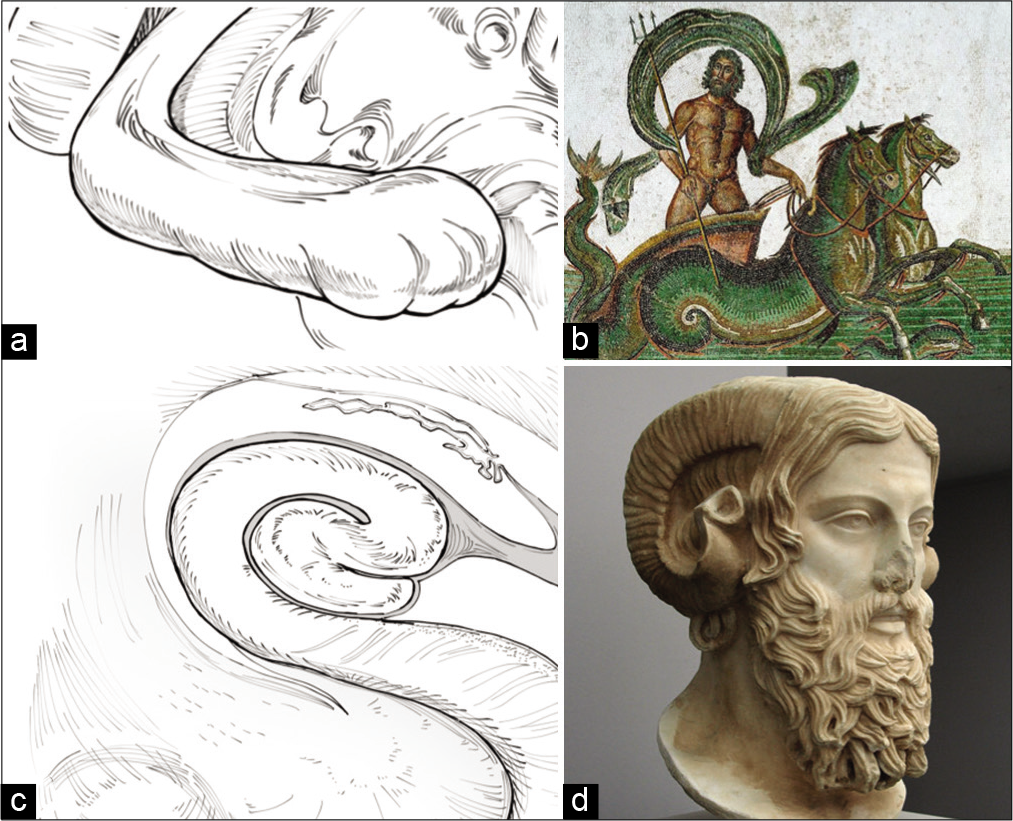
The hippocampal formation is phylogenetically one of the oldest structures of the brain serving critical functions in learning, episodic, and spatial memory. The structure was named by Julius Caesar Arantius (1529–1589) in the 16th century due its gross morphological resemblance to a seahorse, the small marine fish in the genus hippocampus (Greek: hippos for “horse” and kampos for “sea”).[
Figure 1:
Schematic of the right hippocampus (a) and its mythological counterpart, the steed of Poseidon, Greek god of the sea (b), Roman mosaic, 3rd century, courtesy of the Sousse Museum, Tunisia. A coronal section schematic of the body of the hippocampus revealing the hippocampal formation and the cornu ammonis (c). Ammon, the amalgamation of the gods Zeus and Amun-Ra with his distinctive horns (d), marble sculpture, 2nd century, courtesy of Liebieghaus Skulpturensammlung, Frankfurt, Germany.
Hermes and his winged helmet: The pterion
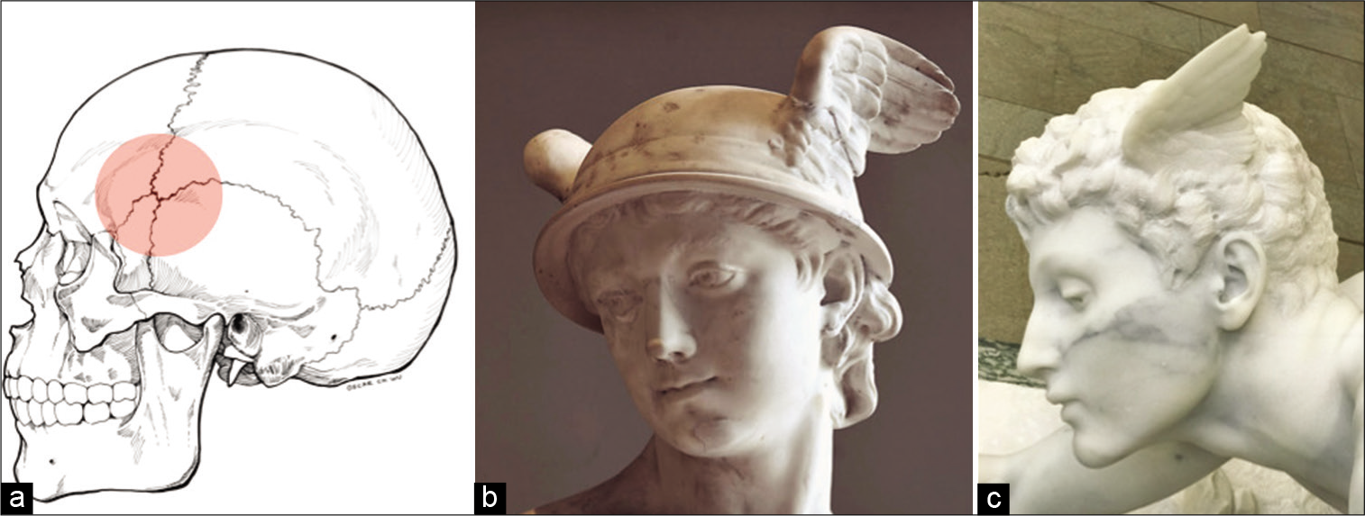
Hermes, the son of Zeus, is considered the herald of the Olympian gods and protector of travelers. He is classically presented wearing the talaria, winged sandals, and a winged petasos, a sun hat or helmet, that bestowed the power of swift travel [
Figure 2:
Schematic of the established neurosurgical landmark, the pterion (a) and Broca’s association with Hermes’s, the messenger god, wings of flight attached to his helmet (b), photograph by the author PW, marble sculpture, 18th century, Louvre Museum, Paris, France or directly from his head at this region (c), photograph by the author PW, marble sculpture, 19th century, courtesy of the Musée d’Orsay, Paris, France.
Atlas the titan and the C1 vertebra
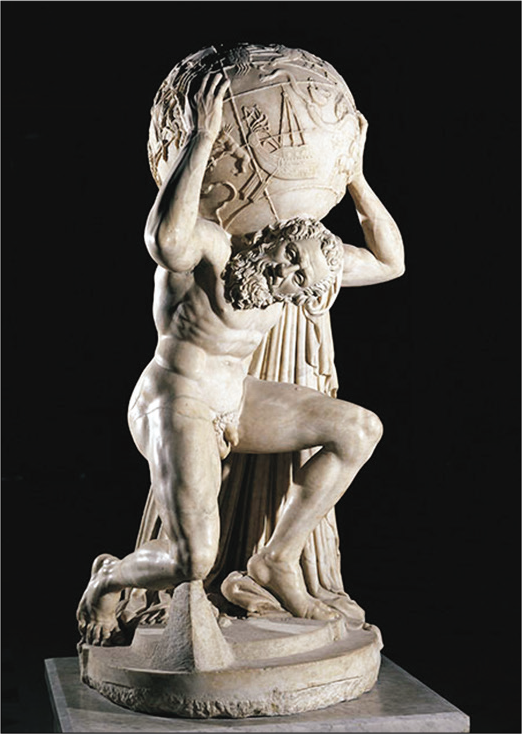
Atlas was a Titan, an older generation of gods that preceded the Olympians, condemned to hold up the sky for eternity on his shoulders according to Hesiod’s Theogeny. His punishment was a result of his defeat in the War of the Titans, the Titanomachy, that was fought against the Olympian gods for dominion of the universe. The first vertebra of the cervical spine takes its name from the Titan for its anatomical location supporting the weight of the skull.[
The thalamus: Resting place of immortals
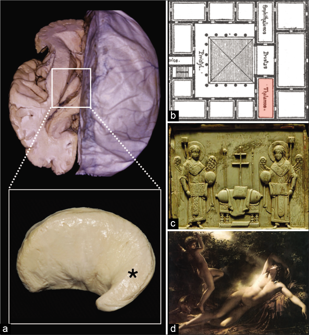
Aelius Galenus (129–216 CE) or Claudius Galenus, commonly known as Galen, was the first to introduce the term “thalamus,” derived from the Greek word thalamos for “sleeping chambers” in the 2nd century CE [
Figure 4:
The thalamus, “sleeping chamber”, within the central core, and the pulvinar, “cushion” (*, inset, a). The architectural plans of a typical Roman villa following Vitruvian design indicating the location of the thalamos, the master bedroom (b), Vitruvius Pollio, De Architectura, 1st century BCE. The cushioned empty throne pulvinus, a ritualistic object often used in religious ceremonies. Note the inscription Hetoimasia, Latin for “prepared throne” for the second coming of Christ (c), Ivory icon, 10th century, courtesy of the Louvre Museum, Paris, France. The final sleeping place of Endymion within Mount Latmos (d), The Sleep of Endymion, oil on canvas by Anne-Louis Girodet de Roucy-Triosson, 18th century, courtesy of the Louvre Museum, Paris, France.
Golgotha and skull hill: The calvaria
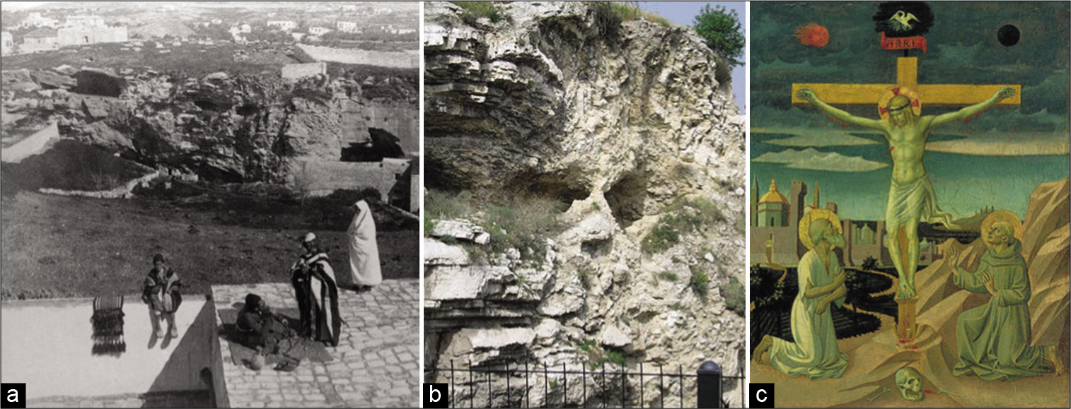
The calvaria was originally described as the “upper part of the head” enclosing the brain. Golgotha (Aramaic for “place of the skull”), also called Calvary in its anglicized form, is the name for the hill of execution situated outside ancient Jerusalem’s walls and the site of Jesus’s crucifixion. The spot is mentioned in all four of the canonical Gospels, for example, in Matthew 23:33 “And when they were come unto place call Golgotha, that is to say a place of a skull.” As an execution site, it would have been strewn with skulls, but others have attributed the hill’s namesake for its topographical shape upon which the Church of the Holy Sepulcher is now located [
Figure 5:
Calvary or Golgotha “Skull Hill”, the believed location of the crucifixion of Jesus as seen in 1901 from the northern walls of Jerusalem’s old city (a), courtesy of The Garden Tomb (Jerusalem) Association. A current day photograph revealing a section of the hill topographically resembling the features of a skull (b), photograph by the author PW. Skull Hill was likely strewn with the skulls of the executed (c), The Crucifixion with Saint Jerome and Saint Francis, tempera on wood by Pesellino, 15th century, courtesy of the National Gallery of Art, Washington, D.C., USA.
The petrous bone, claustrum, fornix, and the thalamus: Cerebral vitruvian architecture
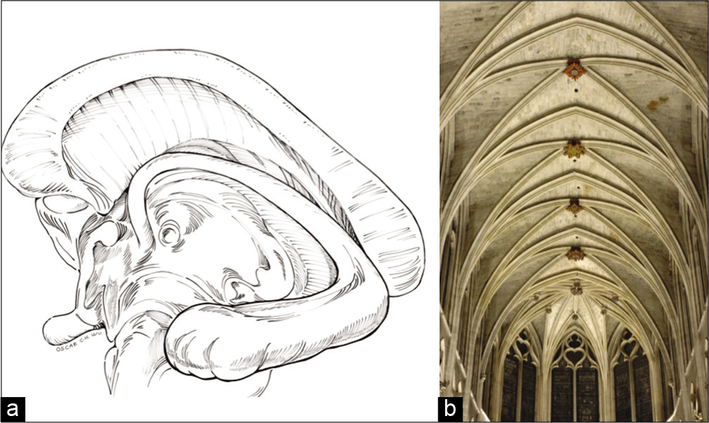
Marcus Vitruvius Pollo (80–15 BCE), commonly known as Vitruvius, was a Roman architect who described in his seminal work De Architectura, the importance of viewing the human body as the principal source of proportion for constructing places of worship.[
The petrous, an important neurosurgical landmark, is a pyramid-shaped portion of the temporal bone located at the skull base between the sphenoid and occipital bones. Petrous is derived from the Greek word pétra meaning “stone” which is also the etymological root for the name Peter (or Pieter, Pierre). It was also the name Jesus bestowed to his apostle Simon, subsequently known as Saint Peter, one of the first leaders of the early Church. Although it is unclear who or why this skull base landmark was named the petrous bone, an intriguing dialogue between Jesus and Simon exists in Matthew 16:13-19: “Blessed are you, Simon son of Jonah, for this was not revealed to you by flesh and blood, but by my Father in heaven. And I tell you that you are Cephas (Peter) (Petros), and on this rock (petra) I will build my church, and the gates of Hades will not overcome it. I will give you the keys of the kingdom of heaven.” The association between this ideology and the naming of the petrous bone as a literal foundation on which the brain rests is compelling.
The English term cloister (Latin: claustrum for “enclosure”) represents a covered walkway or gallery running along the walls of a quadrangular church space. Cloisters effectively separated those looking to lead an ascetic monastic life from the distractions of the city. Anatomically, the claustrum, a thin layer of grey matter lying between the extreme and external capsules, is situated within the equally aptly named insular lobe. Its function has yet to be understood, but it has been proposed to be involved in permitting widespread cross-model transmission of perceptual, cognitive, and motor information contributing to synchronized processing.[
The fornix (Latin: fornix for “arch” or “vault”) is a C-shaped structure forming part of the limbic lobe. It is the major efferent pathway from the hippocampus to the mammillary bodies originating from the mesial temporal lobe arching around the thalamus to the diencephalon and basal forebrain.[
The three crowns: Coronal suture, corona radiata and Saint Stephen
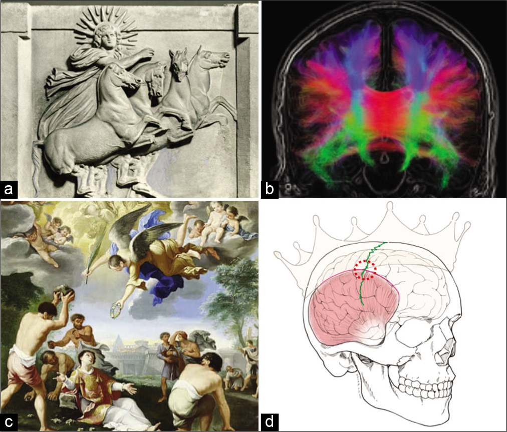
The crown (Latin: corona or ancient Greek: korone) is a traditional head adornment typically worn by monarchs symbolizing their power and dignity. In European Christendom, the royal crown is placed on the new monarch’s head by an ecclesiastical official in a coronation ceremony. The coronal suture and the corona radiata were most likely coined by early Greek anatomists seeking inspiration from Helios, the god of the sun, who was often portrayed adorning a crown embellished with sun rays when viewed on the frontal plane [
Figure 7:
Helios, the sun god of Greco-Roman mythology, riding his chariot across the sky adorned with his corona “crown” (a), marble relief, 4th century BCE, courtesy of the Pergamon Museum, Berlin, Germany. The corona radiata known today, a term used to describe the numerous thalamo-cortical projection fibers running through the internal capsule (b), coronal view of magnetic resonance imaging tractography. Saint Stephen, the first martyr of Christendom, being bestowed a crown as he is stoned to death (c), The Stoning of Saint Stephan, oil on canvas by Luigi Garzi, 18th century. The Stephanion (red encircled area), an important craniometric point representing the intersection of the coronal suture (green) and the superior temporal line. The landmark indicates the location of the intersection between the inferior frontal sulcus and the pre-central sulcus (d).
First systematically documented by Broca in Sur la topographie crânio-cérébrale in the 1870s, craniometric points are superficial landmarks that aid with the localization of important intracranial structures.[
From apoplexy to being stricken by god’s hand
Before the modern utilization of the word “stroke” as a diagnosis, apoplexy (Greek: apoplexia for “to be struck down with violence”) was the predominant term for millennia until the early 20th century.[
The Monsters
A tale of jealousy and piety in the meninges: The arachnoid and pia mater
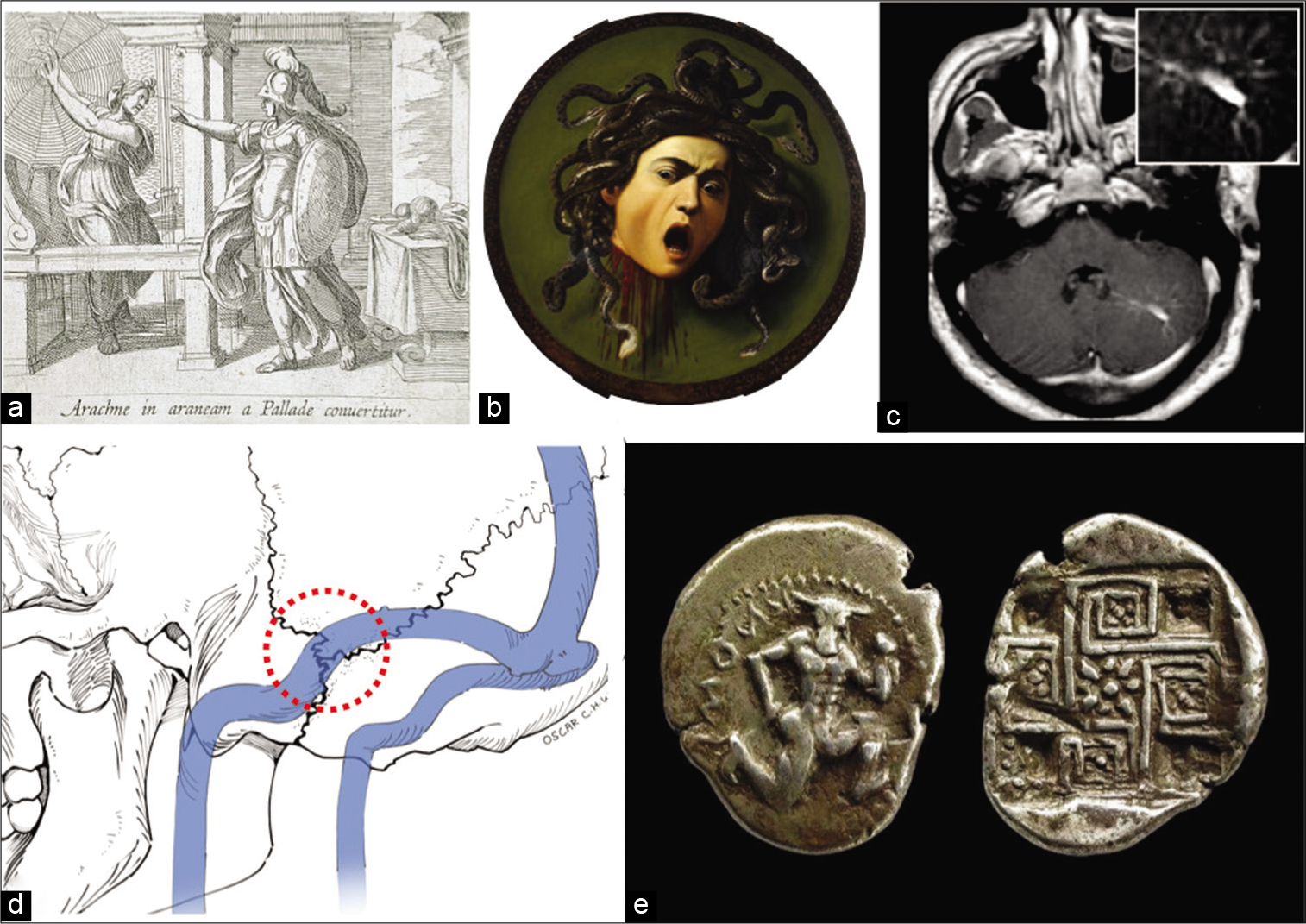
Herophilus of Chalcedon (335–280 BCE), known as the father of anatomy, described the arachnoid trabeculae as reminiscent of a spider’s web and bestowed the name “arachnoid” from the Greek word arachne for “spider” for this layer of the meninges.[
Figure 8:
The monsters gallery. Arachne at the moment of being transformed into a spider by the jealous Athena (a), Athena Changing Arachne into a Spider, plate by Antonio Tempesta, 17th century. The classic portrayal of Medusa at the moment of her beheading (b), Medusa, oil on canvas by Caravaggio, 16th century, courtesy of the Uffizi, Florence, Italy. A magnetic resonance imaging scan revealing an incidental caput medusa of the left cerebellum (c), axial, T1-weight gadolinium contrast-enhanced sequence. The asterion, a craniometric landmark historically used to indicate the location of the sigmoid-transverse dural venous sinus junction (asterion, red-encircled area, d). A silver coin discovered at the ruins of Knossos, Crete, revealing Asterion “the starry one,” the name of the ancient city state’s legendary minotaur, and his dwelling at the center of the labyrinth (e), 5th century BCE.
Use of the Latin word mater, denoting “mother,” to describe the meninges was derived from the Arab term umm aldimāgh meaning “mother of the brain” and was introduced by the medieval Persian physician Haly Abbas (930–994).[
The tragedy of Medusa
The neuroradiological term caput Medusa (Latin for “head of Medusa”) is used to describe cerebral developmental venous anomalies that comprise the commonest form of vascular malformations.[
Asterion: The craniometric landmark and the minotaur
The asterion is the junction of the lambdoid, occipitomastoid, and parietal-mastoid sutures and is an important neurosurgical landmark for lateral approaches to the posterior fossa. It is historically considered a craniometric landmark for the junction between the transverse and sigmoid dural venous sinuses [
Pan and Syrinx’s vengeance
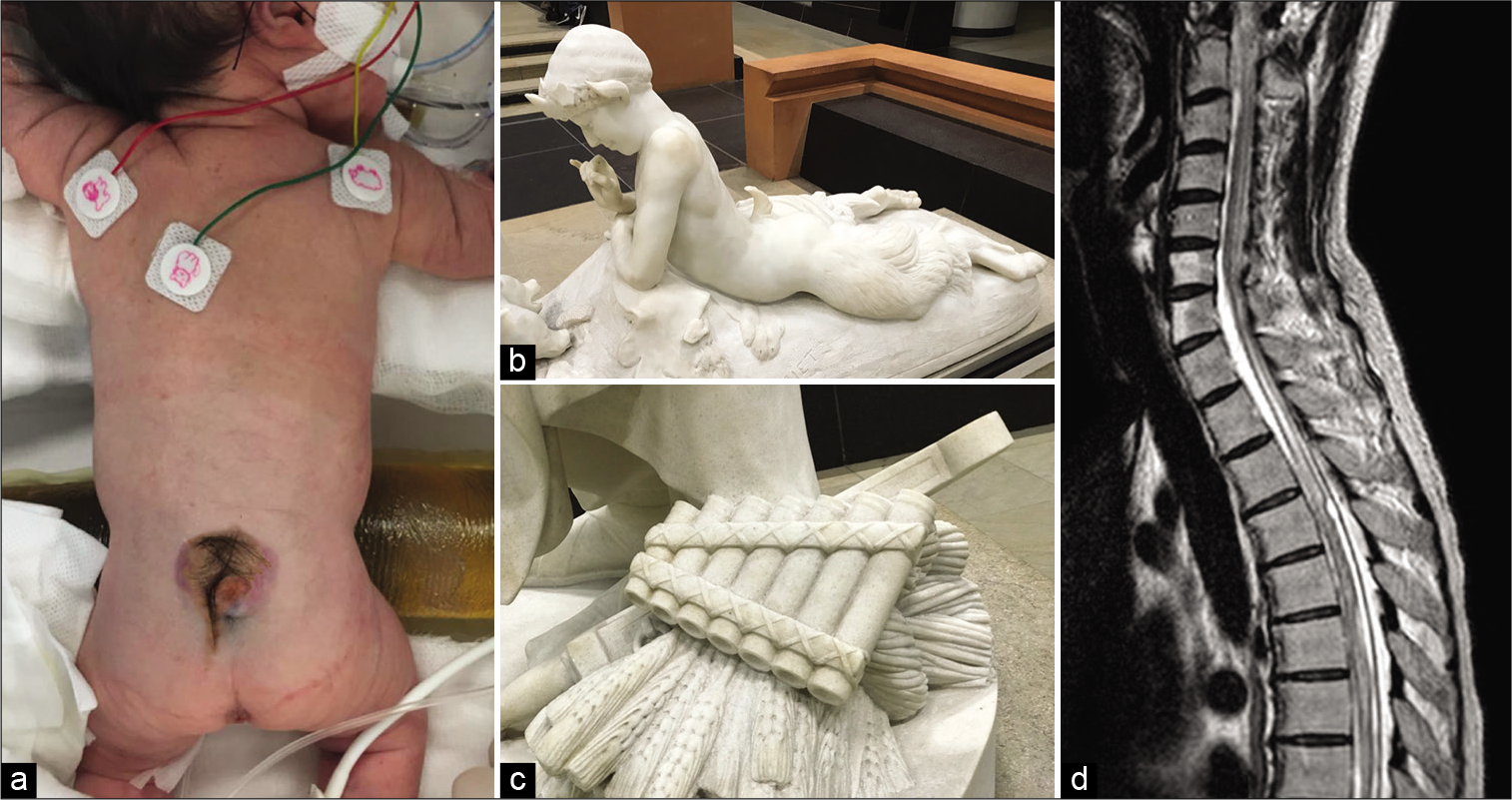
Pan, the son of Hermes, is the lustful deity of the wild and is generally depicted in the male form with the hindquarters and legs of a goat. The majority of artistic representations include a tuft of hair arising from the lumbosacral spine meant to resemble a faun’s tail. One of Pan’s lasting contributions to neurosurgery is his memorable features. For clinicians, the presence of lumbar hypertrichosis coupled with pes cavus deformities would suggest the presence of spinal dysraphism[
Figure 9:
Clinical photograph of a neonate with lumbar hypertrichosis and spina bifida aperta (a). Pan demonstrating his faun tail and clubbed feet (b), photograph by the author PW, marble sculpture 19th century, Musée d’’Orsay, Paris, France. Syrinx’s ultimate fate as a panpipe often seen by Pan’s side (c), photograph by the author PW, marble sculpture 19th century, Musée d’’Orsay, Paris, France. Post traumatic thoracolumbar syringomyelia (d), sagittal T2-weighted sequence magnetic resonance imaging.
Today, the syringe is named in memory of Syrinx’s tale for its hollow tubular structure. In neurosurgery, the diagnosis of a syrinx or syringomyelia refers to the development of a fluid-filled cavity within the spinal cord [
DISCUSSION
Neuroscience was borne from the great ages of human scientific discovery namely, Greek antiquity and the Italian Renaissance.[
One of the limitations of this study was that only English language references, or their translations, were consulted with most being secondary sources. Reviewing the Greek or Latin primary source materials would have enriched our descriptions, but locating and accessing them as well as translating the original text proved challenging. Second, the decision was made to describe only commonly used neurosurgical terms and archaic vocabulary seldom adopted in clinical practice was excluded. For example the hippocampal commissure and the crura of the fornix was once referred anatomically as the psalterium of Kind David, a harp-shaped instrument.[
CONCLUSION
It was the ancient Greeks that first espoused the philosophical school of cephalocentric thought. This may be why the clinical neurosciences are enriched with probably more mythological references than any other medical specialty. This comprehensive exploration of the Greek mythical and Christian roots of neurosurgical terminology serves as a reminder of the evolution of neuroscience and the importance of symbolism in the history of humankind.
Declaration of patient consent
Patient’s consent not required as there are no patients in this study.
Financial support and sponsorship
Nil.
Conflicts of interest
There are no conflicts of interest.
References
1. Abbott A. The man who bared the brain. Nature. 2015. 521: 160
2. Bazopoulou-Kyrkanidou E. Chimeric creatures in Greek mythology and reflections in science. Am J Med Genet. 2001. 100: 66-80
3. Bem Junior LS, Lemos NB, de Lima LF, Dias AJ, Neto OD, de Lira CC. The anatomy of the brain learned over the centuries. Surg Neurol Int. 2021. 12: 319
4. Brasiliense LB, Safavi-Abbasi S, Crawford NR, Spetzler RF, Theodore N. The legacy of hephaestus: The first craniotomy. Neurosurgery. 2010. 67: 881-4
5. Broca P. Instructions craniologiques et craniometriques. Mem Soc Anthrop Paris. 1875. 2: 1-203
6. Camerota F, Gerbino A.editors. A Scientific Concept of Beauty in Architecture Vitruvius Meets Descartes Galileo and Newton. Geometrical Objects: Architecture and the Mathematical Sciences 1400-1800. Switzerland: Springer, Cham; 2014. 38:
7. Clarke E. Apoplexy in the hippocratic writings. Bull Hist Med. 1963. 37: 301-14
8. Coupland AP, Thapar A, Qureshi MI, Jenkins H, Davies AH. The definition of stroke. J R Soc Med. 2017. 110: 9-12
9. Crick FC, Koch C. What is the function of the claustrum?. Philos Trans R Soc Lond B Biol Sci. 2005. 360: 1271-9
10. Crivellato E, Ribatti D. Soul, mind, brain: Greek philosophy and the birth of neuroscience. Brain Res Bull. 2007. 71: 327-36
11. Diab M. Errors of language in orthopaedics. J Bone Joint Surg Am. 2001. 83: 1269-181
12. García-Cabezas M, Pérez-Santos I, Cavada C. The Epic of the Thalamus in Anatomical Language. Front Neuroanat. 2021. 15: 744095
13. Ghosh SK. Human cadaveric dissection: A historical account from ancient Greece to the modern era. Anat Cell Biol. 2015. 48: 153-69
14. Griffin AL. Role of the thalamic nucleus reuniens in mediating interactions between the hippocampus and medial prefrontal cortex during spatial working memory. Front Syst Neurosci. 2015. 9: 29
15. Hakan T. Neurosurgery and a small section from the Greek myth: The God Pan and Syrinx. Childs Nerv Syst. 2009. 25: 1527-9
16. Hamilton E.editors. Mythology. New York: Little, Brown and Company; 1942. p.
17. Karakis I. Neuroscience and Greek mythology. J Hist Neurosci. 2019. 28: 1-22
18. Kothari M, Goel A. Maternalizing the meninges: A pregnant Arabic legacy. Neurol India. 2006. 54: 345-6
19. Lee KS, Lin CL, Chuang CL, Hwang SL, Howng SL, Lieu AS. Odyssey between the constellations and neuromedicine. Surg Neurol. 2006. 65: 99-101
20. Lewis FT. The significance of the term hippocampus. J Comp Neurol. 1923. 35: 213-30
21. Mallucci C, Brodbelt A. The enigma of syringomyelia. Br J Neurosurg. 2007. 21: 423-4
22. Nanda A, Filis A, Kalakoti P. Mythological and prehistorical origins of neurosurgery. World Neurosurg. 2016. 89: 568-73
23. Nanda A, Khan IS, Apuzzo ML. Renaissance neurosurgery: Italy’s iconic contributions. World Neurosurg. 2016. 87: 647-55
24. Okten AI. Mythology and neurosurgery. World Neurosurg. 2016. 90: 315-21
25. Orly R, Haines DE. Cerebral mythology: A skull stuffed with gods. J Hist Neurosci. 1998. 7: 81-3
26. Palacios-Sanchez L, Botero-Meneses JS, Velez-Florez MC. Pan, syrinx and syringomyelia. Arq Neuropsiquiatr. 2017. 75: 890-1
27. Paluzzi A, Fernandez-Miranda J, Torrenti M, Gardner P. Retracing the etymology of terms in neuroanatomy. Clin Anat. 2012. 25: 1005-14
28. Pearce JM. Ammon’s horn and the hippocampus. J Neurol Neurosurg Psychiatry. 2001. 71: 351
29. Pearce JM. The neuroanatomy of herophilus. Eur Neurol. 2013. 69: 292-5
30. Pound P, Bury M, Ebrahim S. From apoplexy to stroke. Age Ageing. 1997. 26: 331-7
31. ReFaey K, Quinones GC, Clifton W, Tripathi S, QuiñonesHinojosa A. The eye of horus: The connection between art, medicine, and mythology in ancient Egypt. Cureus. 2019. 11: e4731
32. Ribas GC, Yasuda A, Ribas EC, Nishikuni K, Rodrigues AJ. Surgical anatomy of microneurosurgical sulcal key points. Neurosurgery. 2006. 59: ONS177-210
33. Sanan A, van Loveren HR. The arachnoid and the myth of Arachne. Neurosurgery. 1999. 45: 152-5
34. Scatliff JH, Clark JK. How the brain got its names and numbers. AJNR Am J Neuroradiol. 1992. 13: 241-8
35. Senova S, Fomenko A, Gondard E, Lozano AM. Anatomy and function of the fornix in the context of its potential as a therapeutic target. J Neurol Neurosurg Psychiatry. 2020. 91: 547-59
36. Sommer W. Disease of Ammon’s horn as an etiological element of epilepsy. Arch Psychiatr. 1880. 10: 631-75
37. Soutis M. Ancient Greek terminology in pediatric surgery: About the word meaning. J Pediatr Surg. 2006. 41: 1302-8
38. Stefanou MI. The footprints of neuroscience in Alexandria during the 3rdcentury BC: Herophilus and erasistratus. J Med Biogr. 2020. 28: 186-94
39. Tubbs RS, Loukas M, Shoja MM, Apaydin N, Salter EG, Oakes WJ. The intriguing history of the human calvaria: sinister and religious. Childs Nerv Syst. 2008. 24: 417-22
40. Tubbs RS, Loukas M, Shoja MM, Cohen-Gadol AA, Wellons JC, Oakes WJ. Roots of neuroanatomy, neurology, and neurosurgery as found in the Bible and Talmud. Neurosurgery. 2008. 63: 156-62
41. Turliuc D, Turliuc Ş, Cucu A, Dumitrescu GF, Cărăuleanu A, Buzdugă C. A review of analogies between some neuroanatomical terms and roman household objects. Ann Anat. 2016. 204: 127-33
42. Vantomme G, Osorio-Forero A, Lüthi A, Fernandez LMJ. regulation of local sleep by the thalamic reticular nucleus. Front Neurosci. 2019. 13: 576
43. Williams GP, Leavitt L. Essay: How pan got his tail: The fusion of medicine, art, and myth. Neurology. 2004. 62: 519-22




Dr. Miguel A. Faria
Posted March 1, 2022, 8:22 am
Fantastic article giving a boost to the much needed and almost forgotten history of medicine, especially history of neuroscience/neurosurgery! The Authors’ working collaboration is also applauded in this endeavor!
Dr. Miguel A. Faria
Posted March 2, 2022, 5:39 am
For those interested in the history of medicine an/ or neurosurgery, especially younger surgeons not aware of these old classics, I recommend:
1. The classic, A History of Neurological Surgery (1967), edited by Dr. A. Earl Walker.
2. The enchanting multivolume, History of Medicine, by Dr. Plinio Prioreschi (several of which volumes I have reviewed for SNI.
3. Great Men with Sick Brains by Dr. Bengt Ljunggren.
4. Devils, Drugs & doctors by Dr. Howard W. Haggard (1929; reprinted 1980.
5. And for a history of medicine and political implications in our time my very small contribution, Vandals at the Gates of Medicine (1994), by Dr. Miguel A. Faria
Mustafa Ismail
Posted October 8, 2022, 3:48 pm
Great Article. Thank you, Dr. Miguel, for the suggested resources.