

- Department of Pediatric Neurosurgery, Tel Aviv Sourasky Medical Center, Dana Children’s Hospital, Tel Aviv University, Tel Aviv, Israel.
DOI:10.25259/SNI_434_2019
Copyright: © 2019 Surgical Neurology International This is an open-access article distributed under the terms of the Creative Commons Attribution-Non Commercial-Share Alike 4.0 License, which allows others to remix, tweak, and build upon the work non-commercially, as long as the author is credited and the new creations are licensed under the identical terms.How to cite this article: Voramol Rochanaroon, Danil A. Kozyrev, Jonathan Roth, Shlomi Constantini. Illustrated dynamic stories behind pediatric neurosurgery. 13-Sep-2019;10:178
How to cite this URL: Voramol Rochanaroon, Danil A. Kozyrev, Jonathan Roth, Shlomi Constantini. Illustrated dynamic stories behind pediatric neurosurgery. 13-Sep-2019;10:178. Available from: http://surgicalneurologyint.com/surgicalint-articles/9641/
Date of Submission
29-Jul-2019
Date of Acceptance
02-Aug-2019
Date of Web Publication
13-Sep-2019
Abstract
Background: The aim of this paper is to provide a depiction of the surgical technique and dynamic story behind the procedures in pediatric neurosurgery.
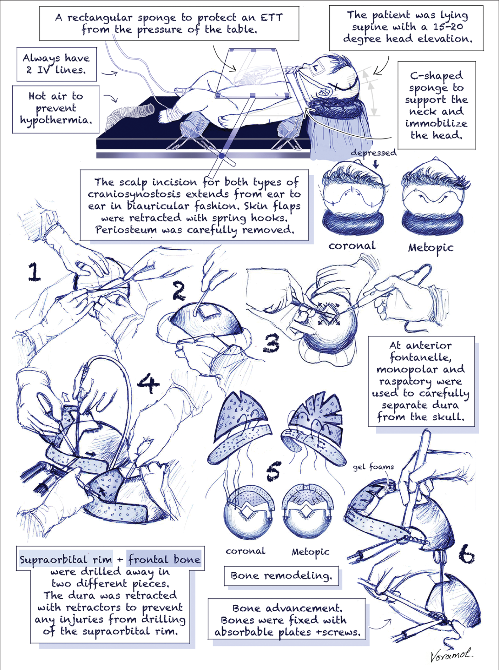
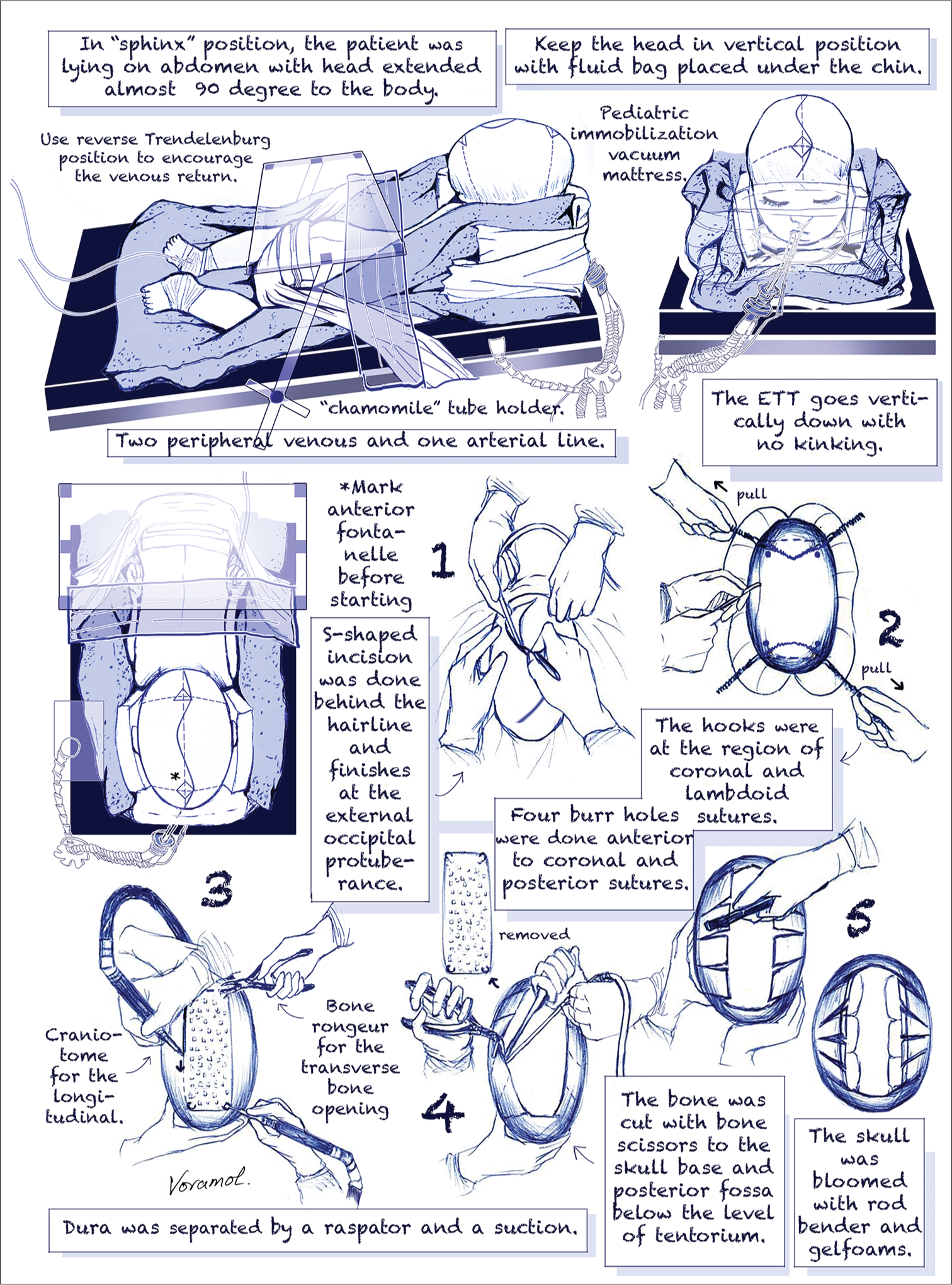
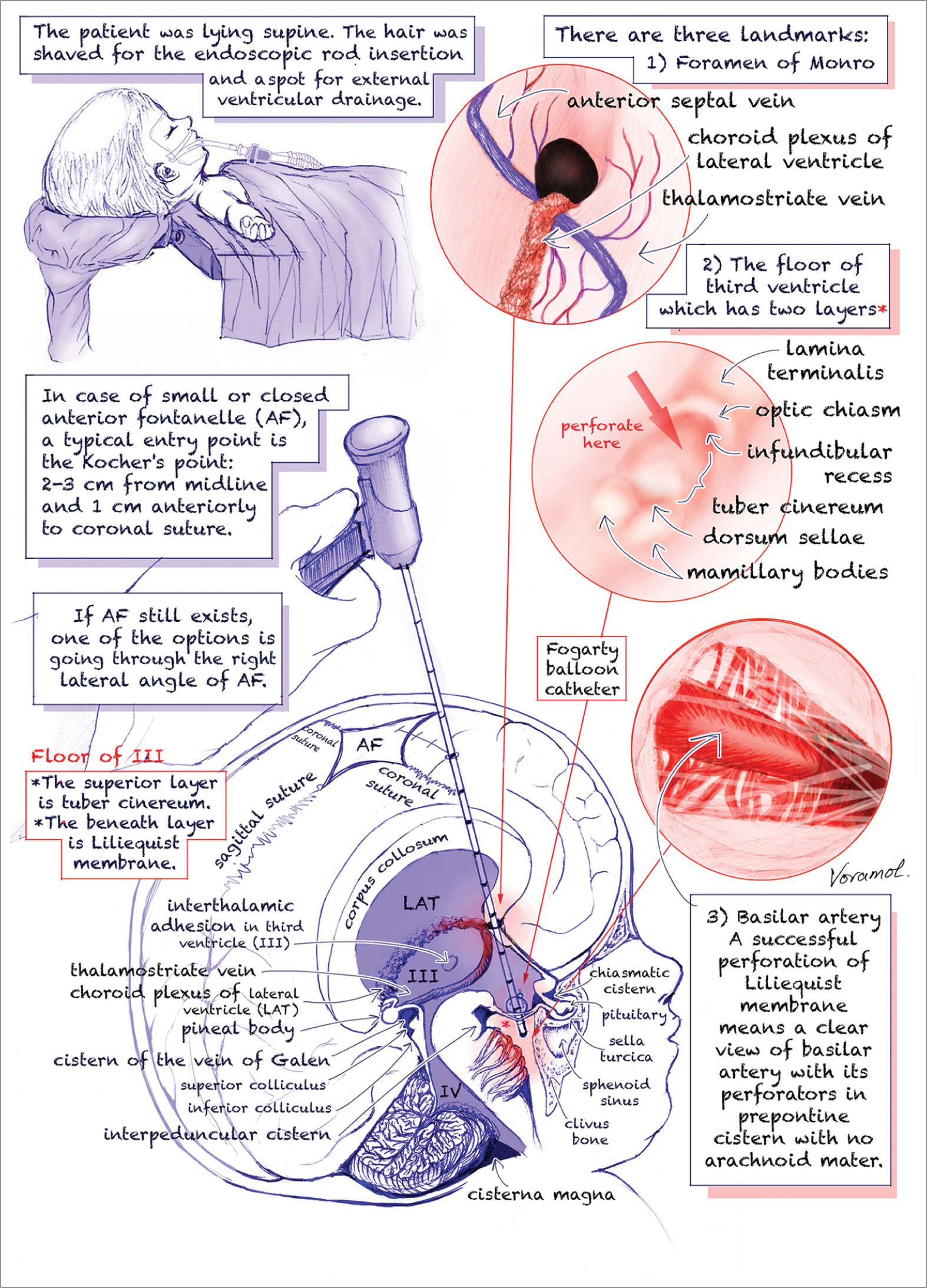
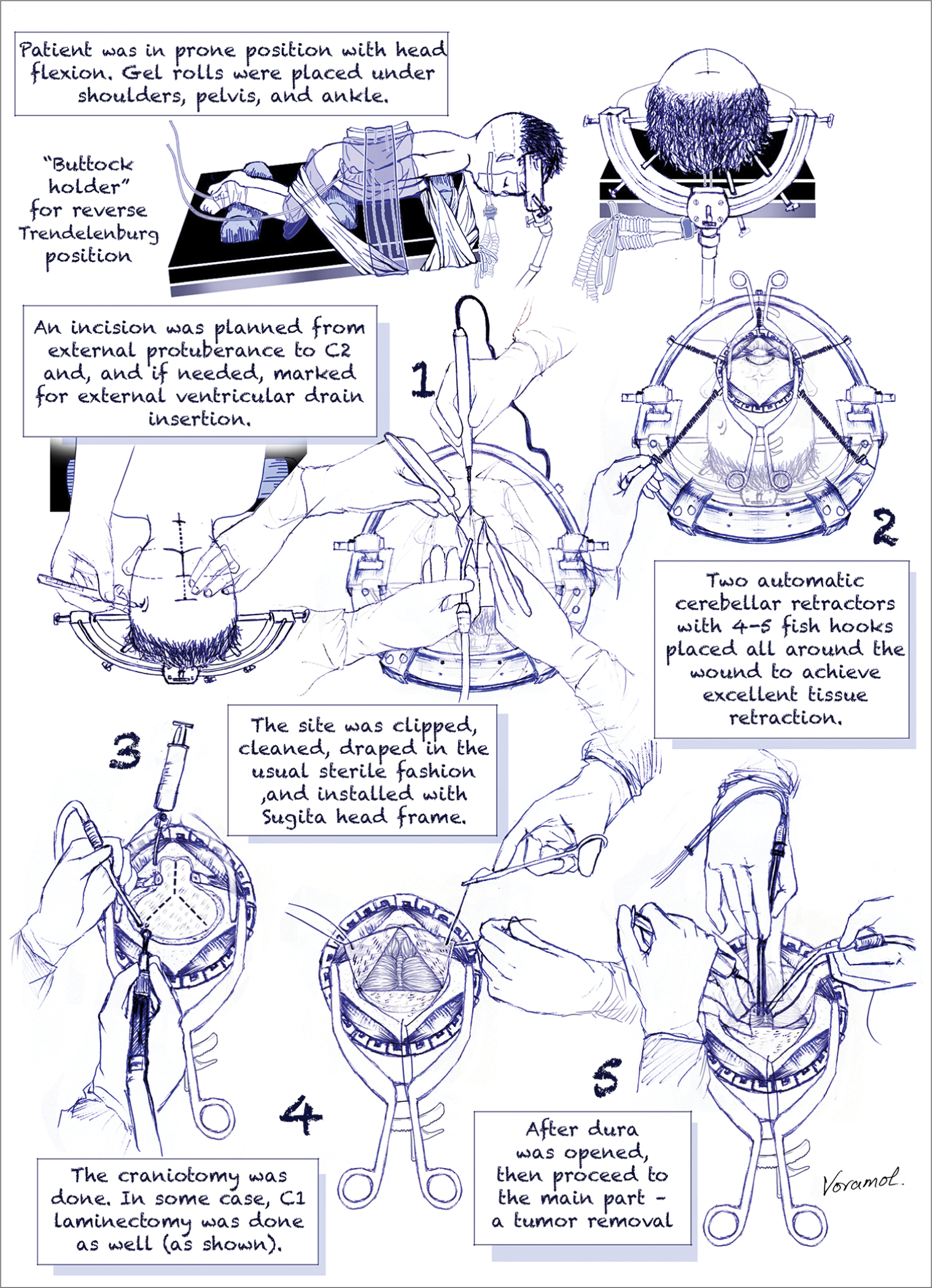
Methods: Five standard common pediatric neurosurgeries: endoscopic third ventriculostomy, fronto-orbital advancement for metopic and coronal craniosynostoses, posterior fossa craniotomy, strip craniectomy for sagittal craniosynostosis, and ventriculoperitoneal shunting were chosen to be exampled in illustrations.
Results: Surgical techniques were depicted in a step-by-step fashion with comic-like style of images. Illustrations enable to highlight specific surgical and anatomical features and also convey surgical procedures in a sequential order from beginning to end as if it is a story.
Conclusion: Surgical illustrations may serve as an educational tool with potentially instructional value for practical application, especially for surgical trainees.
Keywords: Depiction, Neurosurgery, Pediatric, Surgical illustrations
INTRODUCTION
Technology has changed the nature of documentation in medicine. However, despite the realistic quality of photos and videos, illustrations remain a leading position in medical education.[
We selected five common procedures of pediatric neurosurgery, in a series of positioning, surgical intervention, and subintervention [
In our illustrations, we show one of many ways to perform common pediatric neurosurgical procedures. As the famous Latin proverb says “All roads lead to Rome,” procedures might be done differently but with the same result.
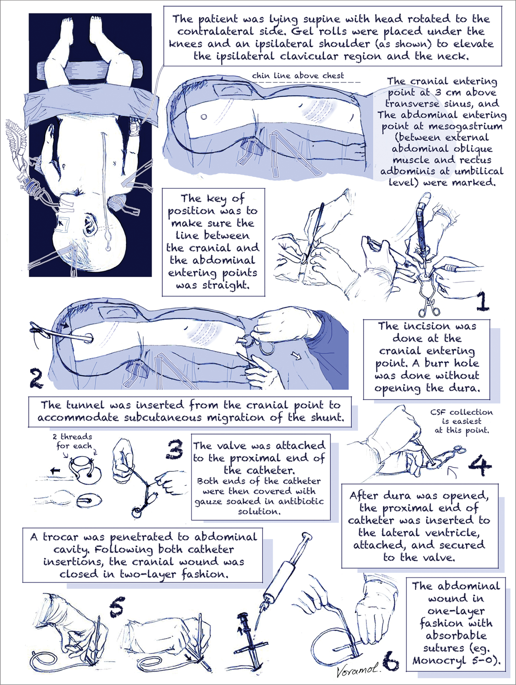
For example, there are several ways to perform a ventriculoperitoneal shunt. As neither anterior or posterior approach for insertion of ventricular catheter has shown significant superiority compare to each other. In most cases, we favor the posterior approach as it requires only two incisions instead of three in the anterior approach. Similarly, to save time and to achieve a better cosmetic result, we prefer insertion of the peritoneal catheter using trocar to minilaparotomy. In our experience, perforation of abdominal organs or missing abdominal cavity by trocar is extremely rare.
A ROOM FOR IMPROVEMENT
Max Brödel was the pioneer who famously created the original angle.[
The notably mentioned Rhoton’s collection has a special place in modern academic neurosurgery.[
Another outstanding example of neurosurgical drawing collection is “Operative Neurosurgery” by Kempe.[
In the world of anatomical and surgical illustrations, it is impossible not to mention the legendary works of Frank Henry Netter. His atlases of normal anatomy and clinical subjects have become the most own for several generations of medical students and physicians.[
In more recent surgical perspective, Michael T. Lawton’s books comprise five different views: surgeon’s view, coronal view, other surgical view, real photo view, and imaging view. It has every viable detail which we need to know about the climax of that particular surgery.[
CONCLUSION
While digitized images can undermine the patient’s confidentiality, illustration continues to be a guardian of a new production. Its transparency allows it to be publicized and may reach the larger scale of audiences, even outside medicine.
Financial support and sponsorship
Dr. Rochanaroon has illustrated and presented this work as a poster and won the Best Student Poster Award at the 46th Annual Meeting of International Society for Pediatric Neurosurgery 2018 in Tel Aviv, Israel.
Conflicts of interest
There are no conflicts of interest.
References
1. Barry ME. Art and the role of the rhoton medical illustrators in his legacy. World Neurosurg. 2016. 92: 637-48
2. Cheng H, Chen BP, Soleas IM, Ferko NC, Cameron CG, Hinoul P. Prolonged operative duration increases risk of surgical site infections: A systematic review. Surg Infect (Larchmt). 2017. 18: 722-35
3. Choque-Velasquez J, Kozyrev DA, Colasanti R, Thiarawat P, Intarakhao P, Jahromi BR. The open access video collection project Hernesniemi’s 1001 and more microsurgical videos of neurosurgery: A legacy for educational purposes. Surg Neurol Int. 2017. 8: 188-
4. Davidson B, Alotaibi NM, Hendricks BK, Cohen-Gadol AA. Popularity of online multimedia educational resources in neurosurgery: Insights from the neurosurgical atlas project. J Surg Educ. 2018. 75: 1615-23
5. Elmaci I, Balak N. Pioneering turkish neurosurgeon hami dilek and the traces of harvey cushing’s legacy in his work. J Neurosurg. 2008. 108: 821-9
6. Groen RJ, Koehler PJ, Kloet A. The role of harvey cushing and walter dandy in the evolution of modern neurosurgery in the netherlands, illustrated by their correspondence. J Neurosurg. 2013. 118: 539-49
7. Jones HR, Netter FH.editors. Netter’s Neurology. Icon Learning Systems. 2005. p.
8. Kempe LG.editors. Operative Neurosurgery: Cranial, Cerebral, and Intracranial Vascular Disease. Vol. 1. New York: Springer Berlin Heidelberg; 1985. p.
9. Lawton MT.editorsSeven Aneurysms: Tenets and Techniques for Clipping. New York: Thieme; 2011. p.
10. Lawton MT.editorsSeven AVMs: Tenets and Techniques for Resection. New York: Thieme; 2014. p.
11. Lawton MT.editorsSeven Bypasses: Tenets and Techniques for Revascularization. New York: Thieme; 2018. p.
12. Mehta VA, Wijesekera O, Pendleton C, Quiñones-Hinojosa A, Jallo GI, Ahn ES. Harvey cushing and “birth hemorrhage”: Early pediatric neurosurgery at the johns hopkins hospital. J Neurosurg Pediatr. 2011. 8: 647-53
13. Patel SK, Couldwell WT, Liu JK. Max brödel: His art, legacy, and contributions to neurosurgery through medical illustration. J Neurosurg. 2011. 115: 182-90
14. Pendleton C, Ahn ES, Quiñones-Hinojosa A. Harvey cushing and pediatric brain tumors at johns hopkins: The early stages of development. J Neurosurg Pediatr. 2011. 7: 575-88
15. Peris-Celda M, Martinez-Soriano F, Rhoton AL.editorsRhoton’s Atlas of Head, Neck, and Brain: 2D and 3D Images. New York: Thieme; 2017. p.
16. Shillito J, Matson DD, Codding MB, Lashbrook GS.editorsAn Atlas of Pediatric Neurosurgical Operations. Philadelphia, PA: W.B. Saunders Co.; 1982. p.
17. Udelsman R. Presidential address: Harvey cushing: The artist. Surgery. 2006. 140: 841-6

