

- Department of Neurosurgery, Christian Medical College,
- Department of Neurosurgery, Dayanand Medical College, Ludhiana, Punjab, India.
Correspondence Address:
Ashish Acharya
Department of Neurosurgery, Christian Medical College,
DOI:10.25259/SNI_442_2020
Copyright: © 2020 Surgical Neurology International This is an open-access article distributed under the terms of the Creative Commons Attribution-Non Commercial-Share Alike 4.0 License, which allows others to remix, tweak, and build upon the work non-commercially, as long as the author is credited and the new creations are licensed under the identical terms.How to cite this article: Ashish Acharya1, Sarvpreet Singh Grewal1, Shivender Sobti2, Paul Sudhakar John1, Ravindra Kumar Bind1, Maneesh Kumar Bhardwaj1, Shefin J. Mathews1. Intramedullary mature teratoma of spinal cord: A rare tumor with review of literature. 29-Aug-2020;11:266
How to cite this URL: Ashish Acharya1, Sarvpreet Singh Grewal1, Shivender Sobti2, Paul Sudhakar John1, Ravindra Kumar Bind1, Maneesh Kumar Bhardwaj1, Shefin J. Mathews1. Intramedullary mature teratoma of spinal cord: A rare tumor with review of literature. 29-Aug-2020;11:266. Available from: https://surgicalneurologyint.com/surgicalint-articles/10228/
Date of Submission
17-Jul-2020
Date of Acceptance
12-Aug-2020
Date of Web Publication
29-Aug-2020
Abstract
Background: Spinal teratomas are rare in adults. The clinical findings are nonspecific, reflecting only in the intramedullary location of these lesions. The potential differential diagnosis for intramedullary spinal teratomas include schwannomas, dermoids, epidermoids, and neurofibromas.
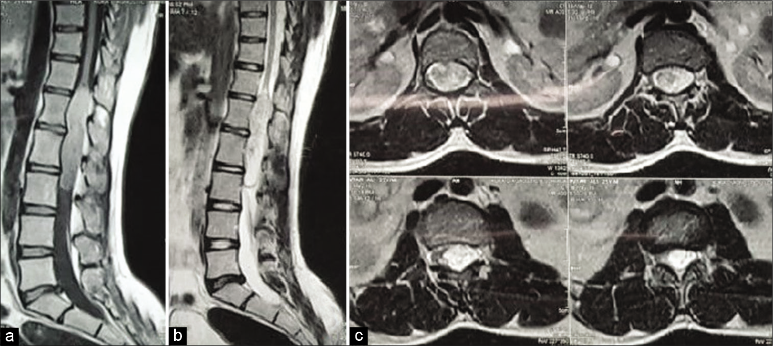
Case Description: A 25-year-old male presented with RLE weakness (iliopsoas/quadriceps [4/5], and extensor hallucis longus/dorsiflexor [0/5]) and urinary incontinence. As the contrast, MR showed a heterogeneous intramedullary lesion with well-defined edges located at the T12-L1 level, the patient underwent a focal laminectomy for gross total tumor excision. Pathologically, it proved to be a mature teratoma.
Conclusion: Teratomas should be considered among the differential diagnostic considerations for intramedullary spinal cord lesions. Although gross total resection is preferred, these lesions have a low recurrence rate, and therefore, partial removal is also valid, where lesions are densely adherent to adjacent neural structures.
Keywords: Intradural intramedullary, Spinal tumor, Teratoma of spinal cord
INTRODUCTION
Spinal teratomas constitute only 0.15–0.18% of all intraspinal tumors and are more commonly found in children. Potential differential diagnoses for these lesions include schwannomas, dermoids, epidermoids, and neurofibromas.[
CASE REPORT
A 25-year-old male presented with backache and a progressive left lower extremity proximal paresis (iliopsoas/quadriceps 4/5) and a distal severe foot drop (extensor hallucis longus/ dorsiflexor 0/5) of 5 months duration. As his bladder was not completely emptying, he required a Foley catheter. The enhanced MRI of the lumbar spine showed a T12-L1 intramedullary lesion involving the conus medullaris/proximal filum terminale [
Surgery
Following a D12-L1 laminectomy, gross total excision of an intramedullary lesion was achieved, including total removal of tumor capsule. Postoperatively, the improved within 1 month (e.g., left iliopsoas/quadriceps 5/5 and the left-sided foot drop (2/5), and the urinary dysfunction resolved. Histopathologically, the tumor was found to be a mature teratoma [
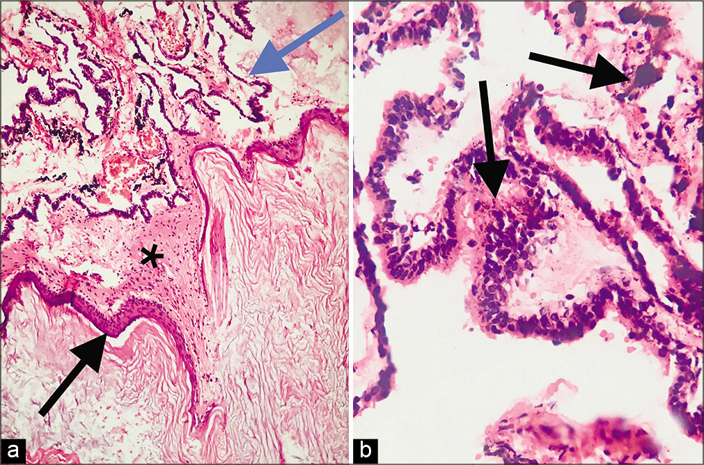
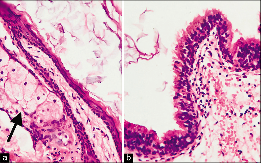
Figure 2:
Histopathology showed mature teratoma comprising of (a) cyst lined by keratinizing stratified squamous epithelium (black arrow) filled with keratin flakes, beneath which was seen glial tissue (starred), and choroidal tissue (blue arrow) comprised convoluted strips of epithelium forming papillae (H&E, 100x). (b) High magnification showing stratified tall columnar epithelium with intracytoplasmic and stromal neuromelanin pigment (black arrows) (H&E, 400×).
DISCUSSION
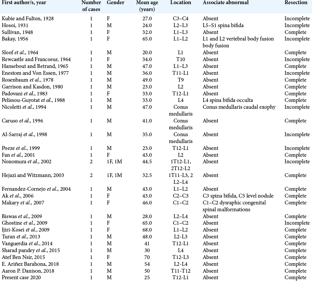
Frequency of spinal teratomas
In pediatric patients, 5–10% of spinal lesions are intraspinal teratomas,[
Treatment options
The treatment for spinal teratomas is primarily surgery, whether ideally gross total or subtotal/partial resection. It was found that intramedullary teratomas could be completely removed in 61.8% of cases.[
The use of intraoperative electrophysiologic monitoring is extremely helpful in the resection of these lesions, as it signals when dissection should be stopped (e.g., for densely adherent lesions) before incurring permanent neurological sequelae.
CONCLUSION
Intradural spinal teratomas are very rare in adults. Once documented on preoperative MR studies, either gross total, subtotal, or partial resections are viable surgical alternatives as all result in good long-term outcomes with a low recurrence rate.
Declaration of patient consent
The authors certify that they have obtained all appropriate patient consent.
Financial support and sponsorship
Nil.
Conflicts of interest
There are no conflicts of interest.
References
1. Agay AK, Garg S, Hedaoo K. Spinal intradural extramedullary mature cystic teratoma in young adult: A rare tumor with review of literature. Int J Res Med Sci. 2016. 4: 5481-3
2. Banna M, Talalla A. Intraspinal dermoids in adults. Br J Radiol. 1975. 48: 28-30
3. Danison AP, Ramanathan D, Matin M, Kim K, Panchal RR. Adult thoracic intradural exophytic mature teratoma: Case report and literature review. Asian J Neurosurg. 2018. 13: 1182-5
4. Garrison JE, Kasdon DL. Intramedullary spinal teratoma: Case report and review of the literature. Neurosurgery. 1980. 7: 509-12
5. Nonomura Y, Miyamoto K, Wada E, Hosoe H, Nishimoto H, Ogura H. Intramedullary teratoma of the spine: Report of two adult cases. Spinal Cord. 2002. 40: 40-3
6. Rosenbaum TJ, Soule EH, Onofrio BM. Teratomatous cyst of the spinal canal. Case report. J Neurosurg. 1978. 49: 292-7
7. Shikata J, Yamamuro T, Mikawa Y, Kotoura Y. Intraspinal epidermoid and dermoid cysts. Surgical results of seven cases. Arch Orthop Trauma Surg. 1988. 107: 105-9

