

- Department of Neurosurgery, Suwa Red Cross Hospital, Kogandori, Suwa, Nagano, Japan.
DOI:10.25259/SNI_59_2020
Copyright: © 2020 Surgical Neurology International This is an open-access article distributed under the terms of the Creative Commons Attribution-Non Commercial-Share Alike 4.0 License, which allows others to remix, tweak, and build upon the work non-commercially, as long as the author is credited and the new creations are licensed under the identical terms.How to cite this article: Masahito Katsuki, Yukinari Kakizawa, Yasunaga Yamamoto, Akihiro Nishikawa, Naomichi Wada, Toshiya Uchiyama. Magnetic resonance angiography with ultrashort echo time evaluates cerebral aneurysm with clip. 11-Apr-2020;11:65
How to cite this URL: Masahito Katsuki, Yukinari Kakizawa, Yasunaga Yamamoto, Akihiro Nishikawa, Naomichi Wada, Toshiya Uchiyama. Magnetic resonance angiography with ultrashort echo time evaluates cerebral aneurysm with clip. 11-Apr-2020;11:65. Available from: https://surgicalneurologyint.com/surgicalint-articles/9956/
Date of Submission
13-Feb-2020
Date of Acceptance
26-Mar-2020
Date of Web Publication
11-Apr-2020
Abstract
Contrast-enhanced computed tomography angiography is usually valuable for the evaluation of clipped cerebral aneurysm, but it has side effects of contrast medium. Time-of-flight magnetic resonance angiography (MRA) is a non-invasive and fast method. However, clip-induced artifact limits assessment of the artery in the vicinity of a clip. MRA with ultrashort echo time (TE) reduces metal artifact. We use MAGNETOM Aera 1.5T (SIEMENS, München, Germany) and perform pointwise encoding time reduction with radial acquisition (PETRA)-MRA using ultrashort TE for the assessment of the cerebral aneurysm after clipping. We, herein, presented two representative cases with a clipped aneurysm which could be evaluated by PETRA- MRA. Especially in one of them, the neck remnant was revealed by PETRA-MRA. PETRA-MRA can reduce the time and the invasiveness and may be helpful for the usual follow-up of the clipped aneurysm with the development of MRA technology in the future.
Keywords: Cerebral aneurysm, Clipping, Less invasive, Magnetic resonance angiography, Ultrashort echo time
We would like to report the usefulness of magnetic resonance angiography (MRA) with ultrashort echo time (TE) for the assessment of the cerebral aneurysm after clipping. Non-invasive methods are better for the assessment of the cerebral aneurysm after clipping. Contrast-enhanced computed tomography angiography (CTA) is usually valuable,[
MRA with ultrashort TE reduces metal artifact.[
Case 1: clipping for the unruptured aneurysm at the right middle cerebral artery using the Sugita titanium clip (Mizuho, Tokyo, Japan). CTA described M1 and M2 portion of the middle cerebral artery and the vicinity of the clip in detail (arrow,
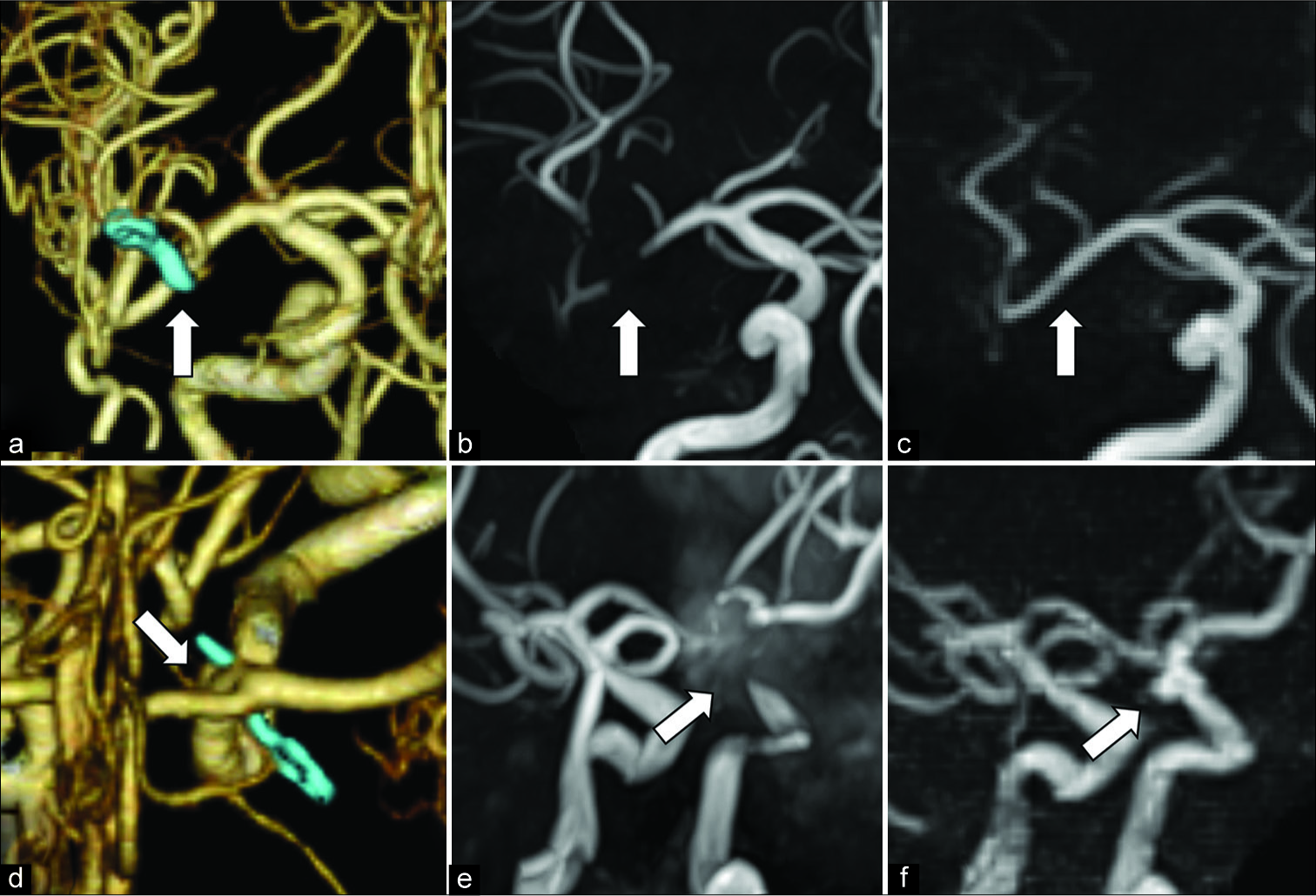
Figure 1:
Case 1: clipping for the unruptured aneurysm at the right middle cerebral artery using the Sugita clip made of titanium (Mizuho, Tokyo, Japan). Computed tomography angiography described M1 and M2 portion of the middle cerebral artery and the vicinity of a clip in detail (arrow, a). Time-of-flight magnetic resonance angiography (TOF-MRA) did not describe them due to clip-induced artifact (arrow, b), but pointwise encoding time reduction with radial acquisition (PETRA)-MRA revealed the arteries (arrow, c). Case 2: clipping for the ruptured aneurysm at the left internal carotid artery-posterior communicating artery bifurcation using the Sugita clip made of titanium. CTA described the aneurysm neck remnant due to incomplete clipping (arrow, d), but it was indistinguishable by TOF-MRA (arrow, e). However, PETRA-MRA clearly revealed the remnant and could morphologically evaluate it (arrow, f).
To the best of our knowledge, this is the first report on the usefulness of PETRA-MRA for the assessment of the cerebral aneurysm and its neck remnant after clipping. PETRA-MRA can reduce the time and the invasiveness and may be helpful for the usual follow-up of the clipped aneurysm with the development of MRA technology in the future.
DECLARATION OF PATIENT CONSENT
The authors certify that they have obtained all appropriate patient consent.
Financial support and sponsorship
Nil.
Conflicts of interest
There are no conflicts of interest.
Acknowledgment
Conception and design: YK, YY. Acquisition of data: YK, YY, TU, AN, NW. Drafting the article: MK, YK. Reviewed submitted version of manuscript: all authors. Approved the final version of the manuscript on behalf of all authors: YK.
References
1. Becker RL, Norfray JF, Teitelbaum GP, Bradley WG, Jacobs JB, Wacaser L. MR imaging in patients with intracranial aneurysm clips. AJNR Am J Neuroradiol. 1988. 9: 885-9
2. Gönner F, Lövblad KO, Heid O, Remonda L, Guzman R, Barth A. Magnetic resonance angiography with ultrashort echo times reduces the artifact of aneurysm clips. Neuroradiology. 2002. 44: 755-8
3. Schmalbrock P, Yuan C, Chakeres DW, Kohli J, Pelc NJ. Volume MR angiography: Methods to achieve very short echo times. Radiology. 1990. 175: 861-5
4. Van Loon JJ, Yousry TA, Fink U, Seelos KC, Reulen HJ, Steiger HJ. Postoperative spiral computed tomography and magnetic resonance angiography after aneurysm clipping with titanium clips. Neurosurgery. 1997. 41: 851-6

