- Department of Neurosurgery, Ochsner Medical Center, New Orleans, USA
- Department of Neurosurgery, University of Montreal, Montreal, Quebec, Canada
- Department of Pain Management, Ochsner Baptist, New Orleans, USA
Correspondence Address:
P. O. Champagne
Department of Neurosurgery, Ochsner Medical Center, New Orleans, USA
DOI:10.4103/sni.sni_279_17
Copyright: © 2017 Surgical Neurology International This is an open access article distributed under the terms of the Creative Commons Attribution-NonCommercial-ShareAlike 3.0 License, which allows others to remix, tweak, and build upon the work non-commercially, as long as the author is credited and the new creations are licensed under the identical terms.How to cite this article: S. Raju, P. O. Champagne, L. Walsh, Daniel J. Denis. Minimally invasive repair of a pseudomeningocele caused by a sheared intrathecal catheter following implantation of a drug delivery system. 06-Dec-2017;8:297
How to cite this URL: S. Raju, P. O. Champagne, L. Walsh, Daniel J. Denis. Minimally invasive repair of a pseudomeningocele caused by a sheared intrathecal catheter following implantation of a drug delivery system. 06-Dec-2017;8:297. Available from: http://surgicalneurologyint.com/surgicalint-articles/minimally-invasive-repair-of-a-pseudomeningocele-caused-by-a-sheared-intrathecal-catheter-following-implantation-of-a-drug-delivery-system/
Abstract
Background:Shearing of an intrathecal catheter during implantation of a drug delivery system is an underreported complication that can be challenging to manage.
Case Description:A 53-year-old man with refractory cancer pain had an intrathecal pump system implanted. The procedure was complicated with catheter shear and retention in the intrathecal space. A second catheter was successfully placed but formation of a painful pseudomeningocele and ineffective pain relief complicated the outcome. A minimally invasive approach through a tubular retractor was employed to access the spinal canal via a laminotomy, the sheared catheter was removed and the dural defect repaired. Complete resolution of the pseudomeningocele and efficient pain control were observed at follow-up.
Conclusion:Minimally invasive approach to the spine is demonstrated as a safe and effective alternative in this case of retained catheter induced cerebrospinal fluid (CSF) leak.
Keywords: Cerebrospinal fluid leak, minimally invasive surgery, pseudomeningocele, retained intrathecal catheter
INTRODUCTION
Percutaneous placement of an intrathecal catheter is commonly performed for drug delivery or cerebrospinal fluid (CSF) diversion. Catheter breakage is an underreported complication that can occur during or after the procedure and may result in CSF leak, pseudomeningocele and failure or drug delivery to the intrathecal space.[
CASE REPORT
Clinical presentation
A 53-year-old man was referred to the pain management clinic for a 7 months history of intractable thoracic back pain and abdominal pain following surgical resection of a duodenal adenocarcinoma (stage IV). His pain was refractory to all treatments previously tried, with a reported maximal pain intensity of 10/10. His pain did not respond to methadone, hydromorphone, cyclobenzaprine, gabapentin, pregabalin nor lidocaine patches. Previous interventional therapies provided only temporary, if any, relief. These included neurolysis of the celiac plexus, neurolysis of the superior epigastric nerve, 12th intercostal nerve, and paraspinal trigger point injections. Given, his lack of response, the decision was made to move forward with a trial of intrathecal medication in preparation for a possible intrathecal drug delivery. At this time, he was receiving oral long acting morphine 100 mg three times daily, oral morphine 30 mg every 6 h as needed, tizanidine 4 mg three times daily as needed, and amitriptyline 25 mg every evening. The intrathecal drug trial was successful leading to the implantation of an intrathecal delivery system. During the implantation, the intrathecal catheter was successfully placed through a Tuohy needle at L3-4. In the course of the Tuohy removal, the epidural portion of the catheter was sheared. The fractured portion could not be removed. The decision was made to leave the sheared catheter in place. A second catheter was successfully placed at the same level and connected to the pump.
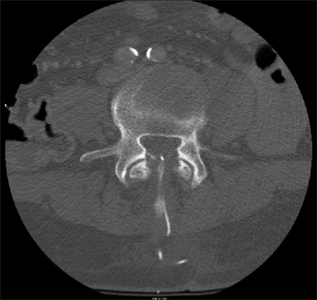
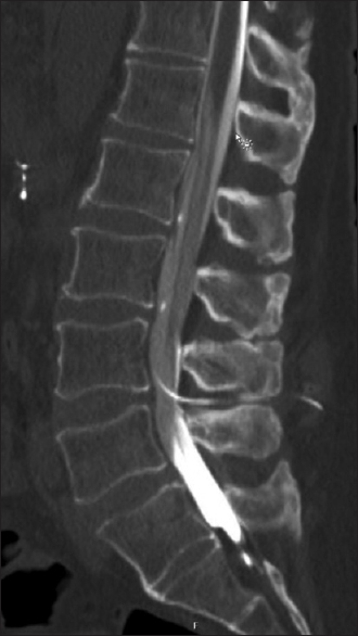
In the immediate postoperative period, the patient reported about 50% pain relief but 10 days after the procedure, he began to experience orthostatic headaches, nausea, vomiting, and the development of a painful lump in his right paraspinal lumbar area. The physical exam was unremarkable for any neurological deficits, but a tender mass was palpable in the left paraspinal area at L3-4. A pseudomeningocele in the subcutaneous lumbar tissue was demonstrated on computed tomographic (CT) imaging. Two intrathecal catheters were noted on CT imaging entering the spinal canal at the L3-4 level and a few millimeters of the proximal tip of the sheared catheter were visualized into the epidural space [
The patient was initially treated conservatively with bed rest for 48 h, but his symptoms did not improve over time. In addition, to the symptomatic pseudomeningocele and intracranial hypotension, the patient presented significant anxiety due to his concerns about the retained drain fragment. Surgical resection of the retained catheter with CSF leak repair was then recommended.
Operation
The patient underwent a minimally invasive repair of symptomatic CSF leak and removal of the sheared intrathecal catheter. Under general anesthesia with the patient prone on a Wilson frame, an 18 mm right-sided longitudinal incision 15 mm of the midline at L3-L4 was planned using fluoroscopy. Serial dilators were inserted and final 18 mm diameter retractor was docked at the junction of the inferior L3 lamina and facet complex.
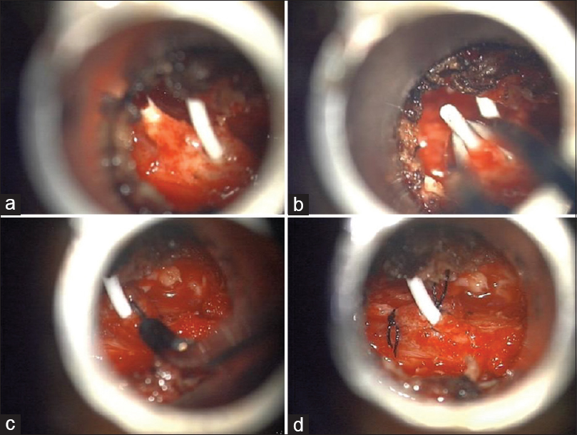
A L3 laminotomy and medial facetectomy were performed and upon removal of the ligamentum flavum, CSF became visible in the surgical field. The functional intact catheter was visible at the midline in the epidural space and some CSF was leaking around its insertion site in the dura. The second sheared catheter was identified following completion of a L4 laminotomy and CSF was freely leaking from its open end [
Figure 3
Intra-operative images showing (a) the functional catheter (right side) and the tip of the non-functional catheter (left side), (b) forceps being used to pull the severed catheter and (c) purse string suture being made around the remaining functional catheter to ensure a watertight closure and (d) before closure with a suture at the site of the sheared catheter and the purse string suture around the functional catheter
The non-functional catheter tip was removed with micro bayonet curved tip forceps [
Follow-up
Postoperatively, the patient complained of nausea and abdominal pain that improved in the weeks following procedure. He further reported complete resolution of his postural headaches and was able to tolerate continued use of his functioning morphine pump. Complete resolution of his presenting symptoms with no evidence of CSF leak was confirmed at 8 weeks postoperatively. The patient continued to benefit from palliative care until his death 5 months after the procedure.
DISCUSSION
Catheter-related complications
Intrathecal catheters are commonly used for CSF diversion or drug delivery. Complications involving the catheter itself such as leakage, fracture, dislodgement/migration, kink/occlusion, disconnection at the pump, granuloma formation, or infection may warrant catheter revision or explanation. Catheter-related complications can occur in 3% to 76% of patients implanted with an intradural drug delivery system (IDDS) with catheter breakage, migration, and obstruction being more frequently reported.[
Indications for surgical extraction of a sheared intrathecal catheter
Management of a retained intrathecal catheter and surgical removal must be decided on an individual basis. Low surgical risk, progressive neurological symptoms, infectious risk, presence of pseudomeningocele, scheduled spine surgery at the same level, migrating catheter, and patient psychological distress are factors that can support surgical removal of a retained catheter fragment.[
The place of MIS for extraction of intradural foreign objects and repair of cerebrospinal fluid leak
MIS of the spine through tubular retractors has gained popularity due to its decreased surgical morbidity and the potential to achieve better postoperative clinical outcome.[
Intradural surgery can be safely performed through tubular retractors using MIS techniques.[
We present the first case of successful removal of a retained intra/extradural catheter and treatment of resulting CSF leak through a minimally invasive approach. We present this approach as an optimal alternative for this population of terminally-ill patients in which more extensive procedures are preferably avoided.
CONCLUSION
MIS technique utilizing commonly available instruments can be successfully and safely used to remove retained sheared intrathecal catheters and to repair persistent spinal CSF leak after implantation of a IDDS. Although more technically demanding when compared to open approaches, MIS offers the advantage of minimizing the surgical morbidity, without creating a significant empty space in which CSF can accumulate, and thus can possibly decrease the incidence of postoperative pseudomeningocele.
Financial support and sponsorship
Nil.
Conflicts of interest
There are no conflicts of interest.
References
1. Abdulla S, Vielhaber S, Heinze HJ, Abdulla W. A new approach using high volume blood patch for prevention of post-dural puncture headache following intrathecal catheter pump exchange. Int J Crit Illn Inj Sci. 2015. 5: 93-8
2. Ahn J, Iqbal A, Manning BT, Leblang S, Bohl DD, Mayo BC. Minimally invasive lumbar decompression-the surgical learning curve. Spine J. 2016. 16: 909-16
3. Aoyama T, Hida K. Spinal intradural granuloma as a complication of an infected cerebrospinal fluid drainage tube fragment--case report. Neurol Med Chir (Tokyo). 2010. 50: 165-7
4. Cheung AT, Pochettino A, Guvakov DV, Weiss SJ, Shanmugan S, Bavaria JE. Safety of lumbar drains in thoracic aortic operations performed with extracorporeal circulation. Ann Thorac Surg. 2003. 76: 1190-
5. Chou D, Wang VY, Khan AS. Primary dural repair during minimally invasive microdiscectomy using standard operating room instruments. Neurosurgery. 2009. 64: 356-
6. Dahlberg D, Halvorsen CM, Lied B, Helseth E. Minimally invasive microsurgical resection of primary, intradural spinal tumours using a tubular retraction system. Br J Neurosurg. 2012. 26: 472-5
7. Dvorak EM, McGuire JR, Nelson ME. Incidence and identification of intrathecal baclofen catheter malfunction. PM R. 2010. 2: 751-6
8. Fluckiger B, Knecht H, Grossmann S, Felleiter P. Device-related complications of long-term intrathecal drug therapy via implanted pumps. Spinal Cord. 2008. 46: 639-43
9. Follett KA, Naumann CP. A prospective study of catheter-related complications of intrathecal drug delivery systems. J Pain Symptom Manage. 2000. 19: 209-15
10. Forsythe A, Gupta A, Cohen SP. Retained intrathecal catheter fragment after spinal drain insertion. Reg Anesth Pain Med. 2009. 34: 375-8
11. Gandhi RH, German JW. Minimally invasive approach for the treatment of intradural spinal pathology. Neurosurg Focus. 2013. 35: E5-
12. Guppy KH, Silverthorn JW, Akins PT. Subarachnoid hemorrhage due to retained lumbar drain. J Neurosurg Spine. 2011. 15: 641-4
13. Hnenny L, Sabry HA, Raskin JS, Liu JJ, Roundy NE, Dogan A. Migrating lumbar intrathecal catheter fragment associated with intracranial subarachnoid hemorrhage. J Neurosurg Spine. 2015. 22: 47-51
14. Hurley RJ, Lambert DH. Continuous spinal anesthesia with a microcatheter technique: Preliminary experience. Anesth Analg. 1990. 70: 97-102
15. Manix M, Wilden J, Cuellar-Saenz HH. Percutaneous retrieval of an intrathecal foreign body: Technical note. J Neurointerv Surg. 2015. 7: e36-
16. Mercaitis OP, Dropkin BM, Kaufman EL, Carl AL. Epidural granuloma arising from epidural catheter placement. Orthopedics. 2009. p. 32-
17. Mobbs RJ, Li J, Sivabalan P, Raley D, Rao PJ. Outcomes after decompressive laminectomy for lumbar spinal stenosis: Comparison between minimally invasive unilateral laminectomy for bilateral decompression and open laminectomy: Clinical article. J Neurosurg Spine. 2014. 21: 179-86
18. Nakaji P, Consiglieri GD, Garrett MP, Bambakidis NC, Shetter AG. Cranial migration of a baclofen pump catheter associated with subarachnoid hemorrhage: Case report. Neurosurgery. 2009. 65: E1212-3
19. Nzokou A, Weil AG, Shedid D. Minimally invasive removal of thoracic and lumbar spinal tumors using a nonexpandable tubular retractor. J Neurosurg Spine. 2013. 19: 708-15
20. O’Toole JE, Eichholz KM, Fessler RG. Surgical site infection rates after minimally invasive spinal surgery. J Neurosurg Spine. 2009. 11: 471-6
21. Olivar H, Bramhall JS, Rozet I, Vavilala MS, Souter MJ, Lee LA. Subarachnoid lumbar drains: A case series of fractured catheters and a near miss. Can J Anaesth. 2007. 54: 829-34
22. Orr RD, Thomas SA. Intradural migration of broken IDET catheter causing a radiculopathy. J Spinal Disord Tech. 2005. 18: 185-7
23. Podichetty VK, Spears J, Isaacs RE, Booher J, Biscup RS. Complications associated with minimally invasive decompression for lumbar spinal stenosis. J Spinal Disord Tech. 2006. 19: 161-6
24. Rosen DS, O’Toole JE, Eichholz KM, Hrubes M, Huo D, Sandhu FA. Minimally invasive lumbar spinal decompression in the elderly: Outcomes of 50 patients aged 75 years and older. Neurosurgery. 2007. 60: 503-
25. Ross DA. Complications of minimally invasive, tubular access surgery for cervical, thoracic, and lumbar surgery. Minim Invasive Surg 2014. 2014. p. 451637-
26. Simmerman SR, Fahy BG. Retained fragment of a lumbar subarachnoid drain. J Neurosurg Anesthesiol. 1997. 9: 159-61
27. Taira T, Ueta T, Katayama Y, Kimizuka M, Nemoto A, Mizusawa H. Rate of complications among the recipients of intrathecal baclofen pump in Japan: A multicenter study. Neuromodulation. 2013. 16: 266-72
28. Tan LA, Takagi I, Straus D, O’Toole JE. Management of intended durotomy in minimally invasive intradural spine surgery: Clinical article. J Neurosurg Spine. 2014. 21: 279-85
29. Vasudevan RR, Galvan G, Pait GT, Villavicencio AT, Bulsara KR. Muscle splitting approach with MetrX system for removal of intrathecal bullet fragment: A case report. J Trauma. 2007. 62: 1290-1
30. Vender JR, Hester S, Waller JL, Rekito A, Lee MR. Identification and management of intrathecal baclofen pump complications: A comparison of pediatric and adult patients. J Neurosurg. 2006. 104: 9-15
31. Vodapally MS, Thimineur MA, Mastroianni PP. Tension pseudomeningocele associated with retained intrathecal catheter: A case report with a review of literature. Pain Physician. 2008. 11: 355-62


