Tools

Nancy E. Epstein1, Marc A Agulnick2
  1. Department of Neurosurgery, Professor of Clinical Neurosurgery, School of Medicine, State University of NY at Stony Brook, and Editor-in-Chief Surgical Neurology International NY and c/o Dr. Marc Agulnick 1122 Frankllin Avenue Suite 106, Garden City, NY 11530, USA,
  2. Assistant Clinical Professor of Orthopedics, NYU Langone Hospital, Long Island, NY, USA. 1122 Franklin Avenue Suite 106 Garden City, NY 11530., USA

Correspondence Address:
Nancy E Epstein, M.D, Clinical Professor of Neurosurgery, State University of New York at Stony Brook, NY c/o Dr. Marc Agulnick, 1122 Franklin Avenue Suite 106, Garden City, NY 11530 and Editor-in-Chief of Surgical Neurology International.

DOI:10.25259/SNI_193_2023

Copyright: © 2023 Surgical Neurology International This is an open-access article distributed under the terms of the Creative Commons Attribution-Non Commercial-Share Alike 4.0 License, which allows others to remix, transform, and build upon the work non-commercially, as long as the author is credited and the new creations are licensed under the identical terms.

How to cite this article: Nancy E. Epstein1, Marc A Agulnick2. Perspective: Early direct repair of recurrent postoperative cerebrospinal (CSF) fluid leaks: No good evidence epidural blood patches (EBP) work. 31-Mar-2023;14:120

How to cite this URL: Nancy E. Epstein1, Marc A Agulnick2. Perspective: Early direct repair of recurrent postoperative cerebrospinal (CSF) fluid leaks: No good evidence epidural blood patches (EBP) work. 31-Mar-2023;14:120. Available from: https://surgicalneurologyint.com/surgicalint-articles/12222/

Date of Submission
28-Feb-2023

Date of Acceptance
01-Mar-2023

Date of Web Publication
31-Mar-2023

Abstract

Background: “Targeted” epidural blood patches (EBP)” successfully treat “focal dural tears (DT)” diagnosed on thin-cut MR or Myelo-CT studies. These DT are largely attributed to; epidural steroid injections (ESI), lumbar punctures (LP), spinal anesthesia (SA), or spontaneous intracranial hypotension (SICH). Here we asked whether “targeted EBP” could similarly treat MR/Myelo-CT documented recurrent post-surgical CSF leaks/DT that have classically been effectively managed with direct surgical repair.

Methods: Utilizing ultrasound, fluoroscopy, or O-arm guidance, “targeted EBP” effectively manage “focal DT” attributed to ESI, LP, SA, or SICH. Here we reviewed the literature to determine whether similar “targeted EBP” could effectively manage recurrent postoperative CSF leaks/DT.

Results: We were only able to identify 3 studies involving just 20 patients that attempted to utilize EBP to control postoperative CSF fistulas/DT. EBP controlled CSF fistulas/DT in 6 patients in the first study, and 9 of 10 patients (i.e. 90%: 2/2 cervical; 7/8 lumbar) in the second study. However, in the third study, 3 (60%) of 5 EBP failed to avert recurrent CSF leaks/DT in 4 patients (i.e. 1 cervical patient (2 EBP failed attempts), 3 lumbar patients (1 failed EBP)).

Conclusion: Early direct surgical repair of recurrent postoperative spinal CSF leaks/DT remains the treatment of choice. Our literature review revealed 3 underpowered studies including just 20 patients where 20% of EBP failed to control recurrent postoperative fistulas (range of failure from 0-60% per study). Although there are likely other studies we failed to identify in this review, they too are likely insufficiently powered to document significant efficacy for performing EBP over direct surgical repair for recurrent postoperative CSF leaks/DT.

Keywords: Spontaneous intracranial hypotension (SICH), Dural tear (DT), Dural repair, Cerebrospinal fluid (CSF) leak, Epidural blood patch (EBP), Fluoroscopy, Targeted EBP, Myelo-CT, MR, Postoperative recurrent lumbar CSF leaks, Insufficient evidence

INTRODUCTION

“Focal cerebrospinal fluid (CSF) leaks/dural tears (DT)” identified on Myelo-CT and/or thin-cut MR studies are largely attributed to; epidural steroid injections (ESI), lumbar punctures (LP), spinal anesthesia (SA), or spontaneous intracranial hypotension (SICH) [ Table 1 ].[ 1 , 3 - 10 , 12 , 13 ] These “focal leaks” have long been successfully treated with “targeted epidural blood patches (EBP)” utilizing ultrasound, fluoroscopy, or O-arm guidance [ Table 1 ]. Here, however, we reviewed the literature to determine whether “targeted EBP”, rather than classically accepted direct surgical repair strategies, could effectively treat recurrent postoperative/post-surgical CSF leaks/DT.[ 2 , 11 , 14 ]


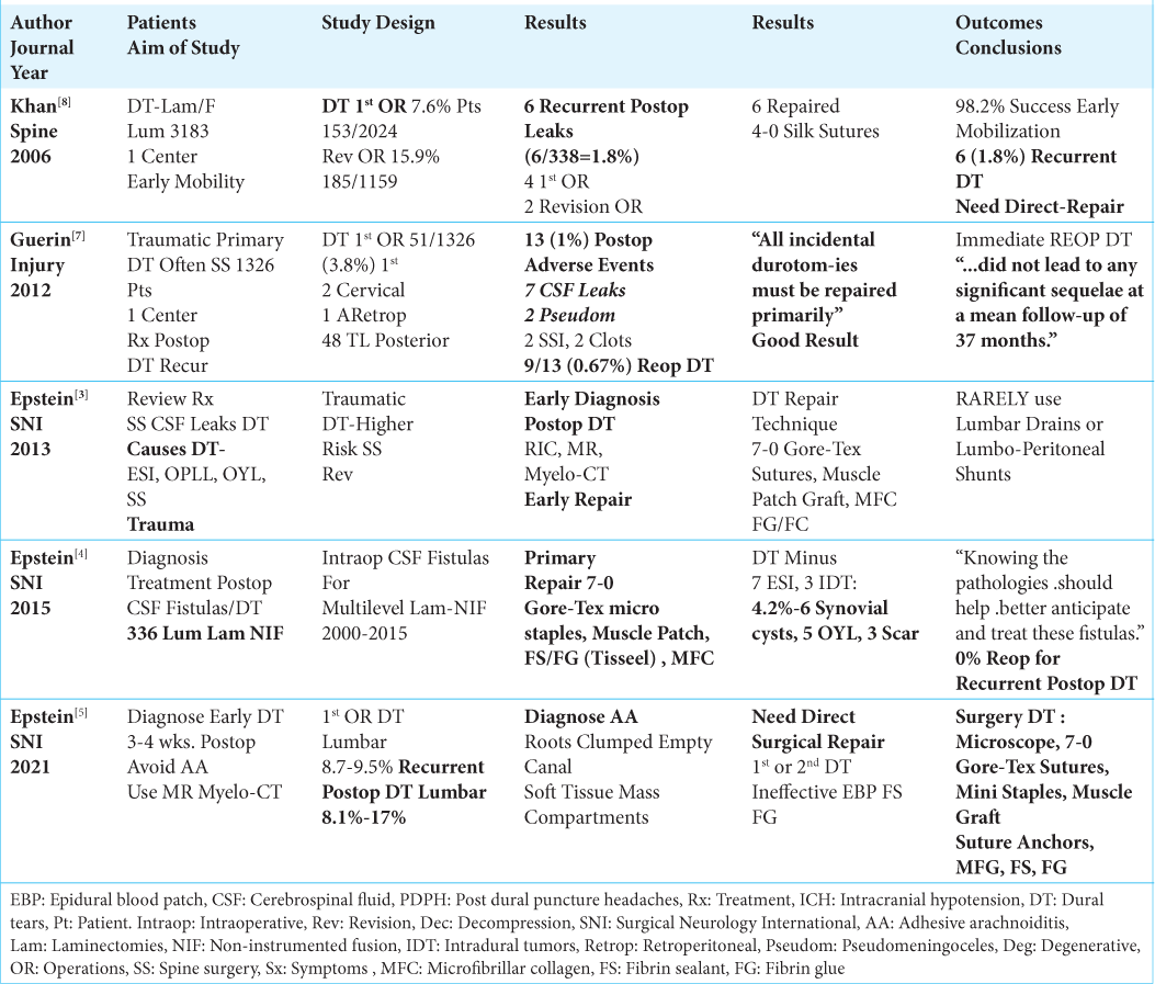
Table 1:

Data for use of epidural blood patches.


Symptoms and Etiology of Intracranial Hypotension

Patients may present with classical symptoms/signs of intracranial hypotension (ICH) due to newly occurrent or recurrent CSF leaks/DT [ Table 1 ].[ 1 - 16 ] These symptoms typically include; postural headaches, nausea/vomiting, visual complaints/double vision, difficulty concentrating, lumbar radiculopathy/cauda equina syndromes, sphincter dysfunction, and/or sexual dysfunction.[ 2 - 9 ] CSF leaks/ DT are largely documented on thin-cut MR or Myelo-CT studies following prior epidural spinal injections (ESI), lumbar punctures (LP), spinal anesthesia (SA), spontaneous intracranial hypotension (SICH), or trauma due to surgery. [ 3 - 5 , 12 - 14 ] Those with “focal DT” attributed to ESI, LP, SA, and SICH are often successfully treated with “targeted EBP” performed under ultrasound, fluoroscopy, or O-arm guidance. [ 7 - 9 , 12 - 14 ] Alternatively, patients with surgically induced initial intraoperative, and/or postoperative recurrent CSF fistulas/DT, that are more variable in size/location, are classically managed with direct surgical repairs [ Table 2 ].[ 3 - 5 , 7 , 8 ] In our review, we were only able to find 3 underpowered studies that failed to adequately document the efficacy of “targeted EBP” in treating recurrent postoperative CSF fistulas/DT [ Tables 1 - 3 ].[ 2 , 11 , 14 ]


Table 2:

Studies advocating repair of intraoperative/postoperative cerebrospinal fluid (CSF) leaks/dural tears (DT).


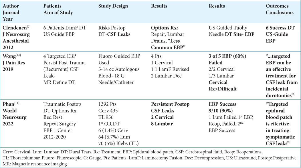
Table 3:

Advocates of epidural blood patches to repair of post-surgical CSF leaks.


Primary or Recurrent Postoperative CSF Fistulas/DT Warranting Direct Surgical Repair

The rates for primary intraoperative (3.0 - 27%) and recurrent postoperative CSF leaks/DT (1.8 - 17.6%) varies [ Tables 1 and 2 ].[ 3 - 5 , 7 , 8 , 11 , 13 , 15 , 16 ] Khan et al. (2006) noted a 7.6% (153/2024 patients) incidence of primary intraoperative DT (i.e. no history of prior surgery) vs. a much higher 15.9% for revision procedures (185/1159 patients); an additional 6 (1.8%) patients developed recurrent postoperative leaks that were successfully surgically repaired.[ 8 ] Primary rates of DT in Guerin et al. (2012) spinal series was 3.8% (i.e. 51/1326: 2 cervical, 1 retroperitoneal, and 48 thoracolumbar); secondary postoperative DT occurred in 9 of 51 (17%) patients (i.e. 7 postoperative recurrent CSF fistulas, and 2 pseudomeningoceles).[ 7 ] In a review article in 2013, Epstein found a 3.0-27% incidence of primary intraoperative CSF fistulas occurring during multilevel lumbar laminectomies with non-instrumented spinal fusions.[ 3 ] Epstein found that other authors further recommended using; “...radioisotope... cisternography (RIC), MR and CT” scans to diagnose the extent/location of postoperative DT.[ 3 ] In Epstein’s 2015 study, she encountered intraoperative DT in 7.14% of 336 patients undergoing multilevel laminectomies/non-instrumented fusions; the rate was reduced to 4.2% (i.e. 3 for scarring, 6 for synovial cysts resection, and 5 for ossification of the yellow ligament penetrating the dura) once the 7 attributed to ESI and 3 to intradural tumors with deliberate durotomies were excluded.[ 4 ] Notably, all patients underwent direct intraoperative surgical repair of DT, and none exhibited recurrent postoperative CSF fistulas/DT. Further, in 2021, Epstein emphasized how critical it is to perform early (i.e. within 3-4 postoperative weeks) direct repair of recurrent postoperative DT to avoid adhesive arachnoiditis, and other adverse events (i.e. acute/subacute/chronic intracranial hypotension (ICH), infection, subdural hematomas, and others).[ 5 ]

Direct Dural Repair Techniques Address Primary/ Recurrent Postoperative CSF Fistulas/DT

In multiple studies, patients underwent primary intraoperative and/or recurrent postoperative direct surgical repair of CSF leaks/DT [ Tables 1 and 2 ].[ 1 , 3 - 5 , 7 , 8 ] Khan et al. (2006) successfully treated intraoperative CSF leaks/DT occurring in 7.6% primary (no prior surgery) vs. 15.9% revision-procedure CSF leaks/DT, and 6 recurrent postoperative DT (1.8%: 2 after primary, and 4 after revision surgery) utilizing 4-0 Silk sutures.[ 8 ] Guerin et al. (2012) advocated direct dural repairs for 51 of 1326 (3.8%) primary surgical, and 9 secondary repairs (i.e. 7 for postoperative recurrent CSF fistulas and 2 with pseudomeningoceles).[ 7 ] They observed; “All incidental durotomies must be repaired primarily, and that following this regimen resulted in no ‘long term sequelae’ (over 37 postoperative months)”.[ 7 ] In 3 studies, Epstein advocated direct “open” dural repair of recurrent postoperative lumbar CSF leaks/DT using 7-0 Gore-Tex sutures (i.e. sutures larger than needles occlude dural punctures), muscle patch grafts, fibrin sealant, and nonsuturable/suturable microfibrillar collagen.[ 3 - 5 ] Further, these procedures should be performed under an operating microscope with intraoperative somatosensory evoked potentials and electromyographic monitoring to avoid inadvertent cauda equina and/or nerve root manipulation. Only rarely should lumbar drains be used to treat complex fistulas as once removed, they leave large dural holes that risk recurrent fistulas; alternatively, lumboperitoneal shunts with horizontal vertical valves should be considered. In 2020, Agulnick, Cohen, and Epstein additionally advocated using mini/micro bone suture anchors to repair difficult lumbar DT with absent lateral dura; it allowed for suturing the fascia, periosteum, muscle grafts and suturable microfibrillar collagen to the lateral laminectomy walls, thus facilitating “water-tight” closures.[ 1 ]

“Targeted” EBP Performed Under Ultrasound, Fluoroscopy, or O-Arm Guidance

EBP involve the epidural injection of a small volume of autologous blood (i.e. 5-20 cc) to occlude a CSF fistulas/ DT [ Tables 1 and 2 ].[ 2 , 6 , 9 , 12 - 14 ] In 1987, Olsen performed EBP using 10-20 cc of autologous blood that they directly injected onto the epidural site of a DT.[ 9 ] They considered EBP: “... an effective and safe method with few and generally mild complications.” Clendenen et al. (2012) used 4-D ultrasound guidance to successfully perform 6 “targeted EBP” in patients with recurrent postoperative DT.[ 2 ] Takai et al. used O-arm stereotactic navigation to perform and monitor the results of their 12 successful “targeted EBP”.[ 13 ] Wong et al. (2019) performed contrast enhanced, fluoroscopically-guided “real time targeted EBP” using 18 gauge needles/catheters and 5-14 cc of autologous blood to treat 4 recurrent postoperative DT (i.e. 1 cervical, and 3 lumbar); here, however, 3 of 5 EBP attempts failed (rate 60%).[ 14 ] Gandhi et al. (2021) considered “EBP, epidural infusions, epidural fibrin glue or surgical repair” as options to treat DT attributed to lumbar punctures, trauma, or surgery.[ 6 ] They further noted; “At this time, epidural blood patches are the first line of treatment.”[ 6 ] Notably, Shin et al. (2022) most recently emphasized the utility of EBP for treating “focal DT” attributed to ESI, SA, LP and SICH; they further utilized MR or ultrasound guidance to directly document their efficacy.[ 12 ] Nevertheless, they also cautioned; “Prophylactic administration of EBP after accidental dural puncture can hardly be substantiated at present.”

Several Studies Documented SICH Were Effectively Treated With “Targeted EBP”

For SICH, Myelo-CT and thin-cut MR studies documented that DT were effectively treated with ultrasound, fluoroscopy, and/or O-arm guided “targeted EBP” [ Tables 1 and 2 ][ 10 , 13 , 15 , 16 ] In Yoshida et al. (2014) 12 patients with SICH, 8 leak sites were documented on Myelo-CT studies, while the remaining 4 were inferred based upon the location of “maximal (epidural) dye extravasation”; following “targeted EBP”, low pressure headaches immediately resolved in 10/12 patients, while they took several months to improve/resolve in 2 patients [ Table 1 ].[ 15 ] Takai et al. (2017) 8 patients with SICH (i.e. defined by Myelo-CT/thin cut MR) underwent O-arm guided “targeted EBP”; 6 of 8 succeeded on the first injection, while it took 2 injections performed under the same anesthesia in the other 2 patients (i.e. O-arm immediately documented inadequate coverage from the 1st injection warranting the second) [ Table 1 ].[ 13 ] Pagani-Estevez (2019) documented for 604 EBP performed in 202 patients with SICH that higher volumes, bilevel, and multilevel “site-directed” EBP were more successful (i.e. lumbar 349 (58%), 75 (12%) bilevel, and 56 (9%) 3 or more level procedures).[ 10 ] Further, Zetaloui et al. (2022) recommended using MR or doppler ultrasound after EBP to document/confirm the accuracy, efficacy, and/or adverse events following these procedures.[ 16 ]

Adverse Events Resulting From Epidural Blood Patches (EBP) to Treat Post LP Dural Punctures

Adverse events (AE) resulting from the application of EBP may include; complications of blood directly entering the CSF pathways, (i.e. sterile meningeal response, adhesive arachnoiditis, and hydrocephalus with cephalad migration amongst others), infection, abscess formation, direct neural compression (i.e. potential for acute cauda equina syndrome), creation of additional sites of dural puncture, and persistent intracranial hypotension [ Table 1 ].[ 9 , 12 , 13 ]

Lack of Evidence that EBP “Effectively” Treat Recurrent Postoperative Lumbar CSF Fistulas/DT

The 3 studies we were able to identify in the literature involving just 20 patients did not document the efficacy for utilizing EBP to treat recurrent postoperative CSF leaks/DT [ Table 3 ].[ 2 , 11 , 12 , 14 ] Clendenen et al. (2012) noted 3 treatment options for patients following lumbar laminectomies/fusions who developed recurrent postoperative CSF fistulas/DT; although these included early direct dural repair, and lumbar subarachnoid drains, they chose the third option - to perform the “less commonly (used) epidural blood patches”.[ 2 ] They then performed 6 “targeted EBP” under 4-D “real-time” ultrasound guidance, and successfully treated recurrent postoperative CSF leaks/DT in all 6 patients. Wong et al. (2019) performed 5 “targeted EBP” for 4 patients with recurrent postoperative CSF leaks/DT under fluoroscopic guidance.[ 14 ] However, they commented that there were “limited data” regarding the efficacy of using EBP in this setting; 3 (60%) of 5 EBP failed (i.e. 2 EBP failed in 1 cervical, 1 EBP failed in 3 lumbar cases). Nevertheless, they ignored their own findings as they wrongly concluded that; “..targeted EBP can be an effective treatment for CSF leaks from incidental durotomies”.[ 14 ] In Phan et al. (2022) 10 of 1392 patients undergoing spinal procedures, developed recurrent postoperative CSF fistulas/DT and were treated with EBP; these successfully resolved following 2 of 2 cervical and 7 of 8 lumbar procedures (i.e. 90% success rate).[ 11 ] They concluded that the; “Targeted epidural blood patch is effective in treating symptomatic CSF leaks.” Nevertheless, Shin et al. recently commented that the; “...prophylactic administration of EBP after accidental dural puncture can hardly be substantiated at present” [ Table 1 ].[ 12 ] In summary, these 3 studies documented a 20% overall failure rate in just 20 patients (i.e. 4/20; up to 60% failure rate in one study) when EBP were used to treat recurrent postoperative CSF leaks/DT.[ 2 , 11 , 12 , 14 ]

CONCLUSION

Epidural blood patches (EBP) successfully treat “focal DT” largely attributed to epidural steroid injections (ESI), lumbar punctures (LP), spinal anesthesia (SA) or spontaneous (SICH). Here, however, as we found insufficient evidence in the 3 studies we identified to support utilizing EBP rather than the classically accepted direct dural repair techniques to treat recurrent postoperative CSF leaks/DT.

Declaration of patient consent

Patient’s consent not required as there are no patients in this study.

Financial support and sponsorship

Nil.

Conflicts of interest

There are no conflicts of interest.

Disclaimer

The views and opinions expressed in this article are those of the authors and do not necessarily reflect the official policy or position of the Journal or its management. The information contained in this article should not be considered to be medical advice; patients should consult their own physicians for advice as to their specific medical needs.

References

1. Agulnick MA, Cohen BR, Epstein NE. Unique Bone suture anchor repair of complex lumbar cerebrospinal fluid fistulas. Surg Neurol Int. 2020. 11: 153

2. Clendenen SR, Pirris S, Robards CB, Leone B, Nottmeier EW. Symptomatic postlaminectomy cerebrospinal fluid leak treated with 4-dimensional ultrasound-guided epidural blood patch. J Neurosurg Anesthesiol. 2012. 24: 222-5

3. Epstein NE. A review article on the diagnosis and treatment of cerebrospinal fluid fistulas and dural tears occurring during spinal surgery. Surg Neurol Int. 2013. 4: S301-17

4. Epstein NE. Incidence and management of cerebrospinal fluid fistulas in 336 multilevel laminectomies with non instrumented fusions. Surg Neurol Int. 2015. 6: S463-8

5. Epstein NE. Perspective: Early diagnosis and treatment of postoperative recurrent cerebrospinal fluid fistulas/dural tears to avoid adhesive arachnoiditis. Surg Neurol Int. 2021. 12: 208

6. Gandhi J, DiMatteo A, Joshi G, Smith NL, Khan SA. Cerebrospinal fluid leaks secondary to dural tears: A review of etiology, clinical evaluation, and management. Int J Neurosci. 2021. 131: 689-95

7. Guerin P, El Fegoun AB, Obeid I, Gille O, Lelong L, Luc S. Incidental durotomy during spine surgery: Incidence, management and complications. A retrospective review. Injury. 2012. 43: 397-401

8. Khan MH, Rihn J, Steele G, Davis R, Donaldson WF, Kang JD. Postoperative management protocol for incidental dural tears during degenerative lumbar spine surgery: A review of 3,183 consecutive degenerative lumbar cases. Spine (Phila Pa 1976). 2006. 31: 2609-13

9. Olsen KS. Epidural blood patch in the treatment of post-lumbar puncture headache. Pain. 1987. 30: 293-301

10. Pagani-Estevez GL, Cutsforth-Gregory JK, Morris JM, Mokri B, Piepgras DG, Mauck WD, editors. Procedural predictors of epidural blood patch efficacy in spontaneous intracranial hypotension. Reg Anesth Pain Med. Jan 13, 2019. p. rapm-2018-000021

11. Phan A, Shah S, Joo P, Mesfin A. Can cervical and lumbar epidural blood patches help avoid revision surgery for symptomatic postoperative dural tears?. World Neurosurg. 2022. 164: e877-83

12. Shin YH. Recent update on epidural blood patch. Anesth Pain Med (Seoul). 2022. 17: 12-23

13. Takai K, Taniguchi M. Targeted epidural blood patch under O-arm-guided stereotactic navigation in patients with intracranial hypotension associated with a spinal cerebrospinal fluid leak and ventral dural defect. World Neurosurg. 2017. 107: 351-7

14. Wong AK, Rasouli MR, Ng A, Wang D. Targeted epidural blood patches under fluoroscopic guidance for incidental durotomies related to spine surgeries: A case series. J Pain Res. 2019. 12: 2825-33

15. Yoshida H, Takai K, Taniguchi M. Leakage detection on CT myelography for targeted epidural blood patch in spontaneous cerebrospinal fluid leaks: Calcified or ossified spinal lesions ventral to the thecal sac. J Neurosurg Spine. 2014. 21: 432-41

16. Zetlaoui PJ, Buchheit T, Benhamou D. Epidural blood patch: A narrative review. Anaesth Crit Care Pain Med. 2022. 41: 101138

Leave a Reply

Your email address will not be published. Required fields are marked *