Tools

Nancy E. Epstein1, Marc A. Agulnick2
  1. Clinical Professor of Neurosurgery, School of Medicine, State University of NY at Stony Brook, and Editor-in-Chief Surgical Neurology International NY and ℅ Dr. Marc Agulnick 1122 Frankllin Avenue Suite 106, Garden City, NY 11530, USA,
  2. Assistant Clinical Professor of Orthopedics, NYU Langone Hospital, Long Island, NY, USA. 1122 Franklin Avenue Suite 106 Garden City, NY 11530.

Correspondence Address:
Nancy E. Epstein, M.D., F.A.C.S. Clinical Professor of Neurosurgery, School of Medicine, State University of NY at Stony Brook, and Editor-in-Chief Surgical Neurology International NY and ℅ Dr. Marc Agulnick 1122 Frankllin Avenue Suite 106, Garden City, NY 11530, USA.

DOI:10.25259/SNI_943_2022

Copyright: © 2022 Surgical Neurology International This is an open-access article distributed under the terms of the Creative Commons Attribution-Non Commercial-Share Alike 4.0 License, which allows others to remix, transform, and build upon the work non-commercially, as long as the author is credited and the new creations are licensed under the identical terms.

How to cite this article: Nancy E. Epstein1, Marc A. Agulnick2. Perspective: Lumbar adhesive arachnoiditis (AA)/ Chronic AA (CAA) are clinical diagnoses that do not require radiographic confirmation. 04-Nov-2022;13:507

How to cite this URL: Nancy E. Epstein1, Marc A. Agulnick2. Perspective: Lumbar adhesive arachnoiditis (AA)/ Chronic AA (CAA) are clinical diagnoses that do not require radiographic confirmation. 04-Nov-2022;13:507. Available from: https://surgicalneurologyint.com/surgicalint-articles/11978/

Date of Submission
13-Oct-2022

Date of Acceptance
13-Oct-2022

Date of Web Publication
04-Nov-2022

Abstract

Background: Our hypothesis was that lumbar adhesive arachnoiditis (AA)/chronic lumbar AA (CAA) are clinical diagnoses that do not require radiographic confirmation. Therefore, patients with these syndromes do not necessarily have to demonstrate significant radiographic abnormalities on myelograms, MyeloCT studies, and/or MR examinations. When present, typical AA/CAA findings may include; central or peripheral nerve root/cauda equina thickening/clumping (i.e. latter empty sac sign), arachnoid cysts, soft tissue masses in the subarachnoid space, and/or failure of nerve roots to migrate ventrally on prone MR/Myelo-CT studies.

Methods : We reviewed 3 articles and 7 clinical series that involved a total of 253 patients with AA/CAA to determine whether there was a significant correlation between these clinical syndromes, and myelographic, Myelo-CT, and/or MR imaging pathology.

Results: We determined that patients with the clinical diagnoses of AA/CAA do not necessarily exhibit associated radiographic abnormalities. However, a subset of patients with AA/CAA may show the classical AA/CAA findings of; central or peripheral nerve root/cauda equina thickening/clumping (empty sac sign), arachnoid cysts, soft tissue masses in the subarachnoid space, and/or failure of nerve roots to migrate ventrally on prone MR/ Myelo-CT studies.

Conclusion: Patients with clinical diagnoses of AA/CAA do not necessary show associated neuroradiagnostic abnormalities on myelograms, Myelo-CT studies, or MR. Rather, the clinical syndromes of AA/CAA may exist alone without the requirement for radiolographic confirmation.

Keywords: Adhesive Arachnoiditis (AA), Chronic Adhesive Arachnoiditis (CAA), Clinical Syndrome, Diagnosis, Lumbar, Magnetic Resonance Imaging (MR), Myelo-CT Scans, Myelography, Mild-Moderate, Severe

INTRODUCTION

Patients with lumbar adhesive arachnoiditis (AA)/chronic AA (CAA) have clinical syndromes characterized by symptoms of pain, paresthesias, and varied motor, sensory, and/or sphincteric deficits. Our hypothesis was that patients with these syndromes do not have to demonstrate any significant radiographic confirmatory pathology on myelograms, Myelo-CT studies, or MR examinations.[ 8 ] To better assess this, we reviewed 3 articles and 7 patient-based studies (i.e. total 253 patients) to determine whether AA/CAA patients did or did not demonstrate central or peripheral nerve root/cauda equina thickening/clumping (i.e. latter empty sac sign), arachnoid cysts, soft tissue masses in the subarachnoid space, and/or failure of nerve roots to migrate ventrally on prone MR, and Myelo-CT studies [ Table 1 ].[ 1 - 5 , 8 - 10 ]


Table 1:

Summary of arachnoiditis literature.


History of AA

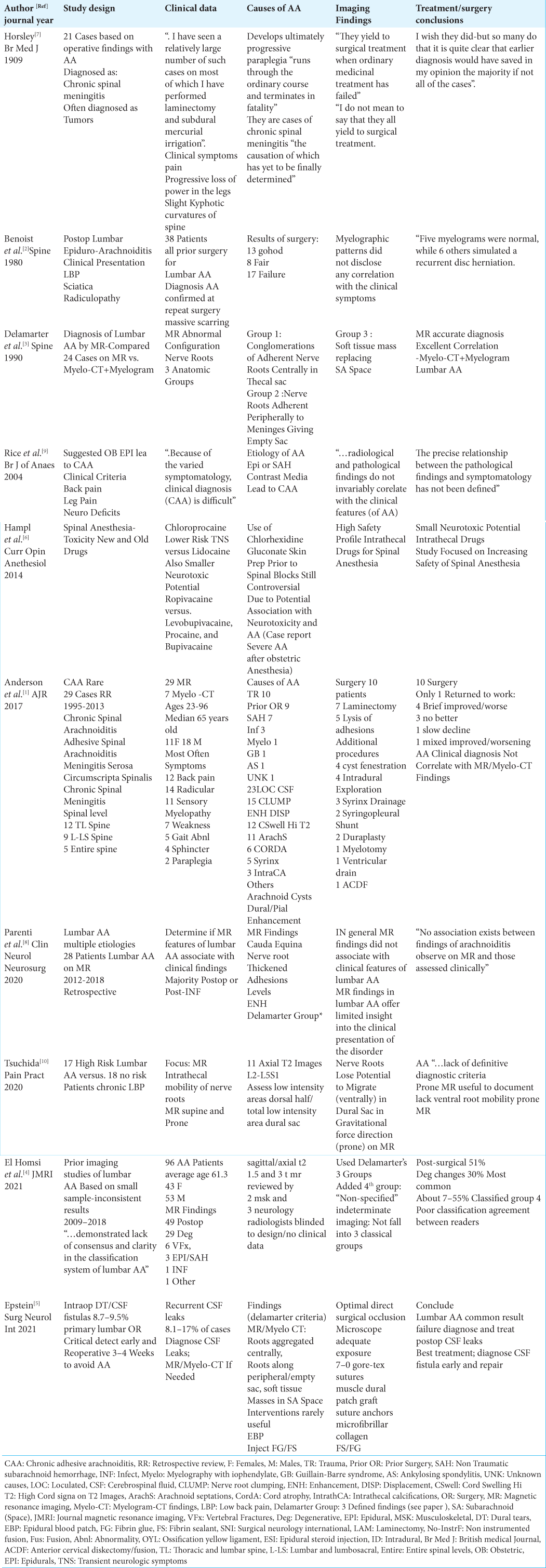
In 1909, Sir Victor Horsely presented 21 cases where he anticipated finding spinal tumors. However, at surgery he encountered AA variously labeled as; chronic spinal meningitis/arachnoiditis, acute myelitis, adhesive spinal arachnoiditis, and meningitis serosa circumscripta spinalis [ Table 1 ].[ 7 ] Later, in 2020, Tsuchida et al. similarly corroborated that lumbar AA could be misinterpreted as; “… tumors of the spinal cord or meninges…” [ Table 1 ].[ 7 , 10 ]

Etiology of adhesive arachnoiditis (AA)/chronic adhesive arachnoiditis (CAA)

The major etiology of AA/CAA typically includes activation of a “…subarachnoid inflammatory cascade…” [ Table 1 ].[ 1 - 10 ] Factors precipitating this cascade include; trauma, myelography, infection/inflammatory reactions, epidural steroid injections/anesthesia, blood in the subarachnoid space (i.e. traumatic/atraumatic), intrathecal catheter placement, and/or prior surgery.[ 1 - 10 ] Horsely could not define a specific etiology for his 21 operated cases: “They are cases of chronic spinal meningitis the causation of which has yet to be finally determined [ Table 1 ].[ 7 ] Benoist et al. (1980) attributed lumbar AA to prior lumbar surgery after reoperating on 38 patients [ Table 1 ].[ 2 ] Rice et al. (2004) attributed severe CAA to epidural injections or the subarachnoid administration of contrast media; “Back pain with or without leg symptoms (e.g. pain, paresthesias or weakness) is typical but a wide range of neurological abnormalities have been associated with CAA” [ Table 1 ].[ 9 ] In 2014, Hempl et al. further reviewed the toxicity of “… new and old drugs and compounds…” for spinal anesthesia as contributors to the clinical syndrome of AA [ Table 1 ].[ 6 ] Specifically, they found that Chloroprocaine resulted in a reduced risk of neurotoxicity vs. Lidocaine, while Ropivacaine exhibited a “…smaller neurotoxic potential…” when compared with Levobupivacaine, Procaine, and Bupivacaine.”[ 6 ] Further, using Chlorhexidine Gluconate for skin disinfection prior to spinal blocks increased the potential risk for neurotoxicity and adhesive arachnoiditis [ Table 1 ].[ 6 ] Anderson et al. (2017), in 29 (14%) patients with severe CAA taken from a total series of 209 AA patients, found that the majority of cases of CAA could be attributed to; trauma (10 patients), prior surgery (9 patients), and non-traumatic subarachnoid hemorrhage (7 patients) [ Table 1 ].[ 1 ] Parent et al. (2020) additionally attributed AA in 28 patients to prior surgery or to prior infections [ Table 1 ].[ 8 ] Tsuchida et al. (2020) further identified 17 “high risk” patients for lumbar AA (i.e. 15 with prior surgery, and 2 after oil-based myelography) who demonstrated: “… fibrous tissue outgrowth into the subarachnoid space… result (ing) in a fibrous tangle, entrapping the lumbar, and sacral nerve roots and cauda equina…”[ Table 1 ].”[ 10 ] In El Homsi et al. (2021) series of 96 patients, lumbar AA was largely attributed to; “… postsurgical findings (49 patients), and/or degenerative disease (29 patients)…” [ Table 1 ].[ 4 ] In 2021, Epstein emphasized the most common reason for AA was prior surgery, the likelihood of which increased with an intraoperative cerebrospinal fluid (CSF) leak (i.e. incidence primary CSF leaks 8.7–9.5%, incidence for subsequent CSF fistulas 8.1–17%) [Table1].[ 5 ] Further, the risk for developing AA was also markedly increased if such dural leaks went undiagnosed and untreated for over 3–4 postoperative weeks [ Table 1 ].[ 5 ]

Typical clinical features of AA

Patients with AA may present with a wide array of neurological symptoms and signs [ Table 1 ].[ 1 , 5 , 8 , 10 ] Anderson et al. (2017) 29 patients with severe CAA displayed significant radiculopathy, back pain, and/or sensory neuropathy [ Table 1 ].[ 1 ] Tsuchida et al. (2020) 17 patients at “high risk” for lumbar AA exhibited; leg pain, neurogenic claudication, motor/sensory deficits including paraparesis/paralysis and hypesthesia/sensory loss, with or without sphincter dysfunction [ Table 1 ].[ 10 ] Parenti et al. (2020) 28 patients with lumbar AA displayed;“… heterogenous (symptoms/ signs) with patients reporting a range of lower extremity neurological symptoms with varying levels of severity”.[ 8 ] Epstein further summarized the neurological findings for AA as including; “…low back pain, lower extremity weakness, cauda equina syndrome, progressive paraparesis, sensory loss/dysesthesias, and sphincter function loss” [ Table 1 ”].[ 5 ]

MR and myelo-CT/plain film myelography (PFM) findings for AA/CAA

Myelograms, Myelo-CT studies, and MR examinations cited multiple imaging findings that may be associated with the clinical syndromes of AA/CAA [ Table 1 ].[ 1 - 5 , 8 - 10 ] These findings often utilized by Delamarter et al. included classical radiological descriptions for AA (i.e. clumping (centrally, peripherally-empty sac sign), and/or soft tissue subarachnoid masses), while other authors like Tsuchida et al. newly added the failure of nerve roots or the cauda equina to migrate ventrally on prone MR studies. [ 1 - 5 , 8 - 10 ]

Benoist et al. findings [ Table 1 ][ 2 ]

In Benoist et al. (1980), 38 patients were diagnosed with lumbar AA attributed to prior lumbar surgery; all findings were confirmed at reoperations. Nevertheless, the authors found no specific correlation between the clinical syndrome of lumbar AA and any specific radiographic abnormalities; “Myelographic patterns did not disclose any correlation with the clinical symptoms…” [ Table 1 ].[ 2 ]

Delamarter et al. findings [ Table 1 ][ 3 ]

In 1990, Delamarter et al., based on a review of 24 MR and 20 Myelo-CT/plain film myelographic (PFM) examinations, divided classical findings for AA into 3 Groups; Group I “… conglomeration of adherent nerve roots resting centrally within the thecal sac; Group II ”… nerve roots adherent peripherally to the meninges giving rise to an ‘empty sac’ appearance”; and Group III ”…showed a soft tissue mass replacing the subarachnoid space” [ Table 1 ].[ 3 ] They also emphasized the close correlation/relationship between MR and Myelo-CT/PFM findings; “MR imaging resulted in accurate diagnosis and had excellent correlation with CT myelography and plain film myelographic findings in the diagnosis of lumbar arachnoiditis”.[ 3 ] Their emphasis on this close correlation between MR and Myelo-CT studies for arachnoiditis remains critical to this day for those attempting to correlate patients with clinical AA/CAA syndromes and neurodiagnostic images.

Rice et al. (2004): Epidural anesthesia for delivery does not lead to CAA [ Table 1 ][ 9 ]

Rice et al. determined that patients who received epidural anesthesia for labor/delivery were not more susceptible to developing CAA as had been previously thought [ Table 1 ].[ 9 ] They further noted that the: “…radiological and pathological findings (i.e. imaging for CAA) do not invariably correlate with the clinical features (of CAA)” [ Table 1 ].[ 9 ]

Anderson et al. findings [ Table 1 ][ 1 ]

Anderson et al. (2017) observed significant imaging findings (i.e. MR 29 patients: Myelo-CT 7 patients) in their 29 patients specifically selected for severe CAA [ Table 1 ].[ 1 ] MR findings included; loculated CSF collections, root clumping (central), peripheralization/empty sac sign, subarachnoid soft tissue masses/pial dural enhancement, cord swelling/ high T2 cord signal, distortion/tethering of the cord, syrinx, arachnoid cysts/webs/septations [ Table 1 ].[ 1 ] Myelo-CT studies demonstrated; arachnoid cysts/septations, thickened/ clumped roots, intrathecal ossification, atrophic cord changes/enlargement, root lack of filling, other subarachnoid (partial/total block) filling defects, and/or soft tissue lesions throughout dural sac [ Table 1 ].[ 1 ] Notably, the majority of nerve root/cauda equina adhesions occurred in the dorsal spinal canal, except for those with the most severe disease, whose adhesions could appear “circumferentially”.[ 1 ]

Parenti et al. (2020) findings [ Table 1 ][ 8 ]

Parenti et al. (2020) noted variable MR findings for their 28 patients with AA; “…cauda equina nerve root clumping/ thickening, adhesion location/levels, enhancement, and (the) Delamarter group (criteria)” [ Table 1 ].[ 8 ] They concluded; “MRI findings in lumbar arachnoiditis offer limited insight into the clinical presentation of the disorder”. Further; “In general, MRI findings did not associate with the clinical features of lumbar arachnoiditis” [ Table 1 ].

Tsuchida et al. findings (2020)[ 10 ]

Tsuchida et al. (2020) compared the MR scans performed in 17 “high-risk” AA vs. 18 “no risk” AA patients looking for failure of nerve roots/cauda equina to migrate ventrally on prone vs. supine studies [ Table 1 ].[ 10 ] Indeed, they found;“… the intrathecal mobility of nerve roots and the cauda equina is reduced in patients at high risk for lumbar AA.” Further, “... conventional MRI which is usually conducted only in the supine position, cannot detect this immobility...” In short, they concluded that many previous studies failed to accurately diagnose AA as they did not include prone MR examinations.

El Homsi et al. findings (2021)[ 4 ]

In 2021, El Homsi et al. diagnosed 96 patients with lumbar AA using Delamarter’s 3 Groups [ Table 1 ].[ 4 ] To these, however, they uniquely added a 4thNon-Specified” Group that was: “…proposed for indeterminate imaging findings that did not fall into the classical groups.” They found that between 7-55% of MR studies were classified into the new 4th Group, and that there was a “… very poor classification agreement between readers…”

Epstein (2021) findings [ Table 1 ][ 5 ]

Epstein (2021) reviewed Delamarter et al. classic 3 Groups based on MR and Myelo-CT studies for AA [ Table 1 ].[ 5 ] They showed; “…nerve root clumping, enhancement/displacement, cord swelling/atrophy, and or syrinx formation.” [ Table 1 ].[ 5 ] Findings for Myelograms/Myelo-CT studies included; Type I: “…empty thecal sac sign (nerve roots adherent to peripheral dura), and Type II: local/diffuse filling defects throughout the thecal sac”. Epstein further recommended incorporating Tsuchida et al. prone MR findings of a; “… a lack of ventral root mobility with prone MR positioning…”, to the analyses of Myelo-CT examinations.

Lack of association between clinical and radiographic AA [ Table 1 ]

Multiple studies have shown that the clinical diagnoses of AA/ CAA are not necessarily corroborated by or associated with “diagnostic” radiographic AA abnormalities [ Table 1 ].[ 1 , 4 , 8 , 9 , 10 ] Rice et al. (2004) found that “The precise relationship between the pathological (radiological) findings and symptomatology has not been defined” [ Table 1 ].[ 9 ] Anderson et al. (2017) observed: “CAA has a variable imaging appearance on both myelography and MRI. CAA does not have a characteristic distribution…”[ 1 ] Further they noted the; “…poorly defined and varying imaging characteristics (of AA) may result in a missed or delayed diagnosis”. Tsuchida et al. concluded; “… it is difficult to diagnose (AA) owing to the lack of definitive diagnostic criteria” [ Table 1 ].[ 10 ] They also observed; “… the precise relationship between the complex clinical symptomatology and pathological imaging findings has not yet been defined and validated.” Further; “Some MR findings (i.e. clumped nerve roots, empty sac appearance, and deformities of the dural sac) have been proposed as the typical presentation of lumbar AA. However these signs variedly emerge in patients with lumbar AA; therefore they are not established and valid as useful diagnostic symptoms”. Parenti et al. (2020) noted that lumbar AA has “… multiple etiologies and a spectrum of imaging and clinical characteristics”, and that: “In general, MR findings did not associate with clinical features of lumbar AA …”[ 8 ] They. concluded: “MR findings in lumbar AA offer limited insight into the clinical presentation of the disorder,” and that; “…the extensive array of clinical and imaging findings exhibited in lumbar AA have been purported to lack association by multiple authors” [ Table 1 ]. Also when El Homsi et al. (2021) evaluated the MR findings for patients with AA, they observed that previous imaging studies … reported, at best, “…inconsistent results.”[ 4 ]

Surgery ineffective for lumbar AA [ Table 1 ]

Surgical intervention typically does not result in sustained neurological improvement for patients with AA/CAA [ Table 1 ].[ 1 , 5 - 8 ] Horsely in 1909 observed; “ I have seen a relatively large number of such cases, on most of which I have performed a laminectomy and subdural mercurial irrigation”.[ 7 ] He goes on to state: “I do not mean to say that they all yield to surgical treatment-I wish they did but so many do that it is quite clear that earlier diagnosis would have saved, in my opinion, the majority of not all of the cases”. In Anderson et al. (2017) series, 10 of 29 CAA patients had surgery; the results were poor as 4 transiently improved then worsened, 3 did not improve, 1 continued to decline, 1 was at first minimally improved and then worsened, and only 1 had improvement allowing him to return to work [ Table 1 ].[ 1 ] They concluded; “Patients who have improvement after surgery often have relapses and progressive symptoms occur later, and their long-term prognosis after surgery remains poor”. Parenti et al. (2020) noted that medical or surgical treatment modalities were typically ineffective for lumbar AA: “Despite surgical and medical advancements, therapeutic options are limited and mainly include microsurgical lysis of adhesions, corticosteroid therapy, and chronic pain control”.[ 8 ] Epstein (2021) summarized the poor results of surgery for AA: “… the majority of clinical studies acknowledge that postoperative lumbar AA is not a surgically remediable lesion.” The best way, therefore to avoid lumbar AA, was for: “. early recognition and repair of such persistent postoperative recurrent CSF fistulas/DT…”[ 5 ]

CONCLUSION

The clinical diagnoses of AA/CAA do not require radiographic confirmation on myelographic, Myelo-CT, and/ or MR studies [ Table 1 ].[ 1 - 5 , 8 - 10 ]

Declaration of patient consent

Patients’ consent not required as patients’ identities were not disclosed or compromised.

Financial support and sponsorship

Nil.

Conflicts of interest

There are no conflicts of interest.

Disclaimer

The views and opinions expressed in this article are those of the authors and do not necessarily reflect the official policy or position of the Journal or its management. The information contained in this article should not be considered to be medical advice; patients should consult their own physicians for advice as to their specific medical needs.

References

1. Anderson TL, Morris JM, Wald JT, Kotsenas AL. Imaging appearance of advanced chronic adhesive arachnoiditis: A retrospective review. AJR Am J Roentgenol. 2017. 209: 648-55

2. Benoist M, Ficat C, Baraf P, Cauchoix J. Postoperative lumbar epiduro-arachnoiditis. Diagnostic and therapeutic aspects. Spine (Phila Pa 1976). 1980. 5: 432-6

3. Delamarter RB, Ross JS, Masaryk TJ, Modic MT, Bohlman HH. Diagnosis of lumbar arachnoiditis by magnetic resonance imaging. Spine (Phila Pa 1976). 1990. 15: 304-10

4. El Homsi M, Gharzeddine K, Cuevas J, Arevalo-Perez J, Rebeiz K, Khoury NJ. MRI findings of arachnoiditis, revisited. Is classification possible?. J Magn Reson Imaging. 2021. 54: 904-9

5. Epstein NE. Perspective: Early diagnosis and treatment of postoperative recurrent cerebrospinal fluid fistulas/dural tears to avoid adhesive arachnoiditis. Surg Neurol Int. 2021. 12: 208

6. Hampl K, Steinfeldt T, Wulf H. Spinal anesthesia revisited: Toxicity of new and old drugs and compounds. Curr Opin Anesthesiol. 2014. 27: 549-55

7. Horsley V. A clinical lecture on chronic spinal meningitis: Its differential diagnosis and surgical treatment. Br Med J. 1909. 1: 513-7

8. Parenti V, Huda F, Richardson PK, Brown D, Aulakh M, Taheri MR. Lumbar arachnoiditis: Does imaging associate with clinical features?. Clin Neurol Neurosurg. 2020. 192: 105717

9. Rice I, Wee MY, Thomson K. Obstetric epidurals and chronic adhesive arachnoiditis. Br J Anaesth. 2004. 92: 109-20

10. Tsuchida R, Sumitani M, Azuma K, Abe H, Hozumi J, Inoue R. A novel technique using magnetic resonance imaging in the supine and prone positions for diagnosing lumbar adhesive arachnoiditis: A preliminary study. Pain Pract. 2020. 20: 34-43

Leave a Reply

Your email address will not be published. Required fields are marked *