

- Department of Neurosurgery, The Center for Advanced Neurology and Neurosurgery, Porto Alegre, Rio Grande do Sul,
- Department of Neurosurgery, University of São Paulo, Faculdade das Clinicas,
- Department of Microsurgical Laboratory, Beneficencia Portuguesa Hospital, São Paulo, Brazil.
Correspondence Address:
Gustavo Rassier Isolan, Department of Neurosurgery, The Center for Advanced Neurology and Neurosurgery, Porto Alegre, Rio Grande do Sul, Brazil.
DOI:10.25259/SNI_75_2022
Copyright: © 2022 Surgical Neurology International This is an open-access article distributed under the terms of the Creative Commons Attribution-Non Commercial-Share Alike 4.0 License, which allows others to remix, transform, and build upon the work non-commercially, as long as the author is credited and the new creations are licensed under the identical terms.How to cite this article: Schlindwein Vaz MA1, Monteiro JM1, Tzu WH2, Neto MR3, Holanda Vd3, Figueiredo EG2, Isolan GR1. Professor Evandro de Oliveira, a guiding light in skull base surgery and vascular neurosurgery. Surg Neurol Int 03-Jun-2022;13:229
How to cite this URL: Schlindwein Vaz MA1, Monteiro JM1, Tzu WH2, Neto MR3, Holanda Vd3, Figueiredo EG2, Isolan GR1. Professor Evandro de Oliveira, a guiding light in skull base surgery and vascular neurosurgery. Surg Neurol Int 03-Jun-2022;13:229. Available from: https://surgicalneurologyint.com/surgicalint-articles/11640/
Date of Submission
18-Jan-2022
Date of Acceptance
28-Apr-2022
Date of Web Publication
03-Jun-2022
Born in 1945, in Santa Catarina, Professor Evandro de Oliveira is a guiding light in skull base surgery that will always be with us. On February 11, 2021, he succumbed to amyotrophic lateral sclerosis in the city of São Paulo. He was one of the greatest names in neurosurgery worldwide and influenced generations of surgeons, especially in Brazil and Latin America [
Professor Oliveira graduated from Federal University of Santa Catarina in 1969 and became a neurosurgery resident at the School of Medicine, University of the Republic in Montevideo, Uruguay, from 1970 to 1972; a research fellow in Microsurgical Anatomy in the Department of Neurosurgery at the University of Florida, Gainesville, United States, from 1981 to 1982, where he was considered the main pupil of Professor Albert Rhoton Jr.; and received a PhD in Morphofunctional Sciences from the University of São Paulo in 2002.
Born in Santa Catarina, a prosperous state in the south region of Brazil, he was the founder of the São Paulo Institute of Neurological Sciences and an associate professor at the Mayo Clinic College of Medicine and Science in Jacksonville, Florida, United States. For decades, he was the head of the neurosurgery team and responsible for the microsurgical laboratory at the Beneficência Portuguesa Hospital in São Paulo. Later, he was a professor emeritus of neurosurgery at the University of Campinas (Unicamp), one of the most respected universities in the country. An excellent teacher, he gave hundreds of lectures on vascular microneurosurgery, skull base surgery, neuro-oncological surgery, and epilepsy surgery and was the director of various microsurgical anatomy courses all over the world.
PROFESSIONAL MERITS
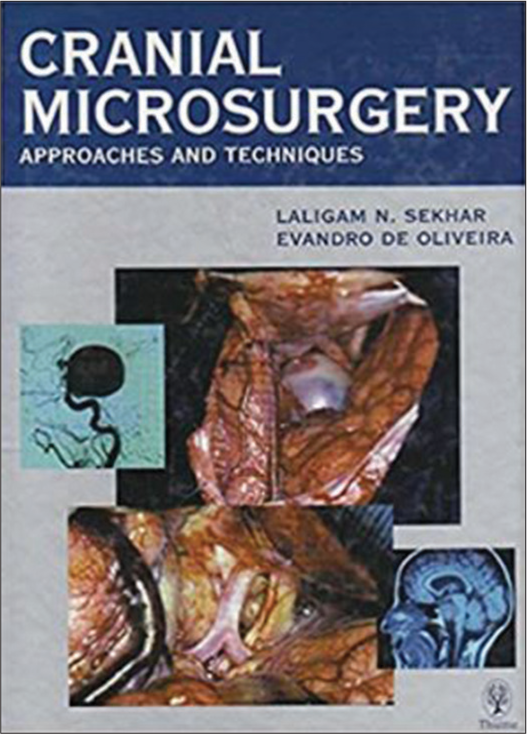
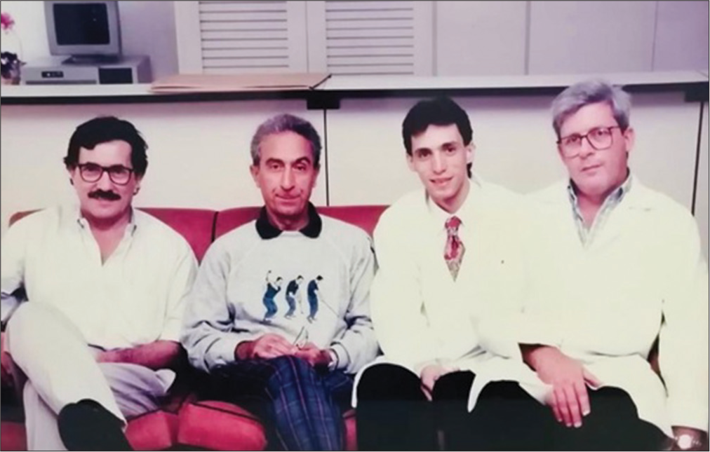
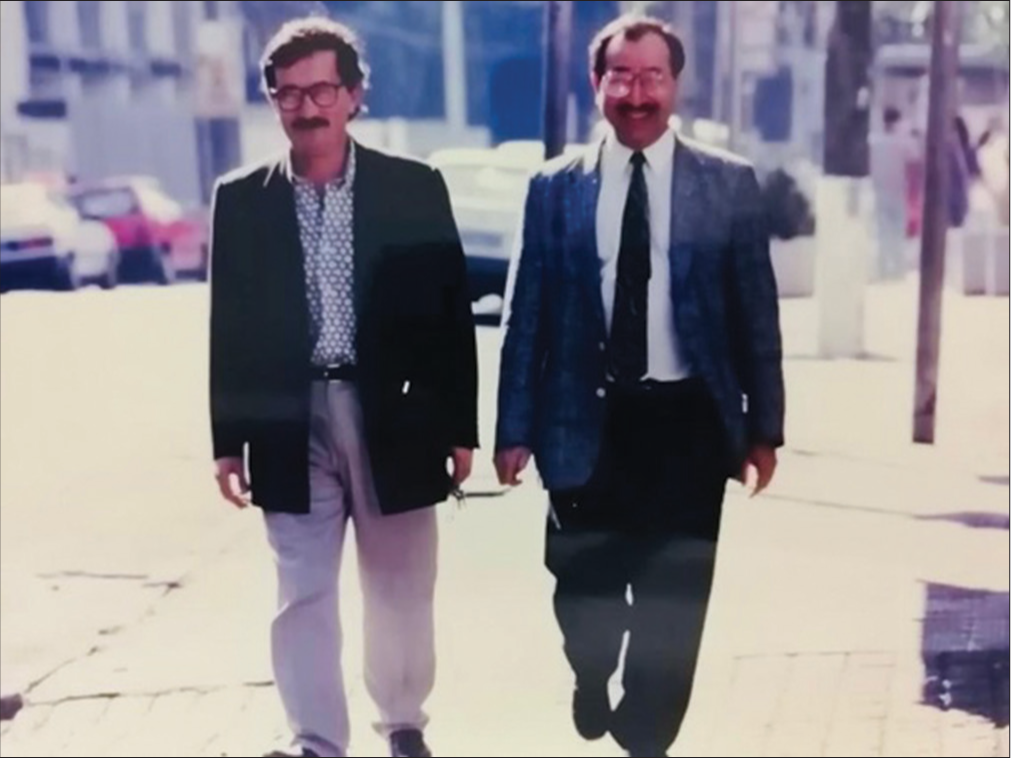
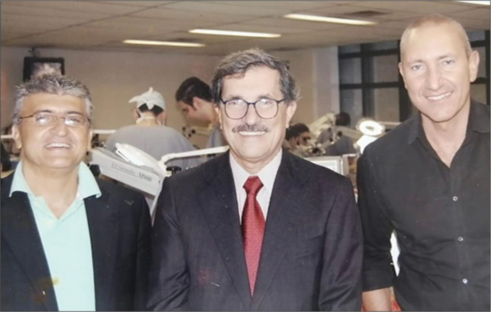
Professor Evandro de Oliveira authored and coauthored hundreds of scientific articles. Some of the world’s most renowned and prominent neurosurgeons of his time, both from Latin America and worldwide, worked with him. Ribas,[
One article which was particularly important was published in 1998.[
For his contribution to the excellence of neurosurgery worldwide, the American Association of Neurological Surgeons named a symposium in his honor that was held in April 2019 in San Diego, California, United States – the Evandro de Oliveira Symposium.
Professionally, he considered his greatest legacy the creation of the Microsurgical Laboratory at the Beneficência Portuguesa Hospital in São Paulo. It became the most active neurosurgery training center in Latin America, where more than 5000 professionals, including neurosurgeons and otolaryngologists, have come to attend a variety of courses on microsurgical anatomy [
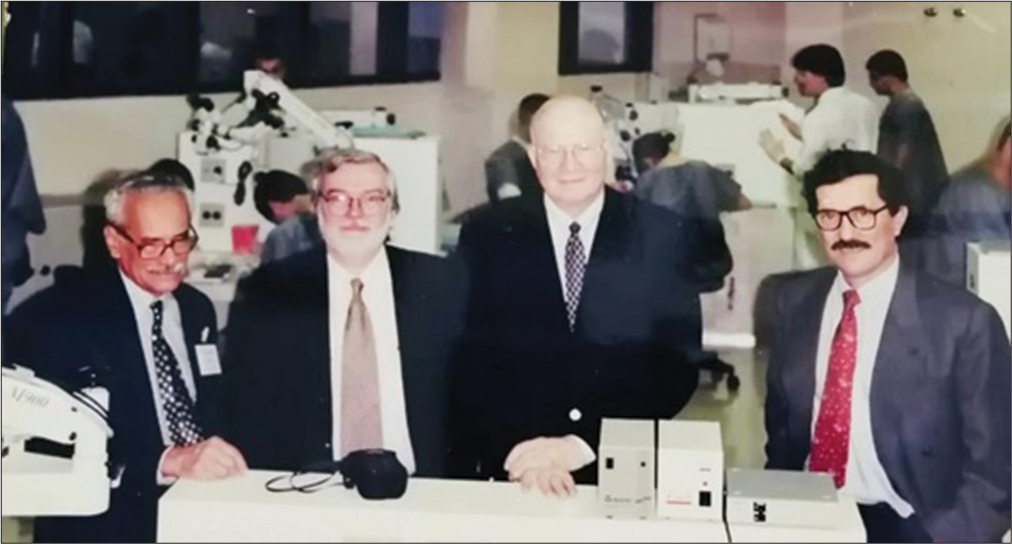
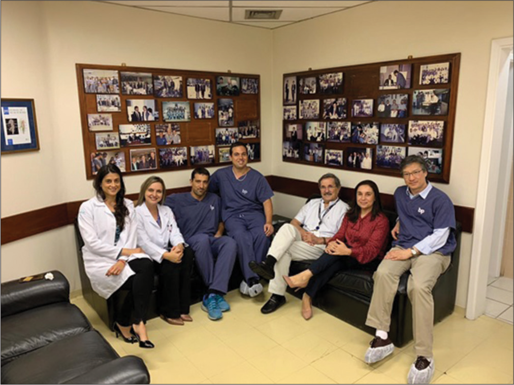
Even before the opening of the microsurgery laboratory, Professor Evandro created and directed continuing education courses since 1989 at the Neurological Institute of São Paulo, with the inaugural seminar “Saccular Intracranial Aneurysms.” The opening of the Microsurgical Laboratory, in 1993, was the first of its kind in the city of São Paulo. It began with five workstations, each consisting of a workbench with a microscope and lighting, and a workbench for the instructor, which thus enabled true hands-on education. Since 1995, continuing education courses have welcomed some of the greatest international figures in the development of neurosurgery, its research, and education.
From 1993 to the present date, the following courses have been given annually for the training and improvement of both neurosurgeons and other specialists:
Anatomy of the cerebral ventricles, sulci, gyri, white matter, and techniques for AVMs and gliomas High-flow extracranial-to-intracranial bypass microsurgery techniques Microsurgical anatomy of the temporal bone and transtemporal approaches to the skull base Microsurgical anatomy and approaches to the sellar and parasellar region/anatomy and endoscopic surgery of the sellar and clival region Peripheral nerve surgery Laboratory training with Midas Rex Vascular hands-on seminar: craniotomy with artificial skull and Sylvian fissure opening simulation, clipping aneurysms, and vascular anastomoses with placenta Sulci, gyri, and ventricles White matter dissection using the Klinger technique Brainstem and cerebellum.
HOW HE STAYS IN OUR MEMORY
According to Professor Evandro de Oliveira, “the Microsurgical Laboratory has a worldwide reputation (being one of the most respected in the world), providing mainly to Brazilians and other Latin Americans, as well as neurosurgeons from various other parts of the world who have also visited, a unique opportunity for learning, training, and development by participating in the continuing education courses offered by the Institute of Neurological Sciences at its facilities.” The principal mission of these hands-on courses is to disseminate throughout Brazil skull base surgery and vascular microsurgery skills using a didactic and rational approach that is based on a laboratory-acquired understanding of surgical anatomy. Professor Evandro de Oliveira had a peculiar way of seeing the world and tolerated nothing less than perfection. He had a unique and serious demeanor.
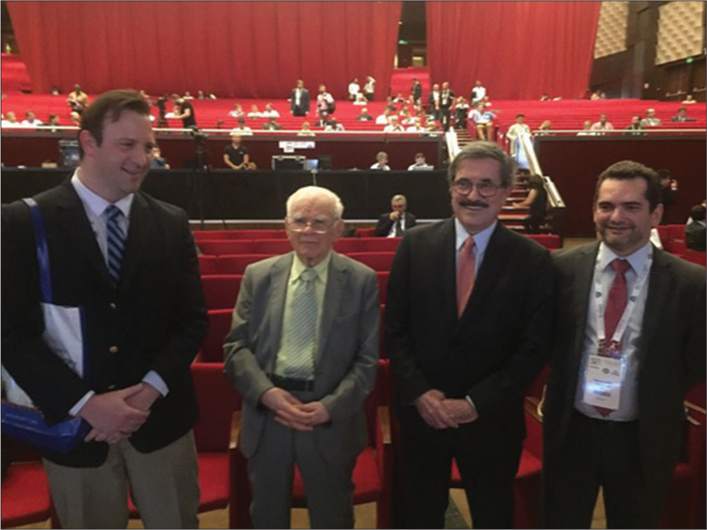
Among the hundreds of fellows who accompanied Professor Evandro de Oliveira, [
References
1. Almeida JP, Neto MR, Neto FC, De Oliveira E. Anatomical considerations in the treatment of intracranial aneurysms. J Neurosurg Sci. 2016. 60: 27-43
2. de Oliveira E, Tedeschi H, Raso J. Comprehensive management of arteriovenous malformations. Neurol Res. 1998. 20: 673-83
3. Fernández-Miranda JC, de Oliveira E, Rubino PA, Wen HT, Rhoton AL. Microvascular anatomy of the medial temporal region: Part 1: Its application to arteriovenous malformation surgery. Neurosurgery. 2010. 67: 237-76
4. Figueiredo EG, Tavares WM, Rhoton AL, de Oliveira E. Nuances and technique of the pretemporal transcavernous approach to treat low-lying basilar artery aneurysms. Neurosurg Rev. 2010. 33: 129-35
5. Figueiredo EG, Tavares WM, Rhoton AL, De Oliveira E. Surgical nuances of giant paraclinoid aneurysms. Neurosurg Rev. 2010. 33: 27-36
6. Frigeri T, Paglioli E, de Oliveira E, Rhoton AL. Microsurgical anatomy of the central lobe. J Neurosurg. 2015. 122: 483-98
7. Isolan GR, Krayenbühl N, de Oliveira E, Al-Mefty O. Microsurgical anatomy of the cavernous sinus: Measurements of the triangles in and around it. Skull Base. 2007. 17: 357-67
8. Joaquim AF, Almeida JP, Dos Santos MJ, Ghizoni E, de Oliveira E, Tedeschi H. Surgical management of intradural extramedullary tumors located anteriorly to the spinal cord. J Clin Neurosci. 2012. 19: 1150-3
9. Joaquim AF, Ghizoni E, Anderle DV, Oliveira Ed, Tedeschi H. Axis instrumentation: Surgical results. Arq Neuropsiquiatr. 2012. 70: 857-63
10. Kadri PA, de Oliveira JG, Krayenbühl N, Türe U, de Oliveira EP, Al-Mefty O. Surgical approaches to the temporal horn: An anatomic analysis of white matter tract Interruption. Oper Neurosurg (Hagerstown). 2017. 13: 258-70
11. Párraga RG, Ribas GC, Welling LC, Alves RV, de Oliveira E. Microsurgical anatomy of the optic radiation and related fibers in 3-dimensional images. Neurosurgery. 2012. 71: 160-71
12. Pastor-Escartín F, García-Catalán G, Holanda VM, Lahirish IA, Quintero RB, Neto MR. Microsurgical anatomy of the insular region and operculoinsular association fibers and its neurosurgical application. World Neurosurg. 2019. 129: 407-20
13. Ribas EC, Yağmurlu K, de Oliveira E, Ribas GC, Rhoton A. Microsurgical anatomy of the central core of the brain. J Neurosurg. 2018. 129: 752-69
14. Spetzler RF, Martin NA. A proposed grading system for arteriovenous malformations. J Neurosurg. 1986. 65: 476-83
15. Wen HT, Rhoton AL, de Oliveira E, Castro LH, Figueiredo EG, Teixeira MJ. Microsurgical anatomy of the temporal lobe: Part 2-sylvian fissure region and its clinical application. Neurosurgery. 2009. 65: 1-35
16. Yasuda A, Campero A, Martins C, Rhoton AL, de Oliveira E, Ribas GC. Microsurgical anatomy and approaches to the cavernous sinus. Neurosurgery. 2008. 62: 1240-63

