- Department of Neurosurgery, Santa Casa de Sao Paulo School of Medical Sciences, Sao Paulo, Brazil.
DOI:10.25259/SNI_105_2020
Copyright: © 2020 Surgical Neurology International This is an open-access article distributed under the terms of the Creative Commons Attribution-Non Commercial-Share Alike 4.0 License, which allows others to remix, tweak, and build upon the work non-commercially, as long as the author is credited and the new creations are licensed under the identical terms.How to cite this article: Charles Alfred Pedrozo, Guilherme Brasileiro de Aguiar, Jose Carlos Esteves Veiga. Report of intradural aneurysm in the cavernous segment of the internal carotid artery presented with subarachnoid hemorrhage and oculomotor palsy. 13-Jun-2020;11:149
How to cite this URL: Charles Alfred Pedrozo, Guilherme Brasileiro de Aguiar, Jose Carlos Esteves Veiga. Report of intradural aneurysm in the cavernous segment of the internal carotid artery presented with subarachnoid hemorrhage and oculomotor palsy. 13-Jun-2020;11:149. Available from: https://surgicalneurologyint.com/surgicalint-articles/10078/
Abstract
Background: Aneurysms of the cavernous segment of the internal carotid artery (ICA) do not usually cause subarachnoid hemorrhage (SAH). We report a patient who presented with this condition due to a ruptured aneurysm located on the posterior genu of the cavernous segment, raising the question of what factors could have led to such evolution.
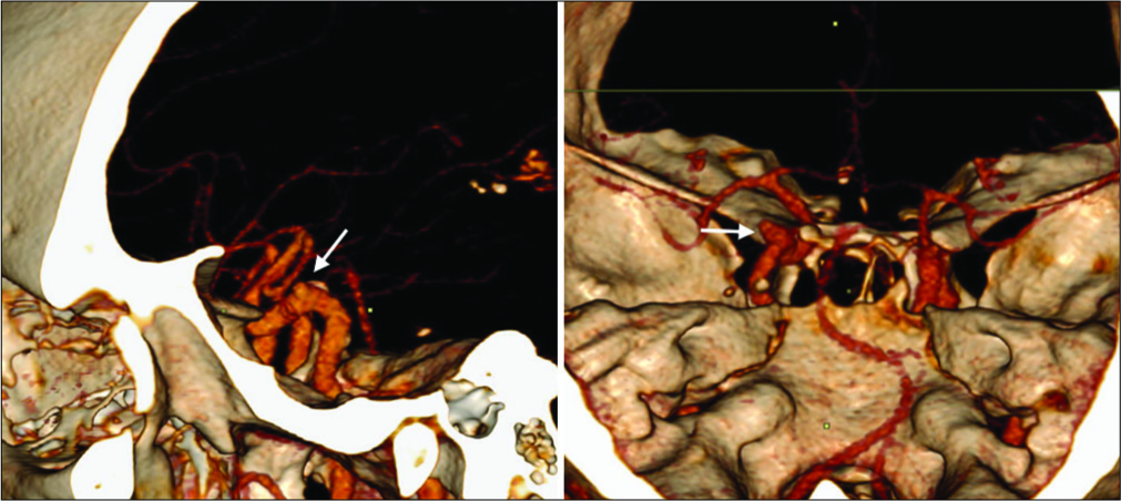
Case Description: A 55-year-old male patient presented with sudden, intense thunderstorm headache, associated with complete palsy of the left oculomotor nerve and neck stiffness. Cranial computed tomography (CT) showed no SAH, but showed an expansive process in the sella turcica, consistent with a pituitary macroadenoma. After that, SAH was confirmed by lumbar puncture (Fisher I). Cranial angio-CT revealed an intradural saccular aneurysm in the cavernous segment of the left ICA. The patient underwent cranial microsurgery for cerebral aneurysm clipping. Unlike the normal anatomic pattern, the cavernous segment of the carotid artery in this patient was located in the intradural compartment.
Conclusion: Intradural rupture of proximal cavernous segment carotid aneurysms is rare. We review the literate for such cases and discuss the possible causes.
Keywords: Aneurysms, Ophthalmoparesis, Pituitary adenoma, Subarachnoid hemorrhage
INTRODUCTION
Subarachnoid hemorrhage (SAH) is a severe pathology with high morbidity and mortality and requires a fast and accurate therapeutic decision. We describe the case of a patient who presented at the emergency unit with acute SAH and left III cranial nerve palsy. Basic computed tomography (CT) was negative, but lumbar puncture was positive for SAH. Angio-CT showed an aneurysm located in the posterior genu of the cavernous segment. Cavernous segment aneurysms are classically extradural and present without SAH, raising the question in this case of the factors that could have led to such evolution. In a review of the literature, there is only one case reported with aneurysm located in the same topography described by Andaluz et al., 2003[
CASE REPORT
A 55-year-old male patient with a medical history of systemic arterial hypertension was admitted to the neuroemergency department, after being transferred from another service, with a report of sudden, intense thunderstorm headache, associated with the left eyelid ptosis and diplopia. During the neurological examination, the patient was awake, lucid, and oriented. He had complete palsy of the left oculomotor nerve, with no other focal neurological deficits. Neck stiffness was present.
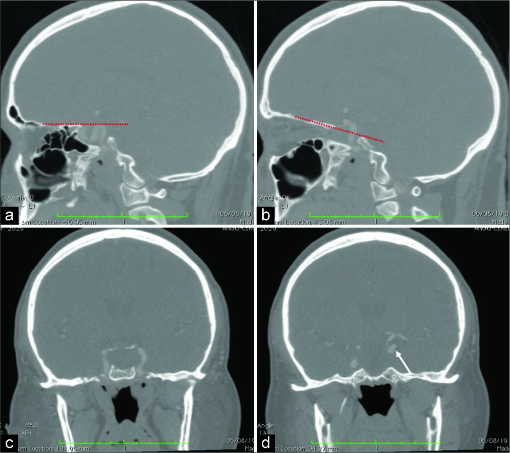
Cranial CT performed on the day following the headache showed no SAH, but showed an expansive process in the sella turcica associated with sellar enlargement [
Figure 1:
Angio-CT scan. (a) Cavernous segment of the right internal carotid artery, below the line of projection of the planum sphenoidale (red dotted line). (b) Cavernous segment of the left internal carotid artery with its posterior genu above the line of projection of the planum sphenoidale (red dotted line). (c) Enlargement of the sella turcica and left carotid artery laterally displaced. (d) Superior displacement of the cavernous segment of the internal carotid artery and an aneurysm close to the communicating segment of the internal carotid artery (white arrow).
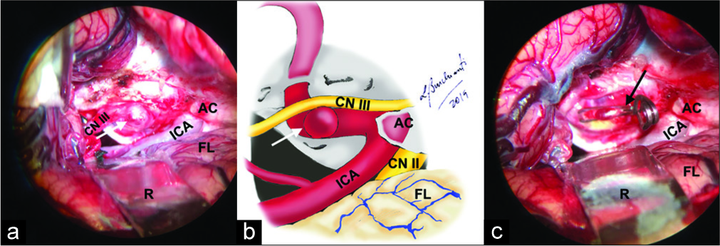
The patient underwent cranial microsurgery for cerebral aneurysm clipping that confirmed the intradural location of the aneurysm, arising from a tortuous cavernous ICA [
Figure 3:
(a) Intraoperative image showing the carotid aneurysm (white arrow), compressing the left oculomotor nerve; (b) schematic representation of the aneurysm (white arrow), compressing the oculomotor nerve; (c) image after microsurgical clipping (black arrow). AC: Anterior clinoid; CN II: Optic cranial nerve; CN III: Oculomotor cranial nerve; FL: Frontal lobe; ICA: Internal carotid artery; R: Fixed retractor.
He underwent control cerebral angiography on the 2nd postoperative day that demonstrated complete aneurysm occlusion [
DISCUSSION
The cavernous segment of the ICA begins at the superior margin of the petrolingual ligament, just after the ICA emerges from the foramen lacerum.[
There are, however, some case reports of SAH from cavernous ICA aneurysms.[
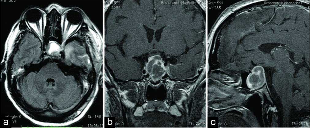
As shown in our patient’s angiographic study, the aneurysm was located at the posterior genu of the cavernous segment that should be inside the cavernous sinus. By definition, the cavernous segment is completely extradural, except for those that become partially intradural by traversing the clinoidal space (between the proximal and distal dural rings); in these cases, they constitute transitional (or clinoidal) aneurysms. During surgery, it was observed that this aneurysm was fully exposed in the intradural compartment. The etiological hypothesis for this finding would be that initially a large adenoma may have eroded the dural boundary of the cavernous sinus. Subsequently, shrinkage of this adenoma following apoplexia could have left a residual dural defect allowing communication between the cavernous sinus and the subarachnoid space and further exposure of the posterior knee of the cavernous segment of the ICA. One of the possible locations for the dural failure might be the virtual ring where the oculomotor nerve enters the cavernous sinus and that had been loosened after the tumor shrank, forming a route for the aneurysm to extend intradurally and compress the third cranial nerve, resulting in the dysfunction of the third nerve, present in our patient, as shown in [
Figure 6:
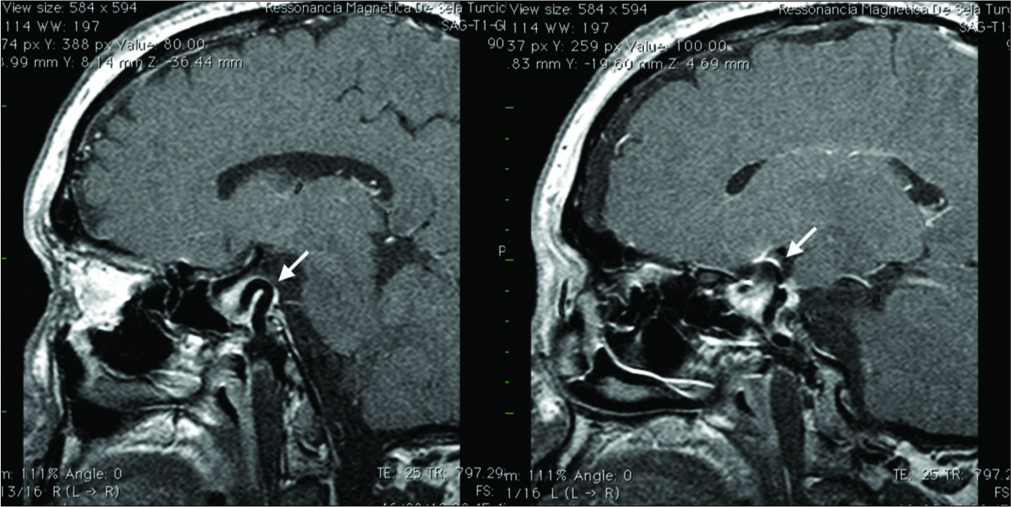
Cranial magnetic resonance imaging (sagittal T1 with gadolinium) showing the presence of an expansive sellar tumor (pituitary macroadenoma) involving the internal carotid arteries. Left: The cavernous segment of the right internal carotid artery (ICA) maintains its usual curvature, being restricted to the cavernous sinus (arrow). Right: The left ICA is elongated and its curvature exceeds the limits of the cavernous sinus (arrow).
CONCLUSION
This case report confirms that anatomical variations produced by disease process can lead to intradural exposure of the proximal cavernous segment of the carotid artery. A detailed analysis of image studies and critical surgical planning is required to avoid surprises or intraoperative technical difficulties and determine the best therapeutic strategy.
Declaration of patient consent
Patient consent not required as patients identity is not disclosed or compromised.
Financial support and sponsorship
Nil.
Conflicts of interest
There are no conflicts of interest.
References
1. Akutsu N, Hosoda K, Ohta K, Tanaka H, Taniguchi M, Kohmura E. Subarachnoid hemorrhage due to rupture of an intracavernous carotid artery aneurysm coexisting with a prolactinoma under cabergoline treatment. J Neurol Surg Rep. 2014. 75: e73-6
2. Andaluz N, Tew JM. Intradural aneurysm arising from the posterior genu of the cavernous carotid artery mimicking a posterior communicating aneurysm: Case report. Neurosurgery. 2003. 53: 432-5
3. Bouthillier A, van Loveren HR, Keller JT. Segments of the internal carotid artery: A new classification. Neurosurgery. 1996. 38: 425-32
4. Hamada H, Endo S, Fukuda O, Ohi M, Takaku A. Giant aneurysm in the cavernous sinus causing subarachnoid hemorrhage 13 years after detection: A case report. Surg Neurol. 1996. 45: 143-6
5. Iwaisako K, Toyota S, Ishihara M, Shibano K, Harada Y, Iwatsuki K. Intracerebral hemorrhage caused by ruptured intracavernous carotid artery aneurysm. Case report. Neurol Med Chir (Tokyo). 2009. 49: 155-8
6. Khalsa SS, Hollon TC, Shastri R, Trobe JD, Gemmete JJ, Pandey AS. Spontaneous subarachnoid hemorrhage due to ruptured cavernous internal carotid artery aneurysm after medical prolactinoma treatment. BMJ Case Rep. 2016. 2016: bcr2016012446-
7. Kupersmith MJ, Stiebel-Kalish H, Huna-Baron R, Setton A, Niimi Y, Langer D. Cavernous carotid aneurysms rarely cause subarachnoid hemorrhage or major neurologic morbidity. J Stroke Cerebrovasc Dis. 2002. 11: 9-14
8. Kupersmith MJ.editors. Neuro-Vascular Neuro-Ophthalmology. Heidelberg: Springer Verlag; 1993. p. 69-108
9. Lee AG, Mawad ME, Baskin DS. Fatal subarachnoid hemorrhage from the rupture of a totally intracavernous carotid artery aneurysm: Case report. Neurosurgery. 1996. 38: 596-8
10. Nishioka T, Kondo A, Aoyama I, Nin K, Takahashi J. Subarachnoid hemorrhage possibly caused by a saccular carotid artery aneurysm within the cavernous sinus. Case report. J Neurosurg. 1990. 73: 301-4
11. Taptas JN. The so-called cavernous sinus: A review of the controversy and its implications for neurosurgeons. Neurosurgery. 1982. 11: 712-7
12. van Loveren HR, Keller JT, el-Kalliny M, Scodary DJ, Tew JM. The Dolenc technique for cavernous sinus exploration (cadaveric prosection). Technical note. J Neurosurg. 1991. 74: 837-44


