- Department of Pharmacology and Clinical Neuroscience, Umeå University, Sweden
- Department of Neurosurgery, Tokyo Women's Medical University, Tokyo, Japan
- UCL Institute of Neurology, Queen Square, London, UK
Correspondence Address:
Patric Blomstedt
UCL Institute of Neurology, Queen Square, London, UK
DOI:10.4103/2152-7806.194061
Copyright: © 2016 Surgical Neurology International This is an open access article distributed under the terms of the Creative Commons Attribution-NonCommercial-ShareAlike 3.0 License, which allows others to remix, tweak, and build upon the work non-commercially, as long as the author is credited and the new creations are licensed under the identical terms.How to cite this article: Patric Blomstedt, Takaomi Taira, Marwan Hariz. Rescue pallidotomy for dystonia through implanted deep brain stimulation electrode. 14-Nov-2016;7:
How to cite this URL: Patric Blomstedt, Takaomi Taira, Marwan Hariz. Rescue pallidotomy for dystonia through implanted deep brain stimulation electrode. 14-Nov-2016;7:. Available from: http://surgicalneurologyint.com/surgicalint_articles/rescue-pallidotomy-dystonia-implanted-deep-brain-stimulation-electrode/
Abstract
Background:Some patients with deep brain stimulation (DBS), where removal of implants is indicated due to hardware related infections, are not candidates for later re-implantation. In these patients a rescue lesion through the DBS electrode has been suggested as an option. In this case report we present a patient where a pallidotomy was performed using the DBS electrode.
Case Description:An elderly woman with bilateral Gpi DBS suffered an infection around the left burr hole involving the DBS electrode. A unilateral lesion was performed through the DBS electrode before it was removed. No side effects were encountered. Burke-Fahn-Marsden (BFM) dystonia movement scale score was 39 before DBS. With DBS before lesioning BFM score was 2.5 points. The replacement of the left sided stimulation with a pallidotomy resulted in only a minor deterioration of the score to 5 points.
Conclusions:In the case presented here a small pallidotomy performed with the DBS electrode provided a satisfactory effect on the patient's dystonic symptoms. Thus, rescue lesions through the DBS electrodes, although off-label, might be considered in patients with Gpi DBS for dystonia when indicated.
Keywords: Deep brain stimulation, dystonia, pallidotomy
INTRODUCTION
Deep brain stimulation (DBS) has revolutionized the treatment of movement disorders and provided an alternative to non-reversible lesions. However, lesions may still constitute a viable option in certain conditions. DBS is prone to a number of various complications that can occur during the remaining lifetime of the patient. Even if DBS might be the preferred initial surgical procedure in a patient at a certain point of time, it is not certain that DBS will remain a preferred option compared to ablative surgery when the patient develops a hardware-related infection years later.
In this case report, we present a patient where a rescue lesion following infection was performed using the DBS electrode.
CASE PRESENTATION
The patient suffereda from tremor of the upper extremities since the age of 10 years. Later, there was a gradual development of dystonia which finally encompassed all extremities, and affected her abdomen and neck as well as her speech. The patient underwent bilateral pallidal DBS at the age of 71 with good effect. Burke-Fahn-Marsden (BFM) dystonia movement scale scores are presented in
The patient was in an advanced age, with a rather poor general condition, and had a very thin scalp. It was, therefore, decided that the patient would not be a candidate for a third DBS procedure. However, considering the patient's deteriorating condition and the suffering caused by her dystonic symptoms in the absence of DBS, it was decided to keep the right electrode and to perform a rescue lesion through the left DBS electrode before extraction.
Before surgery, the exact location of each individual electrode contact was identified from the patient's postoperative computed tomography (CT) fused with the preoperative stereotactic magnetic resonance imaging (MRI). Each individual contact was screened de novo concerning acute stimulation induced effects and side effects. The result indicated that the currently used contacts were optimal. On the left lead, the patient was on bipolar stimulation through contacts 1 and 2 (the two middle contacts), at 3.5 V, 90 uS, and 160 Hz. It was, therefore, decided to use the same contacts for bipolar lesioning.
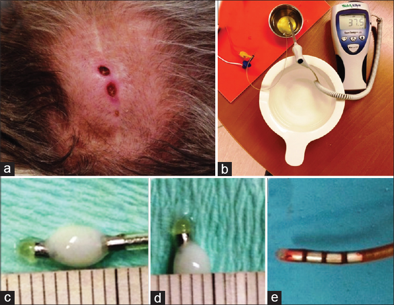
Before surgery, test lesions were performed in egg albumin [
The procedure was performed using local anesthesia. A 1-cm long transverse incision was made over the connector between the intracerebral electrode and the connection cable over the linea temporalis superior. The electrode was disconnected from the connector and its proximal end was visualized. With cessation of stimulation on that side, the patient immediately complained of dystonic cramps in the neck, as well as dystonic speech. Contacts 1 and 2 of the electrode were connected to the lesion generator (Leksell Neuro Generator, Elekta, Stockholm), and a lesion was made using a power of 4 during 30 seconds. During the lesioning, the speech was markedly improved and the dystonic cramps in the neck were resolved.
After the incision over the connector area was closed, the infected wound over the burr hole was revised and the electrode removed. The distal end of the electrode was easily mobilized and showed no adherent tissue [
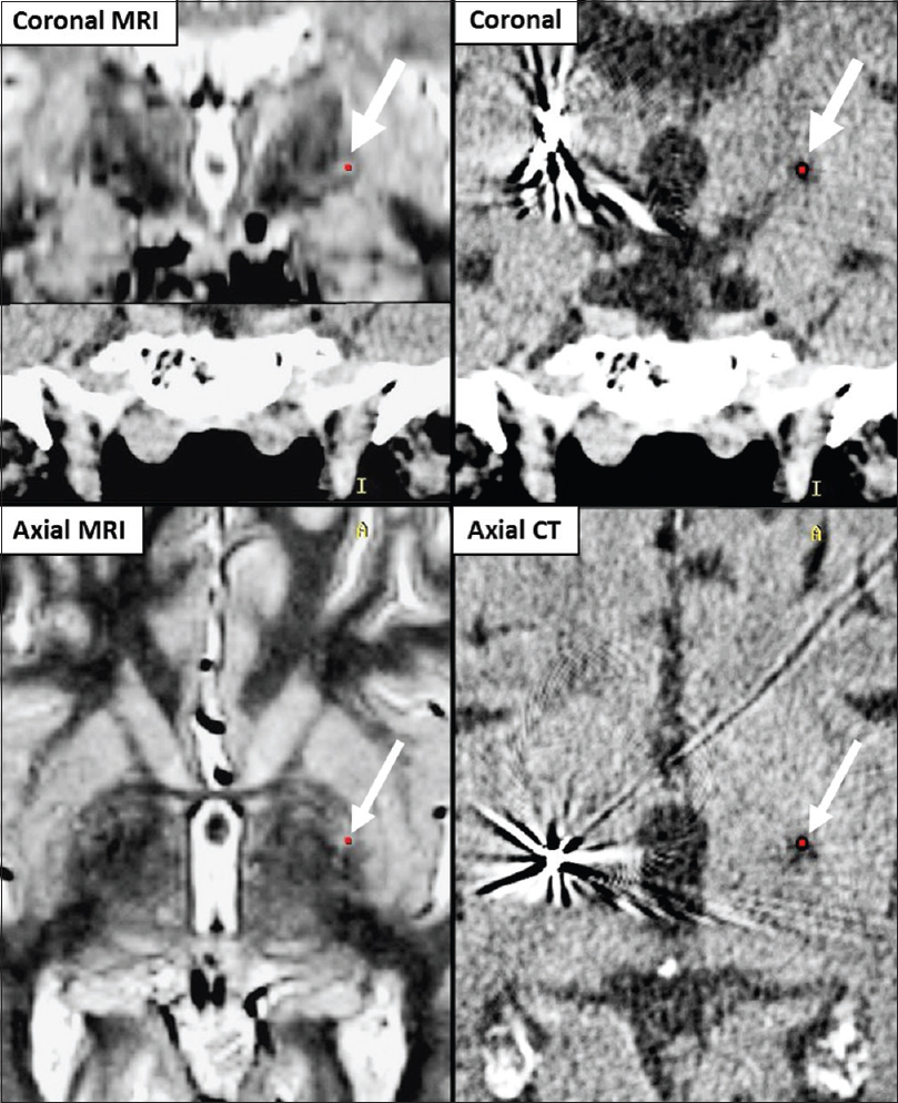
A postoperative CT performed 4 days after surgery and fused with a preoperative MRI demonstrated a well-placed lesion in the posteroventral globus pallidus internus (Gpi) with a height of 4.5 mm and a diameter of 3 mm and surrounded by postoperative edema [
Total BMF movement score was 39 before DBS. With DBS before lesioning, BFM score was 2.5. The replacement of the left-sided stimulation with a pallidotomy resulted in only a minor deterioration of the score to 5, mostly in the form of increased torticollis [
DISCUSSION
In our patient, it was necessary to remove the DBS electrodes due to a repeat hardware-related infection. Because of old age, thin scalp, and frail general condition, it was evident that the patient will not be a candidate for later re-implantation. Cessation of the therapy, even if only on one side, was likely to result in a rebound or at least a re-emergence of the dystonic symptoms, as was evidenced during surgery when stimulation through the left lead was interrupted.[
The technique of lesioning through an implanted DBS electrode has previously been described in two patient with subthalamic nucleus (STN) DBS for Parkinson's disease (PD)[
In the patient described by Raoul et al,[
In our case, because the patient had an infected hardware and the DBS electrode had to be removed, a single lesion proved sufficient to produce a satisfying effect on the dystonic symptoms, albeit not as efficient as the previous stimulation through that electrode.
CONCLUSION
In the case presented here, a small pallidotomy performed with the DBS electrode provided a satisfactory effect on the patient's dystonic symptoms. Thus, rescue lesions through the DBS electrodes, although off-label, might also be considered in patients with Gpi DBS for dystonia when indicated.
Financial support and sponsorship
Nil
Conflicts of interest
There are no conflicts of interest.
References
1. Blomstedt P, Hariz GM, Hariz MI. Pallidotomy versus pallidal stimulation. Parkinsonism Relat Disord. 2006. 12: 296-301
2. Deligny C, Drapier S, Verin M, Lajat Y, Raoul S, Damier P. Bilateral subthalamotomy through dbs electrodes: A rescue option for device-related infection. Neurology. 2009. 73: 1243-4
3. Hariz MI, Hirabayashi H. Is there a relationship between size and site of the stereotactic lesion and symptomatic results of pallidotomy and thalamotomy?. Stereotact Funct Neurosurg. 1997. 69: 28-45
4. Hariz MI, Johansson F. Hardware failure in parkinsonian patients with chronic subthalamic nucleus stimulation is a medical emergency. Mov Disord. 2001. 16: 166-8
5. Hirabayashi H, Hariz M, Wårdell K, Blomstedt P. Impact of parameters of radiofrequency coagulation on volume of stereotactic lesion in pallidotomy and thalamotomy. Stereotact Funct Neurosurg. 2012. 90: 307-15
6. Oh MY, Hodaie M, Kim SH, Alkhani A, Lang AE, Lozano AM. Deep brain stimulator electrodes used for lesioning: Proof of principle. Neurosurgery. 2001. 49: 363-7
7. Raoul S, Faighel M, Rivier I, Verin M, Lajat Y, Damier P. Staged lesions through implanted deep brain stimulating electrodes: A new surgical procedure for treating tremor or dyskinesias. Mov Disord. 2003. 18: 933-8
8. Raoul S, Leduc D, Vegas T, Sauleau P, Lozano AM, Verin M. Deep brain stimulation electrodes used for staged lesion within the basal ganglia: Experimental studies for parameter validation. Laboratory investigation. J Neurosurg. 2007. 107: 1027-35


