Tools

Anders Blach Naamansen1, Carl Christian Larsen2, Bjarni Johannsson1, Sune Munthe1, Troels Halfeld Nielsen1
  1. Department of Neurosurgery, Odense University Hospital, Odense,
  2. Department of Neurosurgery, Rigshospitalet, Copenhagen, Denmark.

Correspondence Address:
Anders Blach Naamansen, Department of Neurosurgery, Odense University Hospital, Odense, Denmark.

DOI:10.25259/SNI_1119_2021

Copyright: © 2022 Surgical Neurology International This is an open-access article distributed under the terms of the Creative Commons Attribution-Non Commercial-Share Alike 4.0 License, which allows others to remix, transform, and build upon the work non-commercially, as long as the author is credited and the new creations are licensed under the identical terms.

How to cite this article: Anders Blach Naamansen1, Carl Christian Larsen2, Bjarni Johannsson1, Sune Munthe1, Troels Halfeld Nielsen1. Small ruptured intracranial aneurysms are overrepresented at the anterior and posterior communicating artery: Results of a multiple regression analysis. 08-Jul-2022;13:288

How to cite this URL: Anders Blach Naamansen1, Carl Christian Larsen2, Bjarni Johannsson1, Sune Munthe1, Troels Halfeld Nielsen1. Small ruptured intracranial aneurysms are overrepresented at the anterior and posterior communicating artery: Results of a multiple regression analysis. 08-Jul-2022;13:288. Available from: https://surgicalneurologyint.com/surgicalint-articles/11710/

Date of Submission
09-Nov-2021

Date of Acceptance
14-Jun-2022

Date of Web Publication
08-Jul-2022

Abstract

Background: Anterior communicating artery (AcomA) represents the most common location for ruptured intracranial aneurysms (rIAs). Approximately 50% of all rIAs are smaller than 7 mm, but factors that lead to rupture are multifactorial. The study investigates whether AcomA location represents an independent risk factor for small size at time of rupture (

Methods: The aSAH cohort was retrospectively searched from our institution charts. The cohort was dichotomized into small aneurysms (

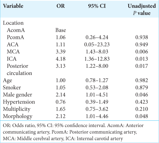
Results: One-hundred and seventy-six patients were included in the study. About 49.4% of the aneurysms were P = 0.006), internal carotid artery (other than PcomA) (P = 0.013), and posterior circulation (P = 0.017), when controlling for risk factors.

Conclusion: Ruptured AcomA and PcomA aneurysms are more frequent smaller than 7 mm compared to other locations. Patients with unruptured UIA at either AcomA or PcomA may be at increased risk of rupture even if the size of the aneurysm is small. Further studies are needed to confirm this finding.

Keywords: Rupture risk, Small aneurysms, Unruptured intracranial aneurysm

INTRODUCTION

Aneurysmal subarachnoid hemorrhage (aSAH) is a devastating event with a mortality of more than 30% and morbidity of around 70%.[ 9 , 11 ] Intracranial aneurysms have a prevalence of 3–4% in the adult population[ 22 ] and are most often found in the anterior circulation.[ 13 ] According to the ISUIA study,[ 25 ] only 0.05% of aneurysms in the anterior circulation smaller than 7 mm rupture. This leads to the widely accepted dogma that size is the single most important predictor of rupture. On the other hand, studies of aSAH cohorts demonstrate that up to 73% of ruptured aneurysms are ≤7 mm, with 37.4% located at anterior communicating artery (AcomA).[ 7 ] The reason for this discrepancy is unknown but it remains clear that other factors than size play an important role in the process that leads to aneurysm rupture. Various scoring systems have been developed to assess and grade the various risk factors for rupture, the most widely used being the unruptured intracranial aneurysm treatment score (UIATS) and PHASES scores.[ 5 , 10 ] These scores incorporate variables such as ethnicity, aneurysm size, and location and comorbidities such as hypertension. However, one recent study suggested that the PHASES score only serves as a weak tool for prediction of aneurysm rupture.[ 16 ] The UIATS is a multidisciplinary consensus, using the Delphi method, indicating how a large group of specialists might manage a specific aneurysm.[ 5 ] The UIATS is based on 29 variables, giving points in favor of either UIA repair or conservative management. Hence, it constitutes a comprehensive set of risk factors associated to aneurysm rupture. In the present study, we retrospective evaluate patients with confirmed aneurysmal subarachnoid hemorrhage (aSAH) and the presence of UIATS defined risk factors, with focus on analyzing discrepancies between small (<7 mm) and large (≥7 mm) aneurysms. The objective of the study was to identify risk factors related to rupture of small aneurysms.

MATERIALS AND METHODS

The study was approved by our Institutional Review Board. According to local and national legislation, patient informed consent was not required for this study. All medical records of patients treated for SAH on our institution between November 2015 and December 2018 were retrospectively reviewed. Patients were identified using the ICD-10 codes I60.1–I60.9. All patients with SAH caused by other etiology than an intracranial aneurysm were excluded along with patients with arteriovenous malformation associated SAH or aneurysms. If the aneurysm causing SAH was not of the saccular type, the patients were excluded and patients with perioperative or postoperative aneurysm rupture were excluded as well. If the medical records lacked adequate information to assess variables included in the UIATS, patients were also excluded from the study. All variables from the original UIATS score were included in the present study except “Fear of rupture,” neurocognitive disease, psychiatric disorder, and life expectancy due to chronic or malignant disorder, as these variables either cannot be assessed retrospectively or does not increase the risk of rupture per se. The relevant medical records were reviewed by a single investigator (AN), scoring all patients according to the UIATS, using all definitions as described in Etminan et al.[ 5 ] Thirty-day mortality was assessed as well. Hypertension was defined as confirmed systolic blood pressure above 140 mmHg prerupture. Alcohol abuse was defined as a confirmed consumption of >14 units/week for women and >21 unit/week for men. A three-dimensional reconstruction of the cerebral angiography was used to determine the size, location, and morphology of the aneurysms. Aneurysm size was defined as the maximum aneurysm diameter. Aneurysm lobulation was defined as irregular daughter sac-like protrusion(s) of the aneurysm wall. Size ratio was calculated as the maximal aneurysm diameter divided by the parent vessel diameter. Aspect ratio was calculated as maximal aneurysm dome diameter divided by neck width. In all cases, the neuroimaging was reviewed by a neurosurgical/neuroradiology specialist. Patients were dichotomized into a “Small Group” and “Large Group.” “Small Group” was defined as patients with a ruptured aneurysm with a maximal aneurysm dome diameter excluding eventual baby sac smaller than 7 mm. The “Large Group” was defined by the same criteria with maximum diameter larger than or equal to 7 mm. Multiple regression analysis was performed to identify variables differentiating the small group aneurysms (<7 mm) from the large group (≥7 mm).

Statistical analysis

Age is presented as median and full range. Variables of the UIATS are presented as count and frequency. Size was dichotomized to <7.0 mm or larger than 7.0 mm. Superior cerebellar artery, basilar artery bifurcation, posterior cerebral artery, vertebral artery, and PICA were consolidated to a single variable labeled “posterior circulation.” Logistic univariate regression analysis was performed with size as the dependent variable and with patient and aneurysm characteristics as independent variables. Variables of interest presenting with P ≤ 0.2 along with patient characteristics were included in a logistic multiple regression. These were location, age (represented as a continuous variable in the analysis, divided by 10 such that odds ratios are fixed in increments of 10 years), gender, hypertension, current cigarette smoking, aneurysm multiplicity, and aneurysm morphology. The variable morphology was designed as a binary composite variable including irregularity or lobulation, size ratio >3, and/or aspect ratio >1.6. Only one of the criteria needed to be present. Aneurysm location was defined as a categorical variable represented by a level each in the model. Statistically significant results are presented as such with respect to an unadjusted α = 0.05 to protect against type-2 errors.

Data availability

All anonymized data will be shared by request from any qualified investigator.

RESULTS

One-hundred and seventy-six patients were included in the study. A flowchart of patient exclusion is given in Figure 1 . The study data were collected from a cohort of 331 patients. The main reason for exclusion was SAH from other etiology than aneurysm, excluding a total of 91 patients.


Figure 1:

Flowchart of exclusion criteria. Top values represent total of eligible patients, decreasing because of the exclusion criteria, which are listed below each top value.


Patient characteristics

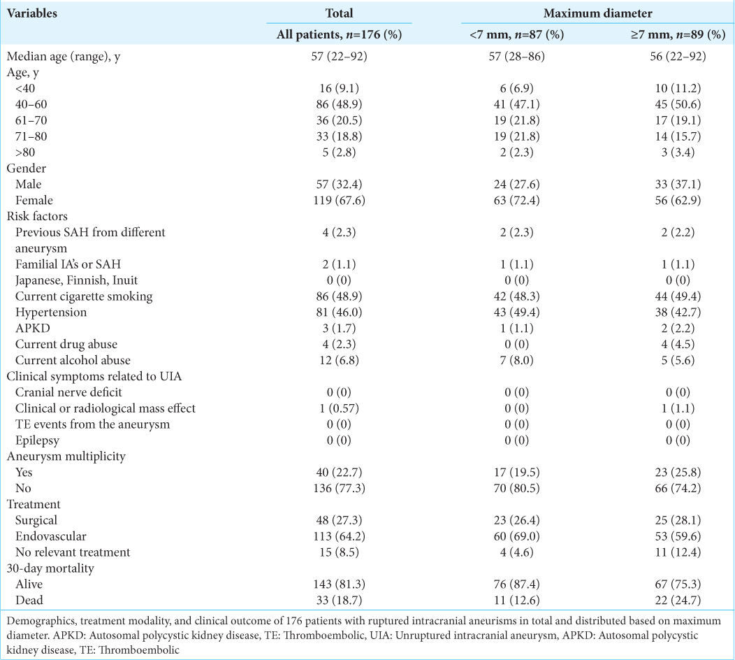
Patient demographics, characteristics, treatment modality, and clinical outcome are given in Table 1 . The 176 eligible patients with aneurysmal SAH had a median age of 57 years (range: 22–92), at time of rupture. Almost half (48.9%) of all patients were between 40 and 60 years old at time of rupture and the majority of all patients were female (67.6%). Eighty-six (48.9%) patients were current cigarette smokers, while hypertension was present in 81 patients (46.0%). These were the two most frequent represented risk factors, with current alcohol abuse at third, found in 12 cases (6.8%). Previous SAH from a different aneurysm occurred in 4 cases (2.3%), while familial intracranial aneurysms or SAH was reported in 2 cases (1.1%). Most ruptured aneurysms were endovascular treated, 113 (64.2%), while surgical clip ligation was chosen in 48 cases (27.3%). In 15 cases (8.5%), no treatment option was relevant and of those, all died within 30 days. Overall, 33 patients died within 30 days of rupture, making the 30-day mortality 18.8%.


Table 1:

Demographics, treatment modality. and clinical outcome of 176 patients with ruptured intracranial aneurisms in total and dichotomized into small (<7 mm) and large (≥7 mm) aneurysms.


Aneurysm characteristics

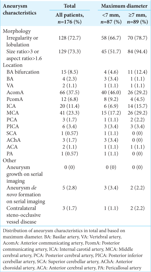
For distribution of aneurysm characteristics see Table 2 . Almost half (49.4%) of the ruptured aneurysms in the analysis were <7 mm. In 69 (39.2%) cases, the maximum diameter of the ruptured aneurysm was between 4.0 and 6.9 mm, while 68 (38.6%) aneurysms were between 7.0 and 12.9 mm. Aneurysms under 3.9 mm were present in 18 cases (10.2%), exactly as frequent as aneurysms between 13.0 and 24.9 mm. Only 3 (1.7%) ruptured aneurysms were found to be or exceed 25 mm. Irregularity or lobulation of the ruptured aneurysm was present in 128 (72.7%) cases, while 129 (73.3%) either had a size ratio above 3.0 or aspect ratio above 1.6. The location of the ruptured aneurysm was in 66 (37.5%) cases, the AcomA and in 41 (23.3%) cases, the middle cerebral artery. The internal carotid artery (other than posterior communicating artery [PcomA]) accounted for 20 (11.4%) ruptured aneurysms, the basilar bifurcation for 15 (8.5%), and the PcomA was the location in 12 (6.8%) cases. In 4 (2.3%) patients, the location was the basilar artery (other than the basilar bifurcation) and the vertebral artery was the location in 2 (1.1%) cases.


Table 2:

Distribution of aneurysm characteristics in total and dichotomized into small (<7 mm) and large (≥7 mm) aneurysms.


Multiple regression analysis

Aneurysm location, gender, and morphology were found to be significantly associated with ruptured aneurysm size in the multiple regression analysis [ Table 3 ]. In the multiple regression analysis with AcomA as the base level, all locations exhibited positive coefficients for size. PcomA and the anterior cerebral artery approached an OR for large size to that of AcomA. A significant size difference in ruptured aneurysms was found between AcomA and the middle cerebral artery (P = 0.006), ICA (P = 0.013), and arteries in the posterior circulation (P = 0.017). Male gender and aneurysm morphology were also found to be significant predictors for rupture at small aneurysm size (P = 0.046 and P = 0.048, respectively).


Table 3:

Results of logistic multiple regression analysis. The dependent variable (aneurysm size) was dichotomized into small (<7 mm) and large (≥7.0 mm) aneurysms.


DISCUSSION

A central dogma in the assessment of intracranial aneurysm rupture risk is that size matter. The larger the aneurysm, the higher the rupture risk. The ISUA study was the first to suggest a cutoff of 7 mm, below which the rupture risk is small. In fact, the study demonstrated a 0.05% 5-year rupture risk for aneurysms <7 mm in the anterior circulation.[ 14 ] Yet, almost half the ruptured aneurysms in our cohort were smaller than 7 mm and 83.8% of these were located in the anterior circulation. This is not surprising and other retrospective studies on mainly ruptured aneurysms support this finding.[ 1 , 7 , 23 ] Hence, factors other than size contribute to aneurysm rupture. Mocco et al. pointed out the importance of morphology for aneurysm rupture when controlling for size. [ 15 ] Wiebers et al. identified PcomA and basilar tip location as risk factors for aneurysm rupture in a multivariate analysis. [ 26 ] In a large meta-analysis, Etminan et al. demonstrated a correlation between decrease in aSAH incidence and decrease in blood pressure and tobacco smoking.[ 6 ] These results indicate that the risk of rupture is multifactorial, and attempts to assess the risk of a single variable should be performed by regression analysis controlling for other known risk factors. In recent years, small AcomA aneurysms have gained attention. One study demonstrated that AcomA aneurysms of 4–7 mm are just as prone to rupture as posterior circulation aneurysms.[ 2 ] However, this study did not control for morphology. Another study demonstrated that ruptured aneurysms <5 mm were significantly more often located in the AcomA in the univariate analysis but the difference was not significant in the multivariate analysis.[ 4 ] Further, Rinaldo et al.[ 18 ] recently observed no difference in size between unruptured and ruptured AcomA aneurysms. Our results suggest that AcomA location, and to a lesser degree PcomA, is significantly associated with small aneurysm size at rupture, compared to ICA, MCA, and arteries located in the posterior circulation (P = 0.006, P = 0.013, and P = 0.017, respectively), when controlling for other predictors of rupture. This implies that these locations should be considered a strong independent predictor for aneurysm rupture even if smaller than 7 mm. As AcomA is the most frequent location for intracranial aneurysms, this finding might explain, together with the recognized selection bias in prospective followed unruptured aneurysm cohorts, the contradiction between the ISUIA study and retrospective studies of ruptured aneurysms. A meta-analysis demonstrated significantly higher rupture risk from posterior circulation aneurysms in univariate analysis.[ 24 ] Further, in the UIATS, higher points for posterior circulation aneurysms are given than for AcomA or PcomA location.[ 5 ] In our cohort, we did not find an association between posterior circulation location and small aneurysm size at rupture. In our view, the discrepancy may suggest that size matters more for posterior circulation aneurysms than for AcomA and PcomA aneurysms. Hence, our data suggest that AcomA and PcomA locations should be weighted high, even at small size, when assessing the risk of rupture. We only dichotomized size into aneurysms larger or smaller than 7 mm. We did not subdivide small aneurysms further. Accordingly, we cannot determine a cutoff value for AcomA aneurysm size under which the rupture risk is negligible. It has been speculated that aneurysms decrease in size after rupture, thereby underestimating the prerupture size of the aneurysm. This has, however, been contradicted by two previous studies.[ 17 , 27 ] Therefore, we believe that the measured size postrupture reflects the prerupture size of the aneurysm. The reason why AcomA and PcomA aneurysms are more prone to rupture at a smaller size is unknown. An association between intrasaccular flow patterns and aneurysm wall degradation and inflammation has been demonstrated in both ruptured and unruptured AcomA aneurysms.[ 3 ] Further, one study has demonstrated that wall shear stress in the parent AcomA artery is significantly associated with AcomA aneurysms growth and rupture.[ 28 ] Hence, we speculate that a lower wall strength to wall shear stress ratio at the AcomA and PcomA site might contribute to increased rupture risk at a smaller size. This might also explain why these aneurysms represent the most frequent location of intracranial aneurysms.[ 8 ] Future studies could focus on histopathological differences and differences in protein composition and inflammation between AcomA and PcomA aneurysms and intracranial aneurysms at other locations. The results of the present study suggest that patients with AcomA and PcomA aneurysms have increased risk of rupture even when the size is regarded as small. This could be of use for clinicians who counsel patients with unruptured intracranial aneurysms to either preventive treatment or conservative management, but further studies are needed to validate this finding.

Limitations

The study has limitations. First, it is a retrospective study and the cohort subject to selection bias. Only patients who presented to hospital and underwent radiologic workup were included in the study. Accordingly, patients with aSAH who never presented to the hospital were not included in the study. It has been estimated that around 12% of aSAH patients die before they reach medical care with the majority of aneurysms located in the posterior circulation.[
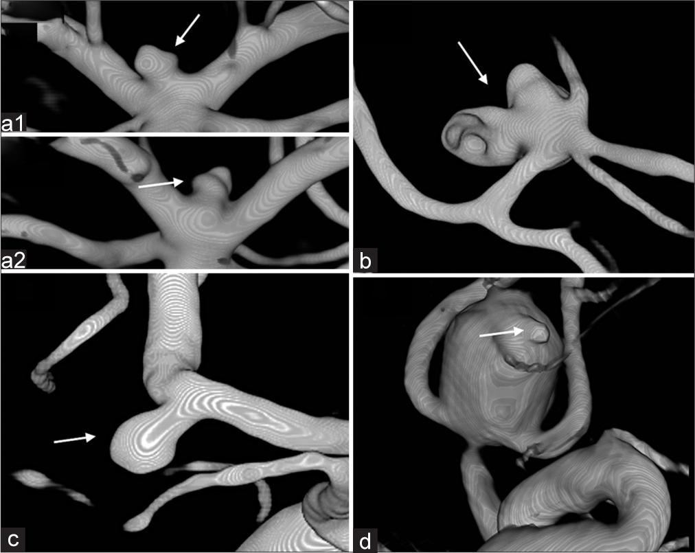
 12 ] Accordingly, patients with posterior circulation aneurysms might be underrepresented in our cohort and might skew our results. On the other hand, only 5 out of 111 patients (4.5%) of a prospectively followed cohort of intracranial aneurysms died of loss of consciousness or sudden death after severe headache without further diagnostic workup.[ 21 ] Only in cases with previous neuroimaging, aneurysm growth/de novo formation was considered. Accordingly, this risk factor is most likely underrepresented in our cohort. Another bias is associated with the aneurysm morphology. We found 72.7% of our aneurysms to be irregular or lobulated, by evaluating the postrupture morphology. It has been suggested that aneurysms might be subject to morphological changes, when they rupture.[ 19 , 20 ] Further, the bleeding point of the aneurysm could be interpreted as lobulation, on the postrupture cerebral angiography. Examples of such cases are presented in Figure 2 . Taking this into account, we might have overestimated this risk factor. Some risk factors are underrepresented or absent in our cohort, that is, ethnicity, cranial nerve deficits, and mass effect. These variables were not included in the regression analysis, and any potential interaction between the absent risk factors and AcomA location, therefore, not considered. Whether such an interaction exists in unknown. The ACA location has not been discussed in this paper. Table 3 demonstrates that this location is also associated with small size at rupture. However, as shown in Table 2 , only two aneurysms in our cohort were in this location. Hence, no conclusion can be made about this location from our dataset. Finally, our dataset lacks a “denominator.” We do not know how many unruptured AcomA aneurysm was present in the background population in the observation period. More important, we do not know how many small AcomA aneurysms were present. However, in the prospective part of the ISUIA study, both ICA (37.8%) and MCA (29.4%) were more frequent than AcomA/ACA location (16.1%). Given that size is evenly distributed among the different locations, it seems unlikely that at higher frequency in the background population could explain our finding that small AcomA aneurysms are overrepresented in an aSAH population.


Figure 2:

Aneurysm morphology examples and in doubt cases. (a1+a2) Aneurysm of doubt regarding irregularity/lobulation (arrows). Scored as irregular/lobulated. (b) Aneurysm evaluated to have significant lobulation (arrow), giving it points for lobulation, but also for high complexity-related risk. (c) Aneurysm with no irregularity or lobulation (arrow). (d) Aneurysm scored to have lobulation. Based on the spherical shape apart from the assumed bleeding point (arrow), it might have been without lobulation prerupture.[ 20]


CONCLUSION

In the present study of an aSAH cohort, small aneurysms <7 mm are significantly more frequently located at the AcomA and PcomA than other locations when controlling for other known risk factors. This could be of use for clinicians when counseling patients regarding preventive treatment or conservative management, but further studies are needed to validate this finding.

Declaration of patient consent

Patient’s consent not required as patient’s identity is not disclosed or compromised.

Financial support and sponsorship

Nil.

Conflicts of interest

There are no conflicts of interest.

References

1. Bender MT, Wendt H, Monarch T, Beaty N, Lin LM, Huang J. Small aneurysms account for the majority and increasing percentage of aneurysmal subarachnoid hemorrhage: A 25-year, single institution study. Neurosurgery. 2018. 83: 692-9

2. Bijlenga P, Ebeling C, Jaegersberg M, Summers P, Rogers A, Waterworth A. Risk of rupture of small anterior communicating artery aneurysms is similar to posterior circulation aneurysms. Stroke. 2013. 44: 3018-26

3. Cebral J, Ollikainen E, Chung BJ, Mut F, Sippola V, Jahromi BR. Flow conditions in the intracranial aneurysm lumen are associated with inflammation and degenerative changes of the aneurysm wall. AJNR Am J Neuroradiol. 2017. 38: 119-26

4. Choi JH, Park HS. The incidence and characteristics of patients with small ruptured aneurysms (<5 mm) in subarachnoid hemorrhage. J Korean Neurosurg Soc. 2017. 60: 424-32

5. Etminan N, Brown RD, Beseoglu K, Juvela S, Raymond J, Morita A. The unruptured intracranial aneurysm treatment score: A multidisciplinary consensus. Neurology. 2015. 85: 881-9

6. Etminan N, Chang HS, Hackenberg K, de Rooij NK, Vergouwen MD, Rinkel GJ. Worldwide incidence of aneurysmal subarachnoid hemorrhage according to region, time period, blood pressure, and smoking prevalence in the population: A systematic review and meta-analysis. JAMA Neurol. 2019. 76: 588-97

7. Froelich JJ, Neilson S, Peters-Wilke J, Dubey A, Thani N, Erasmus A. Size and location of ruptured intracranial aneurysms: A 5-year clinical survey. World Neurosurg. 2016. 91: 260-5

8. Gasparotti R, Liserre R. Intracranial aneurysms. Eur Radiol. 2005. 15: 441-7

9. Greebe P, Rinkel GJ, Hop JW, Visser-Meily JM, Algra A. Functional outcome and quality of life 5 and 12.5 years after aneurysmal subarachnoid haemorrhage. J Neurol. 2010. 257: 2059-64

10. Greving JP, Wermer MJ, Brown RD, Morita A, Juvela S, Yonekura M. Development of the PHASES score for prediction of risk of rupture of intracranial aneurysms: A pooled analysis of six prospective cohort studies. Lancet Neurol. 2014. 13: 59-66

11. Hop JW, Rinkel GJ, Algra A, van Gijn J. Case-fatality rates and functional outcome after subarachnoid hemorrhage: A systematic review. Stroke. 1997. 28: 660-4

12. Huang J, van Gelder JM. The probability of sudden death from rupture of intracranial aneurysms: A meta-analysis. Neurosurgery. 2002. 51: 1101-5

13. Inagawa T, Hirano A. Autopsy study of unruptured incidental intracranial aneurysms. Surg Neurol. 1990. 34: 361-5

14.

15. Mocco J, Brown RD, Torner JC, Capuano AW, Fargen KM, Raghavan ML. Aneurysm morphology and prediction of rupture: An international study of unruptured intracranial aneurysms analysis. Neurosurgery. 2018. 82: 491-6

16. Pagiola I, Mihalea C, Caroff J, Ikka L, Chalumeau V, Iacobucci M. The PHASES score: To treat or not to treat? Retrospective evaluation of the risk of rupture of intracranial aneurysms in patients with aneurysmal subarachnoid hemorrhage. J Neuroradiol. 2020. 47: 349-52

17. Rahman M, Ogilvy CS, Zipfel GJ, Derdeyn CP, Siddiqui AH, Bulsara KR. Unruptured cerebral aneurysms do not shrink when they rupture: Multicenter collaborative aneurysm study group. Neurosurgery. 2011. 68: 155-60

18. Rinaldo L, Nesvick CL, Rabinstein AA, Lanzino G. Differences in Size between unruptured and ruptured saccular intracranial aneurysms by location. World Neurosurg. 2020. 133: e828-34

19. Schneiders JJ, Marquering HA, van den Berg R, VanBavel E, Velthuis B, Rinkel GJ. Rupture-associated changes of cerebral aneurysm geometry: High-resolution 3D imaging before and after rupture. AJNR Am J Neuroradiol. 2014. 35: 1358-62

20. Skodvin TO, Johnsen LH, Gjertsen O, Isaksen JG, Sorteberg A. cerebral aneurysm morphology before and after rupture: Nationwide case series of 29 aneurysms. Stroke. 2017. 48: 880-6

21. Morita A, Kirino T, Hashi K, Aoki N, Fukuhara S. The natural course of unruptured cerebral aneurysms in a Japanese cohort. N Engl J Med. 2012. 366: 2474-82

22. Vlak MH, Algra A, Brandenburg R, Rinkel GJ. Prevalence of unruptured intracranial aneurysms, with emphasis on sex, age, comorbidity, country, and time period: A systematic review and meta-analysis. Lancet Neurol. 2011. 10: 626-36

23. Weir B, Disney L, Karrison T. Sizes of ruptured and unruptured aneurysms in relation to their sites and the ages of patients. J Neurosurg. 2002. 96: 64-70

24. Wermer MJ, van der Schaaf IC, Algra A, Rinkel GJ. Risk of rupture of unruptured intracranial aneurysms in relation to patient and aneurysm characteristics: An updated meta-analysis. Stroke. 2007. 38: 1404-10

25. Wiebers DO, Whisnant JP, Huston J, Meissner I, Brown RD, Piepgras DG. Unruptured intracranial aneurysms: Natural history, clinical outcome, and risks of surgical and endovascular treatment. Lancet. 2003. 362: 103-10

26. Wiebers DO. Unruptured intracranial aneurysms: Natural history and clinical management. Update on the international study of unruptured intracranial aneurysms. Neuroimaging Clin N Am. 2006. 16: 383-90

27. Yi J, Zielinski D, Chen M. Cerebral aneurysm size before and after rupture: Case series and literature review. J Stroke Cerebrovasc Dis. 2016. 25: 1244-8

28. Zhang X, Karuna T, Yao ZQ, Duan CZ, Wang XM, Jiang ST. High wall shear stress beyond a certain range in the parent artery could predict the risk of anterior communicating artery aneurysm rupture at follow-up. J Neurosurg. 2018. 131: 868-75

Leave a Reply

Your email address will not be published. Required fields are marked *