

- Department of Neurosurgery, Pisana University Hospital - University of Pisa, Pisa, Italy.
- Department of Laboratory Medicine, Pisana University Hospital - University of Pisa, Pisa, Italy.
Correspondence Address:
Nicola Montemurro, Department of Neurosurgery, Pisana University Hospital - University of Pisa, Pisa, Italy.
DOI:10.25259/SNI_647_2022
Copyright: © 2022 Surgical Neurology International This is an open-access article distributed under the terms of the Creative Commons Attribution-Non Commercial-Share Alike 4.0 License, which allows others to remix, transform, and build upon the work non-commercially, as long as the author is credited and the new creations are licensed under the identical terms.How to cite this article: Nicola Montemurro1, Daniele Lorenzini1, Valerio Ortenzi2, Jacopo Giorgetti1. Stretched intradural extramedullary tanycytic ependymoma of the thoracic spine. 16-Sep-2022;13:426
How to cite this URL: Nicola Montemurro1, Daniele Lorenzini1, Valerio Ortenzi2, Jacopo Giorgetti1. Stretched intradural extramedullary tanycytic ependymoma of the thoracic spine. 16-Sep-2022;13:426. Available from: https://surgicalneurologyint.com/surgicalint-articles/11864/
Date of Submission
19-Jul-2022
Date of Acceptance
04-Sep-2022
Date of Web Publication
16-Sep-2022
Abstract
Background: Tanycytic ependymoma is a rare variant of ependymoma that commonly affects the cervical and thoracic spinal cord. It usually arises as intramedullary lesions and extramedullary cases are extremely rare.
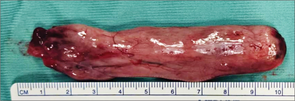
Case Description: We present a 77-year-old woman with the complaints of a 2-year history of progressive paraparesis and sensory loss in her lower extremities. Magnetic resonance imaging revealed a stretched and fusiform intradural extramedullary lesion at T5-T10 level. Gross total removal of the tumor was achieved and a definitive diagnosis of tanycytic ependymoma was established.
Conclusion: This case thus represents a rare case of thoracic intradural extramedullary tanycytic ependymoma and, to the best of our knowledge, it represents the longest intradural extramedullary tanycytic ependymoma in craniocaudal direction ever reported in the literature.
Keywords: Ependymoma, Intradural extramedullary tumor, Tanycytic ependymoma, Thoracic spine
INTRODUCTION
Tanycytic ependymoma is a rare variant of ependymoma that commonly affects the cervical and thoracic spinal cord.[
CASE PRESENTATION
A 77-year-old woman with the complaints of a 2-year history of progressively worsening low back pain and progressive paraparesis and sensory loss in her lower extremities came to our attention. Magnetic resonance imaging (MRI) revealed a stretched and fusiform intradural extramedullary lesion at T5-T10 level with uniform intensity in T2-weighted sagittal and axial images and strongly enhanced after gadolinium administration [
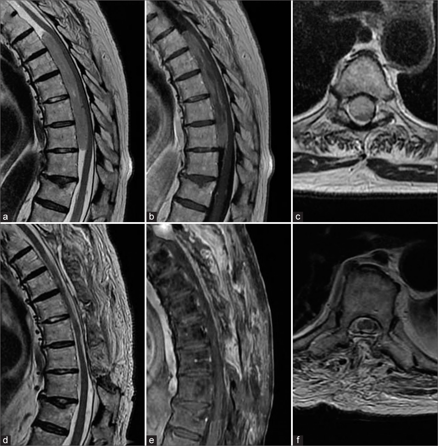
Figure 1:
Preoperative T2-weighted sagittal (a), sagittal gadolinium-enhanced T1-weighted (b) and T2-weighted axial (c) MRI showing a stretched and fusiform intradural extramedullary lesion at T5-T10 level about 13 cm long in craniocaudal direction. Postoperative T2-weighted sagittal (d), sagittal gadolinium-enhanced T1-weighted (e) and T2-weighted axial (f) MRI showing complete resection of the mass lesion.
DISCUSSION
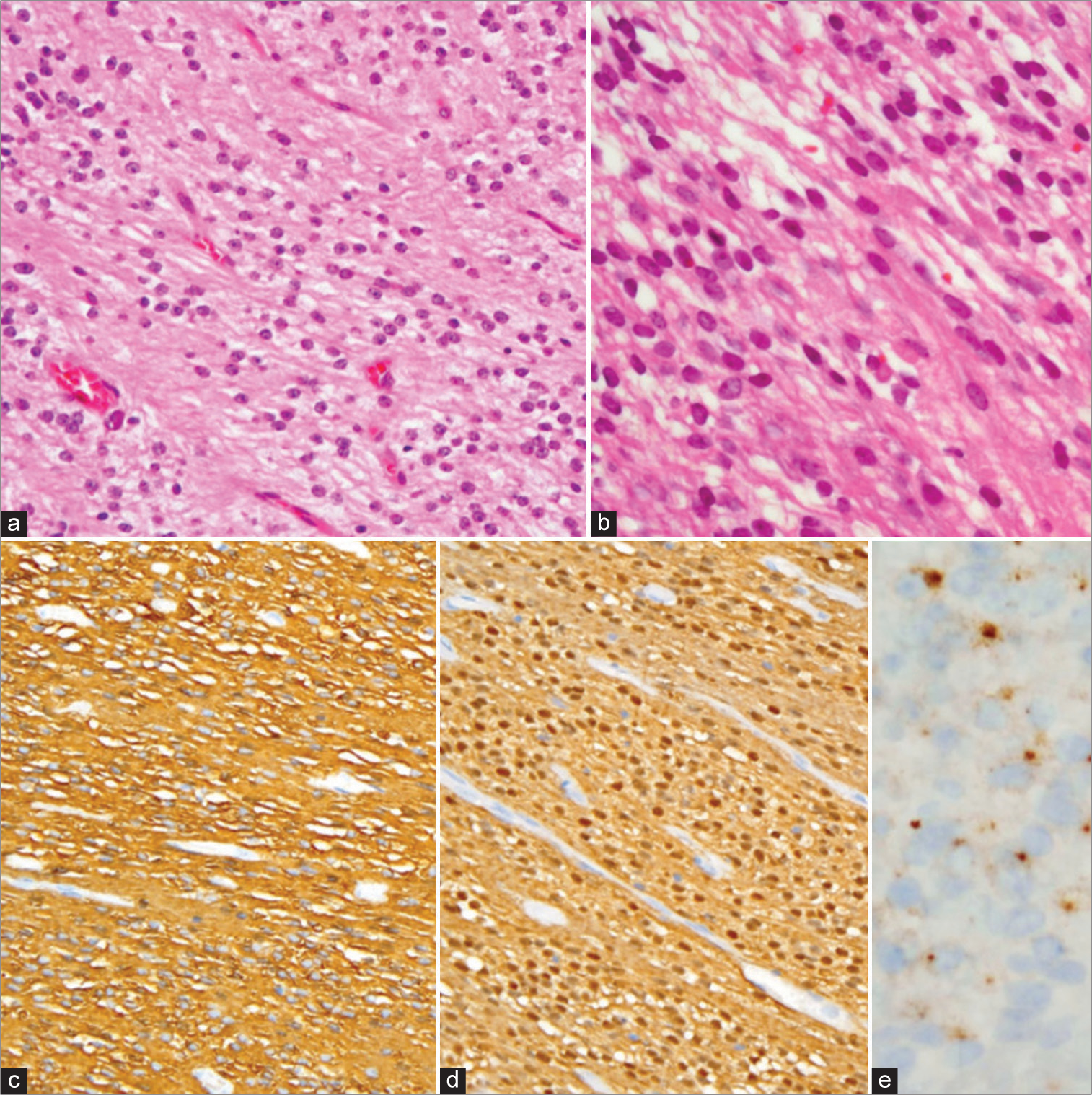
Tanycytic ependymoma is a histologically distinct rare subtype of ependymoma and is recognized as a Grade II tumor in the latest World Health Organization classification in 2021.[
CONCLUSION
This case thus represents a rare case of thoracic intradural extramedullary tanycytic ependymoma and, to the best of our knowledge, it represents the longest intradural extramedullary tanycytic ependymoma in craniocaudal length ever reported in the literature.
Declaration of patient consent
Patient’s consent not required as patient’s identity is not disclosed or compromised.
Financial support and sponsorship
Nil.
Conflicts of interest
There are no conflicts of interest.
References
1. Kim DJ, Han MH, Lee S. Extramedullary tanycytic ependymoma of the lumbar spinal cord. Yeungnam Univ J Med. 2020. 37: 128-32
2. Krisht KM, Schmidt MH. Tanycytic ependymoma: A challenging histological diagnosis. Case Rep Neurol Med. 2013. 2013: 170791
3. Louis DN, Perry A, Wesseling P, Brat J, Cree IA, Hawkins C. The 2021 WHO classification of tumors of the central nervous system: A summary. Neuro Oncol. 2021. 23: 1231-51
4. Tao X, Hou Z, Hao S, Zhang Q, Wu Z, Zhang J. The clinical features and surgical outcomes of spinal cord tanycytic ependymomas: A report of 40 cases. World Neurosurg. 2017. 106: 60-73

