

- Department of Neurosurgery, Suwa Red Cross Hospital, Suwa, Nagano, Japan.
DOI:10.25259/SNI_814_2020
Copyright: © 2021 Surgical Neurology International This is an open-access article distributed under the terms of the Creative Commons Attribution-Non Commercial-Share Alike 4.0 License, which allows others to remix, tweak, and build upon the work non-commercially, as long as the author is credited and the new creations are licensed under the identical terms.How to cite this article: Masahito Katsuki, Yukinari Kakizawa, Akihiro Nishikawa, Yasunaga Yamamoto, Toshiya Uchiyama. Temporal muscle thickness and area are an independent prognostic factors in patients aged 75 or younger with aneurysmal subarachnoid hemorrhage treated by clipping. 14-Apr-2021;12:151
How to cite this URL: Masahito Katsuki, Yukinari Kakizawa, Akihiro Nishikawa, Yasunaga Yamamoto, Toshiya Uchiyama. Temporal muscle thickness and area are an independent prognostic factors in patients aged 75 or younger with aneurysmal subarachnoid hemorrhage treated by clipping. 14-Apr-2021;12:151. Available from: https://surgicalneurologyint.com/surgicalint-articles/10719/
Date of Submission
13-Nov-2020
Date of Acceptance
16-Mar-2021
Date of Web Publication
14-Apr-2021
Abstract
Background: Skeletal muscle mass is an important factor for various diseases’ outcomes. As for its indicators, temporal muscle thickness (TMT) and temporal muscle area (TMA) on the head computed tomography are useful, and TMT and TMA were reported as potential prognostic factors for aneurysmal subarachnoid hemorrhage (SAH). We examined the clinical characteristics, including TMT and TMA, of SAH patients aged 75 or younger.
Methods: We retrospectively investigated 127 SAH patients with all World Federation of Neurosurgical Societies (WFNS) grades and treated by clipping between 2009 and 2019. Clinical outcome was measured with the modified Rankin Scale (mRS) at 6 months, with favorable outcome defined as mRS 0–2. The associations between the clinical variables and the outcomes were analyzed.
Results: The mean age was 60.6 (32–74) years, and 65% were women. The mean ± standard deviation of WFNS grade was 2.8 ± 1.4. TMT and TMA were larger in the favorable outcome group than the poor one. Multivariate analysis revealed that age, smoking, WFNS grade, and TMT or TMA were associated with favorable outcome. Receiver operating characteristic analysis found that the threshold of TMT was 4.9 mm in female and 6.7 mm in male, and that of TMA was 193 mm2 in female and 333 mm2 in male.
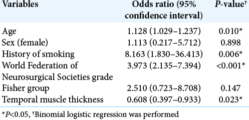
Conclusion: The odds ratios for TMT and TMA related to clinical outcome were lower than for smoking and WFNS grade; however, on multivariate analysis they remained independent prognostic factors in SAH patients aged 75 or younger treated by clipping. Further studies are needed to confirm these findings.
Keywords: Cerebral aneurysm, Clipping, Prognostic factor, Sarcopenia, Subarachnoid hemorrhage, Temporal muscle thickness and area
INTRODUCTION
Low skeletal muscle mass due to low nutrition or aging (sarcopenia in the broadest sense[
Therefore, we focused on the temporal muscle thickness (TMT), and temporal muscle area (TMA) on the head CT, because TMT and TMA are substituted as useful measures of the total body skeletal muscle mass.[
This study was performed to analyze the clinical characteristics of aneurysmal SAH patients aged 75 or younger who were treated by clipping, with a focus on the temporal muscle. To the best of our knowledge, this is the first study, including the young and middle ages, to examine the relationship between TMT and TMA and outcomes of SAH patients treated by clipping.
MATERIALS AND METHODS
Study population
We retrospectively retrieved data from medical records of all the 127 aneurysmal SAH patients who were admitted between 2009 and 2019 and treated by clipping at our institution. All the patients included in this study had been independent in their activities in daily livings (ADLs) before the onset of SAH. The diagnosis of SAH was based on the clinical history and the presence of SAH on CT. The hospital’s research ethics committee approved this study, and we gained written informed consent for this study from all of the patients, the legally authorized representative of the patients, or next of kin of the deceased patients. All methods were carried out in accordance with relevant guidelines and regulations (Declaration of Helsinki).
General management of SAH was similar in all cases: all patients were first treated with nicardipine and kept normovolemic with normal blood pressure (systolic blood pressure <140 mmHg). Indication for surgery was established according to the Japanese Guidelines for the Management of Stroke 2009[
All SAH patients who underwent aneurysm clipping received fasudil, cilostazol, and statin as appropriate after the operation. Rehabilitation and nutritional support were started as soon as possible after the operation, and prophylaxis and treatment of complications were also ensured. Intra-arterial infusion of fasudil was performed when necessary for the treatment of symptomatic vasospasm. In addition, a ventriculoperitoneal shunt was performed when hydrocephalus was observed.
Clinical variables
We collected data regarding physiological symptoms at admission for patients included in this study, that is, age, sex, WFNS grade, systolic blood pressure, administration of antithrombotic drugs, history of smoking and drinking, hypertension, diabetes mellitus, dyslipidemia, symptomatic vasospasm, ventriculoperitoneal shunt, and postoperative complications (except for symptomatic vasospasm and hydrocephalus). Postoperative complications included infectious diseases, heart failure, rerupture, seizure, and disuse syndrome. We also measured albumin, lymphocyte, triglycerides, total cholesterol, high-density lipoprotein cholesterol, low-density lipoprotein (LDL) cholesterol, glucose, and hemoglobin A1c levels at admission. Albumin, lymphocyte, and total cholesterol are known factors for controlling nutritional status score to assess the nutritional status of the patients.[
We determined the location and size of the aneurysm, Fisher CT scale, TMT, and TMA based on the results of the CT or CT angiography at admission. We used the Aquilion ONE (Canon Medical Systems Corporation, Tochigi, Japan) to take CT and CT angiography images of 0.5 × 0.5 × 1.0 mm voxels. The slice thickness was reconstructed to 5 mm [
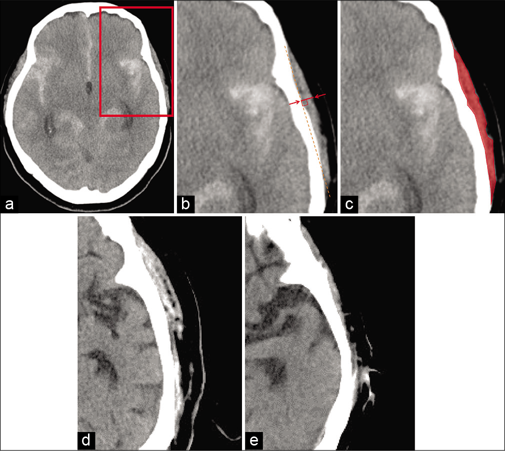
Figure 1:
Head CT image measuring temporal muscle thickness (TMT) and area (TMA). The slice is 5 mm above the superior wall of the orbit (a). The rectangular part in (a) is enlarged in (b) and (c). CT image representing TMT. The line between the arrows indicates TMT (b). CT image representing TMA (c). Patient with a large TMT and TMA (d); patient with a small TMT and TMA (e).
To evaluate the outcomes, modified Rankin Scale (mRS) scores at 6 months after the operations of all 127 patients were collected by either personal outpatient interviews, reports from the rehabilitation hospital or home doctor, or interviews over the telephone, once the ethical approval was obtained for the study. We dichotomized mRS scores into favorable (mRS 0–2) or poor (mRS 3–6).
Statistical analysis
Intraclass correlation coefficients were used to test inter-rater reliabilities of TMT and TMA. The associations between TMT and TMA and other factors were investigated by the Mann–Whitney U-test, Fisher’s exact test, or Spearman’s coefficient correlation. R > 0.3 was defined that there was a significant correlation. The results are described as mean ± standard deviation (SD). Associations and outcomes between the clinical variables were analyzed using the Mann–Whitney U-test and Fisher’s exact test. Binomial logistic regression analysis was performed using the preoperative factors with P < 0.05 extracted through the univariate analysis described above. Age and sex were also used for the multivariate analysis to adjust the difference of TMT and TMA related to age or sex. Variables with small sample numbers under 80 were excluded from the multivariate analysis. We also performed receiver operating characteristic (ROC) analysis by sex and analyzed the thresholds of the TMT and TMA related to the outcome. The minimum distance point to (0,1) determined the thresholds. A two-tailed p < 0.050 was considered statistically significant. We conducted this calculation using SPSS software version 24.0.0. (IBM, New York, USA).
RESULTS
Clinical characteristics
Clinical characteristics of the 127 SAH patients (82 women and 45 men) treated by clipping are summarized in [

TMT and TMA and sex, age, laboratory data, or outcome
The intraclass correlation coefficients (2, 2) measuring TMT and TMA were 0.803 and 0.724, respectively. TMT and TMA were significantly larger in men than in women [P < 0.001 for both,

Figure 2:
Temporal muscle thickness (TMT) was significantly larger in the favorable outcome groups than in the poor outcome groups (P = 0.001 in both sex, P = 0.007 in female, P = 0.007 in male, respectively) (a). Temporal muscle area (TMA) was also significantly larger in the favorable outcome groups (P = 0.005 in both sex, P = 0.007 in female, P = 0.040 in male, respectively) (b). Receiver operating characteristic (ROC) analysis was performed to determine the threshold of the TMT and TMA for outcomes. The threshold of TMT was 4.9 mm in female (sensitivity = 0.661, specificity = 0.750. AUC = 0.703, 95%CI 0.523–0.811, P = 0.003) and 6.7 mm in male (sensitivity = 0.647, specificity = 0.909. AUC = 0.767, 95%CI 0.602–0.933, P = 0.008) (c). The threshold of TMA was 193 mm2 in female (sensitivity = 0.729, specificity = 0.700. AUC = 0.699, 95%CI 0.536–0.813, P = 0.024) and 333 mm2 in male (sensitivity = 0.735, specificity = 0.727. AUC = 0.699, 95%CI 0.521–0.878, P = 0.049) (d).
Relationship between variables and outcome
Age, WFNS grade, smoking, Fisher group, LDL cholesterol, ventriculoperitoneal shunt, and postoperative complications were also significantly associated with poor outcomes in the univariate analysis (P = 0.014, P < 0.001, P = 0.030, P < 0.001, P = 0.04, P = 0.001, P = 0.016, respectively) [

Threshold of TMT and TMA for outcome
ROC analysis revealed that the threshold of TMT was 4.9 mm in female (sensitivity = 0.661, specificity = 0.750. area under the ROC curve [AUC] = 0.703, 95% CI 0.523–0.811, P = 0.003) and 6.7 mm in male (sensitivity = 0.647, specificity = 0.909. AUC = 0.767, 95%CI 0.602–0.933, P = 0.008) [
DISCUSSION
Our findings show that TMT and TMA would be ones of the prognostic factors of SAH patients aged 75 or younger treated by clipping, and our study found that the threshold for the outcome of TMT was 4.9 mm in female and 6.7 mm in male, and that of TMA was 193 mm2 in female and 333 mm2 in male.
Skeletal muscle mass and neurosurgical diseases
In addition to our report, several reports on the association between skeletal muscle mass and neurosurgical diseases are reported. Higher skeletal muscle mass reduces the risk of intracranial arterial stenosis and may protect against ischemic stroke.[
These are some speculations of why low skeletal muscle mass is related to the poor prognosis of SAH. Patients with neurosurgical diseases or after surgical treatment are usually bedridden for some time due to impaired consciousness, sedation, or surgical invasion. The skeletal muscle would be further catabolized during such acute treatment and hospitalization, so those with low skeletal muscle mass may have difficulty performing rehabilitation. In addition, low skeletal muscle mass and sarcopenia are related to malnutrition.[
As a potential explanation, the skeletal muscle is an endocrine organ and secretes myokines. One of the myokines, interleukin-6 (IL), is associated with inflammation, and elevated plasma concentrations of IL-6 are seen in patients with lower muscle mass or sarcopenia.[
Usefulness of TMT and TMA as indicators of skeletal muscle mass in neurosurgical diseases
Many studies on the association between skeletal muscle mass and outcomes used psoas muscle cross-sectional area at the level of the third lumbar vertebra,[
Previously, thresholds of TMT in the magnetic resonance imaging (MRI) for the outcomes in various diseases were investigated. TMT in MRI over the median was associated with favorable outcomes in brain metastasis.[
Clinical application
Awareness of the relationship between loss of muscle mass and outcome may lead to new therapeutic targets for patients with unruptured aneurysms before developing SAH. Low skeletal muscle mass is related to malnutrition and aging,[
Limitations
First, the sample size was small. Second, previous studies reported that sex and age affect temporal muscle mass,[
Furthermore, accurate and standard methods of TMT and TMA measurements were not established. Ranganathan[
CONCLUSION
TMT and TMA were independent prognostic factors for mRS 6 months after aneurysm clipping in aneurysmal SAH patients 75 years of age or younger of all WFNS grades. Our study found that the threshold for the outcome of TMT was 4.9 mm in female and 6.7 mm in male and that of TMA was 193 mm2 in female and 333 mm2 in male. Further studies are needed to confirm these findings.
Declaration of patient consent
The authors certify that they have obtained all appropriate patient consent.
Financial support and sponsorship
Nil.
Conflicts of interest
There are no conflicts of interest.
References
1. Bayram S, Akgül T, Adıyaman AE, Karalar Ş Dölen D, Aydoseli A. Effect of sarcopenia on mortality after percutaneous vertebral augmentation treatment for osteoporotic vertebral compression fractures in elderly patients: A retrospective cohort study. World Neurosurg. 2020. 138: e354-60
2. Benatti FB, Pedersen BK. Exercise as an anti-inflammatory therapy for rheumatic diseases-myokine regulation. Nat Rev Rheumatol. 2015. 11: 86-97
3. Bian AL, Hu HY, Rong YD, Wang J, Wang JX, Zhou XZ. A study on relationship between elderly sarcopenia and inflammatory factors IL-6 and TNF-α. Eur J Med Res. 2017. 22: 25
4. Binay Safer V, Safer U. Comment on. “Clinical characteristics of aneurysmal subarachnoid hemorrhage (SAH) in the elderly over 75: Would temporal muscle be a potential prognostic factor as an indicator of sarcopenia?. ” Clin Neurol Neurosurg. 2020. 188: 105600
5. Chen LK, Liu LK, Woo J, Assantachai P, Auyeung TW, Bahyah KS. Sarcopenia in Asia: Consensus report of the Asian working group for sarcopenia. J Am Med Dir Assoc. 2014. 15: 95-101
6. Costa TM, Costa FM, Moreira CA, Rabelo LM, Boguszewski CL, Borba VZC. Sarcopenia in COPD: Relationship with COPD severity and prognosis. J Bras Pneumol. 2015. 41: 415-21
7. Cruz-Jentoft AJ, Baeyens JP, Bauer JM, Boirie Y, Cederholm T, Landi F. Sarcopenia: European consensus on definition and diagnosis: Report of the european working group on sarcopenia in older people. Age Ageing. 2010. 39: 412-23
8. Cruz-Jentoft AJ, Bahat G, Bauer J, Boirie Y, Bruyère O, Cederholm T. Sarcopenia: Revised European consensus on definition and diagnosis. Age Ageing. 2019. 48: 16-31
9. Furtner J, Berghoff AS, Albtoush OM, Woitek R, Asenbaum U, Prayer D. Survival prediction using temporal muscle thickness measurements on cranial magnetic resonance images in patients with newly diagnosed brain metastases. Eur Radiol. 2017. 27: 3167-73
10. Furtner J, Berghoff AS, Schöpf V, Reumann R, Pascher B, Woitek R. Temporal muscle thickness is an independent prognostic marker in melanoma patients with newly diagnosed brain metastases. J Neurooncol. 2018. 140: 173-8
11. Furtner J, Genbrugge E, Gorlia T, Bendszus M, Nowosielski M, Golfinopoulos V. Temporal muscle thickness is an independent prognostic marker in patients with progressive glioblastoma: Translational imaging analysis of the EORTC 26101 trial. Neuro Oncol. 2019. 21: 1587-94
12. Garcia JM, Stillings SA, Leclerc JL, Phillips H, Edwards NJ, Robicsek SA. Role of interleukin-10 in acute brain injuries. Front Neurol. 2017. 8: 244
13. Hasegawa Y, Yoshida M, Sato A, Fujimoto Y, Minematsu T, Sugama J. Temporal muscle thickness as a new indicator of nutritional status in older individuals. Geriatr Gerontol Int. 2019. 19: 135-40
14. Höllig A, Remmel D, Stoffel-Wagner B, Schubert GA, Coburn M, Clusmann H. Association of early inflammatory parameters after subarachnoid hemorrhage with functional outcome: A prospective cohort study. Clin Neurol Neurosurg. 2015. 138: 177-83
15. Hsieh K, Hwang M, Estevez-Inoa G, Saraf A, Spina CS, Smith D. Temporalis muscle width as a measure of sarcopenia independently predicts overall survival in patients with newly diagnosed glioblastoma. Int J Radiat Oncol. 2018. 102: e225
16. Ignacio de Ulíbarri J, González-Madroño A, de Villar NG, González P, González B, Mancha A. CONUT: A tool for controlling nutritional status. First validation in a hospital population. Nutr Hosp. 2005. 20: 38-45
17. Ishihara H, Oka F, Goto H, Nishimoto T, Okazaki K, Sadahiro H. Impact of frailty on medium-term outcome in asymptomatic patients after carotid artery stenting. World Neurosurg. 2019. 127: e396-9
18. Jones KI, Doleman B, Scott S, Lund JN, Williams JP. Simple psoas cross-sectional area measurement is a quick and easy method to assess sarcopenia and predicts major surgical complications. Color Dis. 2015. 17: 20-6
19. Jung HJ, Jung H, Lee T, Kim J, Park J, Kim H. Decreased muscle mass in Korean subjects with intracranial arterial stenosis: The Kangbuk Samsung Health Study. Atherosclerosis. 2017. 256: 89-93
20. Katsuki M, Suzuki Y, Kunitoki K, Sato Y, Sasaki K, Mashiyama S. Temporal muscle as an indicator of sarcopenia is independently associated with Hunt and Kosnik grade on admission and the modified Rankin scale score at 6 months of patients with subarachnoid hemorrhage treated by endovascular coiling. World Neurosurg. 2020. 137: e526-34
21. Katsuki M, Suzuki Y, Kunitoki K, Sato Y, Sasaki K, Mashiyama S. Temporal muscle thickness and area with various characteristics data of the patients with aneurysmal subarachnoid hemorrhage who underwent endovascular coiling. Data Brief. 2020. 31: 105715
22. Katsuki M, Suzuki Y, Sato Y, Sasaki K, Shingai Y, Mashiyama S.editors. In reply to the letter to the editor regarding “temporal muscle as an indicator of sarcopenia is independently associated with Hunt and Kosnik grade on admission and the modified rankin scale at 6 months of patients with subarachnoid hemorrhage treated by endovascular coiling. World Neurosurg. 2020. p. 140-433
23. Katsuki M, Yamamoto Y, Uchiyama T, Nishikawa A, Wada N, Kakizawa Y. Temporal muscle thickness and area with various characteristics data of the elderly patients over 75 with aneurysmal subarachnoid haemorrhage whose World Federation of Neurosurgical Societies grade were I to III. Data Brief. 2020. 28: 104832
24. Katsuki M, Yamamoto Y, Uchiyama T, Wada N, Kakizawa Y. Clinical characteristics of aneurysmal subarachnoid hemorrhage in the elderly over 75; would temporal muscle be a potential prognostic factor as an indicator of sarcopenia?. Clin Neurol Neurosurg. 2019. 186: 105535
25. Lang T, Streeper T, Cawthon P, Baldwin K, Taaffe DR, Harris TB. Sarcopenia: Etiology, clinical consequences, intervention, and assessment. Osteoporos Int. 2010. 21: 543-59
26. Leitner J, Pelster S, Schöpf V, Berghoff AS, Woitek R, Asenbaum U. High correlation of temporal muscle thickness with lumbar skeletal muscle cross-sectional area in patients with brain metastases. PLoS One. 2018. 13: e0207849
27. Malmstrom TK, Morley JE. SARC-F: A simple questionnaire to rapidly diagnose sarcopenia. J Am Med Dir Assoc. 2013. 14: 531-2
28. Matsushita T, Nishioka S, Taguchi S, Yamanouchi A, Nakashima R, Wakabayashi H. Sarcopenic obesity and activities of daily living in stroke rehabilitation patients: A cross-sectional study. Healthcare (Basel). 2020. 8: 255
29. Minn YK, Suk SH. Higher skeletal muscle mass may protect against ischemic stroke in community-dwelling adults without stroke and dementia: The PRESENT project. BMC Geriatr. 2017. 17: 45
30. Ni HJ, Pudasaini B, Yuan XT, Li HF, Shi L, Yuan P. Exercise training for patients pre-and postsurgically treated for non-small cell lung cancer: A systematic review and meta-analysis. Integr Cancer Ther. 2017. 16: 63-73
31. Nishiguchi S, Hino K, Moriya K, Shiraki M, Hiramatsu A, Nishikawa H.editors. Assessment criteria for sarcopenia in liver disease (first edition): Report from the working group for creation of sarcopenia assessment criteria in the Japan Society of Hepatology. Kanzo. 2016. 57: 353-68
32. Nozoe M, Kanai M, Kubo H, Kobayashi M, Yamamoto M, Shimada S. Quadriceps muscle thickness changes in patients with aneurysmal subarachnoid hemorrhage during the acute phase. Top Stroke Rehabil. 2018. 25: 209-13
33. Paccagnella A, Morassutti I, Rosti G. Nutritional intervention for improving treatment tolerance in cancer patients. Curr Opin Oncol. 2011. 23: 322-30
34. Ranganathan K, Terjimanian M, Lisiecki J, Rinkinen J, Mukkamala A, Brownley C. Temporalis muscle morphomics: The psoas of the craniofacial skeleton. J Surg Res. 2014. 186: 246-52
35. Rinkinen J, Zhang P, Wang L, Enchakalody B, Terjimanian M, Holcomb S. Novel temporalis muscle and fat pad morphomic analyses aids preoperative risk evaluation and outcome assessment in nonsyndromic craniosynostosis. J Craniofac Surg. 2013. 24: 250-5
36. Rong YD, Bian AL, Hu HY, Ma Y, Zhou XZ. Study on relationship between elderly sarcopenia and inflammatory cytokine IL-6, anti-inflammatory cytokine IL-10. BMC Geriatr. 2018. 18: 308
37. Safer U, Tasci I, Binay Safer V.editors. Letter to the editor regarding “effect of sarcopenia on mortality after percutaneous vertebral augmentation treatment for osteoporotic vertebral compression fractures in elderly patients: A retrospective cohort study”. World Neurosurg. 2020. 139: 710
38. Shibahashi K, Sugiyama K, Kashiura M, Hamabe Y. Decreasing skeletal muscle as a risk factor for mortality in elderly patients with sepsis: A retrospective cohort study. J Intensive Care. 2017. 5: 8
39. Someya Y, Tamura Y, Suzuki R, Kaga H, Kadowaki S, Sugimoto D. Characteristics of glucose metabolism in underweight Japanese women. J Endocr Soc. 2018. 2: 279-89
40. Sousa AS, Guerra RS, Fonseca I, Pichel F, Ferreira S, Amaral TF. Financial impact of sarcopenia on hospitalization costs. Eur J Clin Nutr. 2016. 70: 1046-51
41. Steindl A, Leitner J, Schwarz M, Nenning KH, Asenbaum U, Mayer S. Sarcopenia in neurological patients: Standard values for temporal muscle thickness and muscle strength evaluation. J Clin Med. 2020. 9: 1272
42. Teasdale GM, Drake CG, Hunt W, Kassell N, Sano K, Perat B. A universal subarachnoid hemorrhage scale: Report of a committee of the world federation of Neurosurgical societies. J Neurol Neurosurg Psychiatry. 1988. 51: 1457
43. .editors. Japanese Guidelines for the Management of Stroke 2009. Tokyo: Kyowa Kikaku; 2009. p.
44. .editors. Japanese Guidelines for the Management of Stroke 2015. Tokyo: Kyowa Kikaku; 2015. p.
45. Tsuchida K, Fujihara Y, Hiroki J, Hakamata T, Sakai R, Nishida K. Significance of sarcopenia evaluation in acute decompensated heart failure. Skeletal muscle mass index versus fat-free mass index. Int Hear J. 2018. 59: 143-8
46. Uhlich R, Hu P. Sarcopenia diagnosed using masseter muscle area predictive of early mortality following severe traumatic brain injury. Neural Regen Res. 2018. 13: 2089-90
47. Vlak MH, Rinkel GJ, Greebe P, Van Der Bom JG, Algra A. Trigger factors and their attributable risk for rupture of intracranial aneurysms: A case-crossover study. Stroke. 2011. 42: 1878-82

