Tools

Ntenis Nerntengian1, Levent Tanrikulu2, Michael Anthony Manoussos1, Nikolaos Barettas1, Grigorios Gkasdaris1, Theodosios Birbilis1
  1. Department of Neurosurgery, University Hospital of Alexandroupolis, Medical School of Democritus University of Thrace, Dragana, Alexandroupolis, Greece,
  2. Department of Neurosurgery, Universitätsmedizin Göttingen, Georg-August-Universität Göttingen, Göttingen, Germany.

Correspondence Address:
Ntenis Nerntengian, Department of Neurosurgery, University Hospital of Alexandroupolis, Medical School of Democritus University of Thrace, Dragana, Alexandroupolis, Greece.

DOI:10.25259/SNI_158_2022

Copyright: © 2022 Surgical Neurology International This is an open-access article distributed under the terms of the Creative Commons Attribution-Non Commercial-Share Alike 4.0 License, which allows others to remix, transform, and build upon the work non-commercially, as long as the author is credited and the new creations are licensed under the identical terms.

How to cite this article: Ntenis Nerntengian1, Levent Tanrikulu2, Michael Anthony Manoussos1, Nikolaos Barettas1, Grigorios Gkasdaris1, Theodosios Birbilis1. The evaluation of the usefulness of CO2 laser in microsurgical resection of brain tumors. 31-Mar-2022;13:118

How to cite this URL: Ntenis Nerntengian1, Levent Tanrikulu2, Michael Anthony Manoussos1, Nikolaos Barettas1, Grigorios Gkasdaris1, Theodosios Birbilis1. The evaluation of the usefulness of CO2 laser in microsurgical resection of brain tumors. 31-Mar-2022;13:118. Available from: https://surgicalneurologyint.com/surgicalint-articles/11500/

Date of Submission
09-Feb-2022

Date of Acceptance
13-Mar-2022

Date of Web Publication
31-Mar-2022

Abstract

Background: Since its introduction to surgery, the CO2 laser has been used in the treatment of various neurosurgical pathologies as it combines cutting, vaporizing, and coagulating properties in one tool and has a safe penetration depth. In this case series of 29 patients, we present the evaluation of the usefulness of the closed system type – sealed tube surgical CO2 laser in the surgical removal of brain tumors.

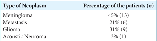
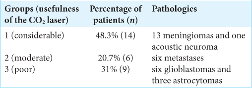
Methods: The Sharplan 40C model SurgiTouch, sealed tube type CO2 laser, was used in the resection of 29 brain tumors; 13 meningiomas, six metastases, nine gliomas, and one acoustic neuroma. The same senior surgeon (BT) assessed and classified the benefit provided by the CO2 laser in the resection of the neoplasms to considerable (Group 1), moderate (Group 2), and poor (Group 3).

Results: Group 1 included 14 patients with 13 meningiomas and one acoustic neuroma, Group 2 included six patients, all of whom had metastases, and Group 3 included nine patients of which six had glioblastoma and three astrocytoma. No complications or technical problems occurred due to the use of the CO2 laser.

Conclusion: The CO2 laser is a valuable complementary tool in brain tumor surgery displaying high efficacy and practicality in the resection of neoplasms which are fibrous and have hard consistency. It has high acquisition and maintenance cost and cannot replace the bipolar diathermy. The newest generation of flexible CO2 laser fiber provides more ergonomy and promises new perspectives of its neurosurgical use in the modern era.

Keywords: Brain tumor surgery, CO2 laser, Laser neurosurgery, Laser surgery, Sealed tube surgical CO2 laser

INTRODUCTION

The term LASER is an acronym for light amplification by stimulated emission of radiation.[ 2 ] The theoretical basis for the laser began in 1916 when A. Einstein published on his paper “on the Quantum Theory of Radiation” that excited atoms can be stimulated by photons and emit photon.[ 8 ] After further scientific research and technical developments, the first laser was finally built in 1960 by Maiman.[ 24 ] Laser beam combines high power, directionality, and increased ability to focus.[ 1 , 24 ] Due to the aforementioned properties of the laser, it began to be used in various fields as dermatology, otorhinolaryngology, urology, general surgery, and ophthalmology.[ 10 , 14 , 16 , 20 , 22 ] The mechanism by which the laser works is through the conversion of the light energy absorbed by the tissue to heat with subsequent coagulation or vaporization of the target and in extremely high energies its photomechanical destruction.[ 1 ] The first CO2 laser was created by Patel[ 21 ] and it is the most widely used in neurosurgery today[ 3 , 15 ] as brain tissue contains approximately 80% water[ 19 ] and CO2 laser energy gets highly absorbed in water thus providing minimal thermal damage to surrounding tissue.[ 3 , 23 ] Furthermore, CO2 laser provides a safe penetration depth[ 18 ] when operating among delicate neurovascular structures. After the early 90s, there has been relatively sparse literature on the use of the CO2 laser in neurosurgery in comparison to other surgical specialties. The aim of this work is the evaluation of the closed system type – sealed tube surgical CO2 laser in microsurgical removal of brain tumors.

MATERIALS AND METHODS

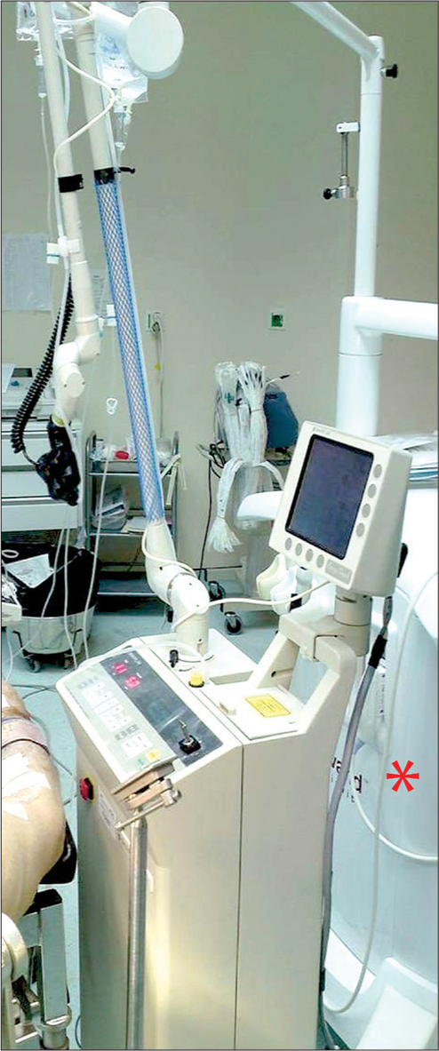
Technical description of the CO2 laser device

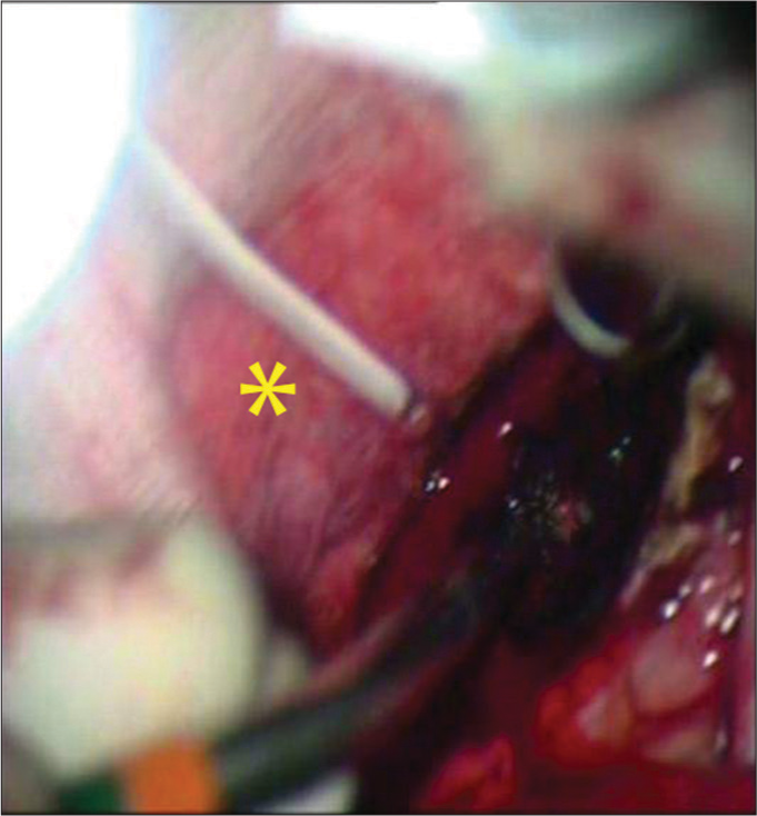
In the neurosurgical department of the Democritus University of Thrace, the CO2 laser sealed tube type was used during surgery of the here reported brain neoplasms. It is a Sharplan 40C model SurgiTouch MS 780 Flash scan 40 Watt, with a wavelength of 10.6 μm and energy output of 750 mJ [ Figures 1 and 2 ]. The beam is manipulated by an articulated arm [ Figure 3 ] utilizing a system of mirrors (length of 1.2 m), to the joystick (a hand-guided focusing lens) that has focusing ability of 125–200 mm [ Figure 4 ] and lastly the beam is transferred to the target. By modifying the power level, an adjustable penetration depth can be achieved varying from 0.03 mm to 2 mm.[ 9 , 17 , 18 ] For combined use with the surgical microscope, the articulated arm can be coupled with the microscope and a micromanipulator which focuses from 200 to 400 mm. The device comes equipped with a He-Ne laser that is on the same axis with the invisible beam of the CO2 laser and functions to aid aiming of the laser during surgery. Safety protocols are followed during the duration of the surgery, due to the risk of fire, burns, and damage to the eyes. To avoid these dangers during a surgical procedure, the use of protective eyewear is used by the faculty. The foot pedal that allows for the emission of the beam covered, and each time that the surgeon activates the device, cautionary lights are triggered which warn those within the room and outside the room that the device is in use. Precaution was taken to avoid reflective surfaces.


Figure 1:

The Sharplan 40C model SurgiTouch MS 780 Flash scan, sealed tube type CO2 laser in the operating room. Red asterisk: neuronavigation device. Also see Figure 2.


Figure 2:

The size of the laser device provides enough space and ergonomy for the concomitant use of the neuronavigation which is shown with the red asterisk.


Figure 3:

The articulated arm of the CO2 laser.


Figure 4:

The hand-guided focusing lens – joystick which transfers the beam to the target.


Patient characteristics and usefulness criteria

Twenty-nine patients with brain tumors were treated with the use of the CO2 laser between 2009 and 2011. Tumors operated with the conventional technique in this time period (bipolar, ultrasonic aspirator, and microsurgical instruments) were not included in this study. The patients had various types of neoplasms of the brain and are represented in [ Table 1 ]. The average age of the patients was 59 and of the 29 patients, 13 were male and 16 were female. The same senior surgeon (BT) empirically evaluated the usefulness of the system, in microsurgical removal of all the above tumors. For this evaluation, the following criteria were considered: (1) precision during surgical manipulation, (2) efficiency of vaporization and cutting, (3) hemostatic capacity (creation of a dry surgical field), (4) the ease with which target was reached, (5) tissue selectivity, (6) duration of surgery, (7) the ease of use, and (8) ergonomics of space. The surgeries, with regards to the histological diagnosis and the above criteria, were categorized into three groups by the same senior surgeon (BT): Group 1: CO2 laser offered a considerable amount of assistance, Group 2: CO2 laser offered a moderate amount of assistance, and Group 3: CO2 laser offered a limited amount of assistance.


Table 1:

Types of neoplasms that where treated by the CO2 laser.


RESULTS

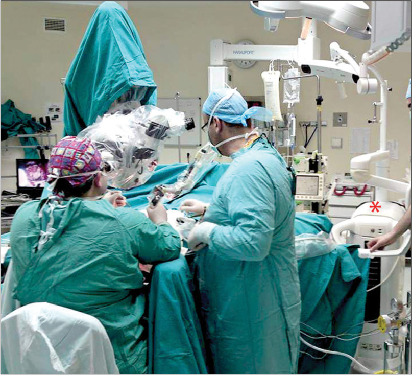
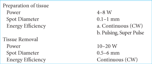
In 26 (90%) of the patients, the joystick was used (f = 125–200 mm); and for three patients, the micromanipulator (f = 200–400 mm) was used. For the preparation and removal of tissues, the CO2 laser was used according to the parameters as described in [ Table 2 ]. Group 1 included 14 patients (48.3%); 13 patients with meningiomas [ Figure 5 ] and one with acoustic neuroma [ Figure 6 and Table 3 ]. From these tumors, six were found to be at the skull base and four were found next to vital structures. Nine of the tumors had a solid and fibrous consistency. The characteristics of this group that contribute to the large benefit provided by the use of the CO2 laser were as follows: the creation of a relatively dry dissection plane with remarkable accuracy. The achievement of surgery “from a distance” with the combined use of the micromanipulator reduced the trauma when accessing the affected area. There was a “cleaner” surgical field due to the fact that a small number of surgical tools were used. In addition, the surgery was finished in less time in relation to surgeries using only bipolar diathermy. Total removal of the tumor was accomplished in 13 (12 meningiomas and the one case of acoustic neuroma) patients (92.9%) and in 1 (7.1%) patient only partial removal of the meningioma was achieved. In a 4-year follow-up period, the 12 totally resected meningiomas and the one acoustic neuroma did not show any evidence of recurrence and the small residuum of the one partially resected meningioma did not show any evidence of progress in the serial MRI controls. Group 2 included six patients (20.7%) all of who had metastatic tumors [ Figure 7 and Table 3 ]. Two of these metastases were located in the cerebellum, four were found to be in the hemispheres, and one was near vital structures. Even though a relatively bloodless dissection plane was achieved in this group, the duration of the surgery was increased without relative benefit with regard to total tumor removal when compared to the use of bipolar diathermy. Total removal of the tumors was possible for 100% of the patients (n = 6), even though the CO2 laser offered a moderate amount of assistance in this group. Group 3 included nine patients (31%); six with glioblastomas and three with astrocytomas [ Table 3 ]. Eight of these tumors had considerable vascularity and one was found to be near vital structures. In this group, the CO2 laser offered limited surgical assistance due to the fact that there was recurring hemorrhaging in the dissection plane and frequent alternation between the laser and bipolar diathermy. The result was an increased surgical time during which the laser was cumbersome and inconvenient. The contrast-enhanced MRI of the brain within 48 h after resection of the glioblastomas and the astrocytomas showed a gross total resection. In a 4-year follow-up period, the mean overall survival of the glioblastoma patients, who all received adjuvant radio-chemotherapy according to Stupp protocol, was approximately 13 months demonstrating no difference from the cases where the tumor was resected with conventional microsurgery using the bipolar diathermy. Overall, there were no complications from the use of CO2 laser in any of the procedures or technical problems during any of the surgeries.


Table 2:

Parameter settings used.


Figure 5:

Utilization of the CO2 laser in the microsurgical resection of a meningioma. The red focusing dot is visible in the center of the image.


Figure 6:

Intraoperative image during microsurgical resection of an acoustic neuroma with the CO2. Here, almost half of the tumor is already sublimated – resected. The black arrow shows the red focusing dot of the CO2 laser cutting the arachnoidal adhesions.


Table 3:

Characteristics of Group 1, 2, and 3.


Figure 7:

Microsurgical resection of a metastasis with the CO2 laser. Note: the generous use of wet cottons (yellow asterisk) to reduce reflective surfaces.


DISCUSSION

The CO2 laser emits an invisible beam of radiation in the field of infrared light, with a wavelength of 10.6 μm, which is absorbed to a great degree by molecules of water[ 23 ] in the interstitial space of the brain tissue. The mechanism by which the CO2 laser functions is by the change of the energy, which is absorbed by the molecules of water, into heat, which results in the rapid local temperature increase.[ 1 , 2 ] The above change produces the coagulation of proteins, necrosis, evaporation of water molecules, charring, and ablation of the tissue.[ 1 , 2 ] When the beam is focused, cutting grade sublimation is achieved like a “light knife.”[ 25 , 26 ] This provides precision in handling and reduced thermal trauma to the surrounding healthy tissue, as opposed to when the beam is not focused and stromatic sublimation occurs. The use of focused beam on an artery with a diameter bigger than 0.3 mm should be avoided due to the danger of wall perforation.[ 11 ] The use of the CO2 laser has benefits during neurosurgical operations such as the combination of sublimation and slight hemostasis during sectioning. In addition, it is a tool that utilizes “non-contact” and “surgery from a distance” methods. These have the advantage of minimizing mechanical trauma and produce modest thermal damage to adjacent tissue, resulting in a reduction of postsurgical edema. The CO2 laser provides speed in debulking of the tumor,[ 13 ] and accesses to hard-to-reach places such as the skull base and areas near vital structures, with good visibility of the surgical field and allows for subtle maneuvers. The use of the micromanipulator allows for an even greater increase in precision due to the fact that the axis of the beam is in line with the surgeon’s axis of visibility.[ 6 ] Thus, the laser is particularly useful for removal of meningiomas and other fibrous lesions that are located at the skull base and less useful for the removal of gliomas. Flexible CO2 laser fiber combines the advantages of this surgical tool with more ergonomy in comparison to the device with articulated arm system used in our department although the cost of the consumable laser fiber of approximately 1500$ may make its use prohibitive when resources are limited.[ 4 , 12 , 23 ] Consiglieri et al. described the use of CO2 laser in resection of cavernomas and performing corticectomies in eloquent neural structures such as brainstem, thalamus, and spinal cord.[ 5 ] Choudhri et al. reported 2015 the first utilization of the CO2 laser in corpus callosotomy in pediatric population.[ 4 ] Zhong et al. published an experimental study showing the suitability of CO2 laser in soldering autologous dural graft in reconstructing dural defects in minipigs with no evidence of thermal injury.[ 27 ] Application of the last can save precious time when large duroplasty is required after resection of large meningiomas where the CO2 laser can also be used in the tumor resection phase. In contrast to the allure of the above benefits, there are disadvantages of the laser that cannot be ignored. There is no hemostatic ability when used on a vessel with a lumen that has a diameter larger than 1 mm (up to 0.5 mm for arteries and up to 1 mm for veins).[ 6 , 11 ] In addition, the surgeon uses the device, as well as the personnel assisting in the surgery require special training. Furthermore, there is a risk of accidents when using the laser and therefore it requires the installation of special fire safety measures. Finally, the laser also has a significant monetary cost regarding both its acquisition and maintenance, which further adds to the disadvantages of the device. Due to its better coagulation properties and less absorption rate in water, the Nd: YAG laser is considered to be more suitable than the CO2 laser in resection of highly vascularized lesions and in intraventricular laser surgery.[ 7 , 11 , 18 ] On the basis on the findings presented above, we conclude that the CO2 laser is neither a “panacea” nor a “surgical technique,” but rather a valuable auxiliary surgical tool that combines the abilities of sublimation and coagulation. In no way does it replace dipolar diathermy or any other microsurgical tools. In our case series, the use of CO2 laser proved to be a valuable asset in resecting fibrous tumors such as meningiomas and acoustic neuromas, benefiting in terms of short operating time, clean operating field, and minimal thermal and mechanical damage to surrounding tissue. In cases of metastatic tumors especially in those with somehow harder consistency, CO2 laser and bipolar diathermy exhibited comparable efficacy. In resection of softer tumors such as gliomas, the CO2 laser failed to demonstrate superiority to the bipolar diathermy mainly due to hemorrhages that could not be managed by laser alone necessitating the frequent utilization of the bipolar diathermy. There were some limitations to our study: (a) the criteria describing the feasibility of tumor resection with this method were evaluated empirically by one surgeon, therefore, causing a possible reproducibility bias. However, not all the faculty members were familiar with the CO2 laser or had the special training/certification to use laser medical devices. Therefore, the performing of the operations and the evaluation of the usefulness were limited to the senior author to preserve homogeneity. (b) The brief duration and small patient cohort. Following 2011 the use of the laser became gradually less frequent, restricted to selected cases of meningiomas and acoustic neuromas, as despite the significant assistance of the CO2 laser in resection of these fibrous tumors, a substantial advantage in the vast majority of the tumor operations compared to conventional methods was not observed. Consequently, not all the consultants felt its use to be necessary. In addition, the lack of specialized neurosurgical nursing staff, made the process of preparing the operating theatre to a “laser compatible” state, somewhat laborious. Furthermore, given the financial crisis in Greece and subsequent underfunding of the public health system, the operating and maintenance costs became prohibitive so that in combination with the aforementioned factors, we were able to use the CO2 laser regularly only in the time period between 2009 and 2011. Overall, our data suggest that the CO2 laser is a valuable tool in the armamentarium for brain tumor surgery for fibrous lesions. The amount of the literature on the use of CO2 laser in brain tumor surgery of the past 10 years has become considerably sporadic compared with its “golden era” from the late 70s until early 90s. The introduction of the flexible CO2 laser fiber may be able to revive its more frequent employment in neurosurgery as it provides more ergonomy.

CONCLUSION

The CO2 laser is a versatile tool that combines the abilities of tissue vaporization, cutting and in a lesser degree of coagulation with minimal thermal and no mechanical trauma to the surrounding tissue. Its uttermost advantage in brain tumor surgery is exhibited in resection of brain tumors with fibrous and hard consistency. The high acquisition and maintenance cost may restrict its use in resource-limited settings and cannot replace bipolar diathermy. The newest generation of flexible CO2 laser fiber provides more ergonomy and unlocks new perspectives of its utilization in neurosurgery.

Declaration of patient consent

Patient’s consent not required as patients identity is not disclosed or compromised.

Financial support and sponsorship

Nil.

Conflicts of interest

There are no conflicts of interest.

Acknowledgments

The authors thank Mrs. Sophia Coelho for her technical support in the editing of the images.

References

1. Allemann IB, Kaufman J. Laser principles. Curr Probl Dermatol. 2011. 42: 7-23

2. Andersen K. Laser technology--a surgical tool of the past, present, and future. AORN J. 2003. 78: 794-802

3. Cerullo LJ, Burke LP. Use of the laser in neurosurgery. Surg Clin North Am. 1984. 64: 995-1000

4. Choudhri O, Lober RM, Camara-Quintana J, Yeom KW, Guzman R, Edwards MS. Carbon dioxide laser for corpus callosotomy in the paediatric population. J Neurosurg Pediatr. 2015. 15: 321-7

5. Consiglieri GD, Killory BD, Germain RS, Spetzler RF. Utility of the CO2 laser in the microsurgical resection of cavernous malformations. World Neurosurg. 2013. 79: 714-8

6. Deruty R, Pelissou-Guyotat I, Mottolese C, Amat D. Routine use of the CO2 laser technique for resection of cerebral tumours. Acta Neurochir (Wien). 1993. 123: 43-5

7. Devaux BC, Joly LM, Page P, Nataf F, Turak B, Beuvon F. Laser-assisted endoscopic third ventriculostomy for obstructive hydrocephalus: technique and results in a series of 40 consecutive cases. Lasers Surg Med. 2004. 34: 368-78

8. Einstein A. Zur Quantentheorie der Strahlung [On the Quantum Theory of Radiation]. Phys Z. 1917. 18: 121-8

9. Gamache FW, Morgello S. The histopathological effects of the CO2 versus the KTP laser on the brain and spinal cord: A canine model. Neurosurgery. 1993. 32: 100-4

10. Graudenz K, Raulin C. Von einsteins quantentheorie zur modernen lasertherapie. Historie des lasers in der dermatologie und ästhetischen medizin [From Einstein’s quantum theory to modern laser therapy. The history of lasers in dermatology and aesthetic medicine]. Hautarzt. 2003. 54: 575-82

11. Jain KK. Lasers in neurosurgery: A review. Lasers Surg Med. 1983. 2: 217-30

12. Killory BD, Chang SW, Wait SD, Spetzler RF. Use of flexible hollow-core CO2 laser in microsurgical resection of CNS lesions: Early surgical experience. Neurosurgery. 2010. 66: 1187-92

13. Koivukangas J, Heikkinen ER. Preliminary experiences with CO2laser and ultrasonic aspirator in brain surgery. Ann Clin Res. 1986. 18: 85-95

14. Lanzafame RJ. Laser safety programs in general surgery. J Laser Appl. 1994. 6: 111-4

15. Lovrić D, Negovetić L, Lupret V, Vidović D, Gnjidić Z. Evaluacija primjene CO2lasera na 134 bolesnika s ekspanzivnim procesima CNS-a [Evaluation of the use of the CO2 laser in 134 patients with expansive processes in the central nervous system]. Acta Chir Iugosl. 1989. 36: 27-43

16. Marks AJ, Teichman JM. Lasers in clinical urology: State of the art and new horizons. World J Urol. 2007. 25: 227-33

17. Massad M, LoCicero J, Matano J, Oba J, Greene R, Gilbert J. Endoscopic thoracic sympathectomy: evaluation of pulsatile laser, non-pulsatile laser, and radiofrequency-generated thermocoagulation. Lasers Surg Med. 1991. 11: 18-25

18. Oh H, Kim J.editors. Clinical application of CO2 laser. CO2 Laser - Optimisation and Application. London, UK: IntechOpen; 2012. p.

19. Oros-Peusquens AM, Loução R, Abbas Z, Gras V, Zimmermann M, Shah NJ. A single-scan, rapid whole-brain protocol for quantitative water content mapping with neurobiological implications. Front Neurol. 2019. 10: 1333

20. Palanker D. Evolution of concepts and technologies in ophthalmic laser therapy. Annu Rev Vis Sci. 2016. 2: 295-319

21. Patel CK. High power carbon dioxide lasers. Sci Am. 1968. 219: 22-3

22. Rebeiz EE. Lasers in otorhinolaryngology-head and neck surgery. J Med Liban. 1994. 42: 242-9

23. Ryan RW, Wolf T, Spetzler RF, Coons SW, Fink Y, Preul MC. Application of a flexible CO(2) laser fibre for neurosurgery: Laser-tissue interactions. J Neurosurg. 2010. 112: 434-43

24. Saleh B, Al-Amri M, El-Gomati M, Zubairy M.editors. The laser. Optics in Our Time. Cham: Springer; 2016. p.

25. Verschueren RC, Koudstaal J, Oldhoff J. The carbon dioxide laser; some possibilities in surgery. Acta Chir Belg. 1975. 74: 197-204

26. Verschueren RJ, Oldhoff J. The carbon-dioxide laser. A new surgical tool. Arch Chir Neerl. 1975. 27: 199-207

27. Zhong H, Wang Z, Yang Z, Zhao F, Wang B, Liu P. CO2 laser soldering for the reconstruction of dural defects in the minipig model. Turk Neurosurg. 2016. 26: 240-5

Leave a Reply

Your email address will not be published. Required fields are marked *