- Department of Neurosurgery, Saitama Medical Center, Saitama Medical University, Kawagoe, Saitama, Japan.
DOI:10.25259/SNI_734_2020
Copyright: © 2020 Surgical Neurology International This is an open-access article distributed under the terms of the Creative Commons Attribution-Non Commercial-Share Alike 4.0 License, which allows others to remix, tweak, and build upon the work non-commercially, as long as the author is credited and the new creations are licensed under the identical terms.How to cite this article: Shunya Hanakita, Soichi Oya, Toru Matsui. Trigeminal neuralgia caused by an arachnoid cyst in Meckel’s cave: A case report and literature review. 10-Feb-2021;12:45
How to cite this URL: Shunya Hanakita, Soichi Oya, Toru Matsui. Trigeminal neuralgia caused by an arachnoid cyst in Meckel’s cave: A case report and literature review. 10-Feb-2021;12:45. Available from: https://surgicalneurologyint.com/?post_type=surgicalint_articles&p=10587
Abstract
Background: We present a rare case of trigeminal neuralgia (TN) caused by an arachnoid cyst (AC) in Meckel’s cave (MC).
Case Description: A 35-year-old man presented with facial pain in the left maxillary and mandibular regions. Since the initial magnetic resonance (MR) imaging showed no apparent offending vessels or tumors, the patient was diagnosed with idiopathic TN, for which carbamazepine was initially effective. When his pain worsened, he was referred to our hospital. A slightly asymmetric shape of MC and distorted course of the trigeminal nerve was confirmed on the initial and repeat MR images. His pain was characterized as electric-shock-like pain, which was triggered by touching the face. Under the tentative diagnosis of an AC confined to MC compressing the trigeminal nerve, the exploration of MC through suboccipital craniotomy was performed. Intraoperatively, the AC was identified in the rostral portion of MC. The indentation of the trigeminal nerve was also observed at the orifice of MC, indicating severe compression by the AC. The wall of the AC was fenestrated. The patient’s pain was relieved immediately after surgery. Postoperative MR images showed that the course of the trigeminal nerve was straightened. Although our literature review found five similar cases, the size of the AC was the smallest in our case.
Conclusion: Although it is rare, the AC confined to MC can cause TN. The findings of this study emphasize the importance of evaluating subtle radiological findings of compression on the trigeminal nerve in cases of TN seemingly without neurovascular compression.
Keywords: Arachnoid cyst, Meckel’s cave, Trigeminal neuralgia
INTRODUCTION
Trigeminal neuralgia (TN) is known as facial pain with a wide range of severity. Although it can be idiopathic, TN also occurs secondary to the compression of the trigeminal nerve by vascular structures or tumor masses at the cerebellopontine angle extending to areas adjacent to the trigeminal nerve. It is widely accepted that microvascular decompression confers favorable surgical outcomes for TN with vascular compression of the trigeminal nerve.[
In the present case, we present a rare case of TN caused by an arachnoid cyst (AC) confined to Meckel’s cave (MC). Preoperative magnetic resonance (MR) imaging demonstrated a severe torsion of the trigeminal nerve due to the AC. We emphasize the importance of careful evaluation of the subtle laterality of MC and the distorted course of the trigeminal nerve in cases without apparent vascular compression or tumors.
CASE PRESENTATION
Patient history and examinations
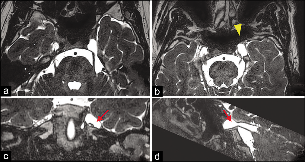
A 35-year-old man presented with facial pain of the left cheek that was similar to that of TN with neurovascular compression, that is, electric-shock like pain with a trigger point. Initial MR images showed no apparent vascular compression of the trigeminal nerve. Medical treatment with carbamazepine allowed gradual improvement of TN; however, the pain worsened within 1 year and remained for 6 months. He was, therefore, referred to our department for further investigation. Repeat constructive interference in steady state MRI revealed slightly expanded MC on the left side with a diameter of 11 × 11 mm compared to that on the right side (6 × 7 mm) [
Figure 1:
Preoperative constructive interference in steady state MRI. (a) No visible vascular compression on the trigeminal nerve. (b) Enlarged Meckel’s cave (MC, yellow arrow head) detected with the isointense cerebrospinal fluid. (c and d) Stretched trigeminal nerve detected at the floor of MC (red arrow), on coronal and sagittal view.
Surgery
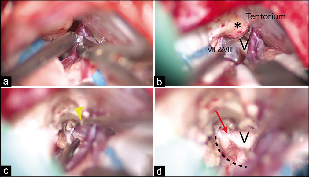
A standard retrosigmoid approach was performed. The trigeminal nerve was found behind the superior petrous vein and its tributaries [
Figure 2:
Intraoperative photographs. (a) The arachnoid sleeve enveloping the petrosal vein was cut. (b) The suprameatal tubercle (SMT; asterisk) obstructed the trigeminal nerve root (v) in direction to MC. (c) After resection of the SMT, the transparent wall of arachnoid cyst (yellow arrow head) was detected above the trigeminal nerve and fenestrated using a micro dissector. (d) The trigeminal nerve was transposed rostrally, an indentation (red arrow) was detected.
Postoperative course
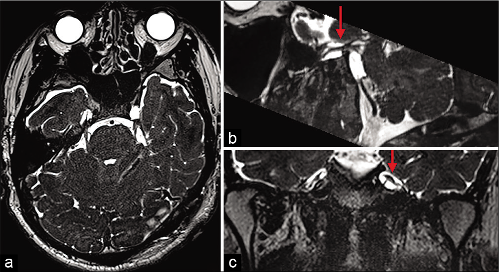
The patient’s facial pain resolved immediately after the surgery. Postoperative MR imaging revealed that the course of the trigeminal nerve in MC was straightened with no compression [
DISCUSSION
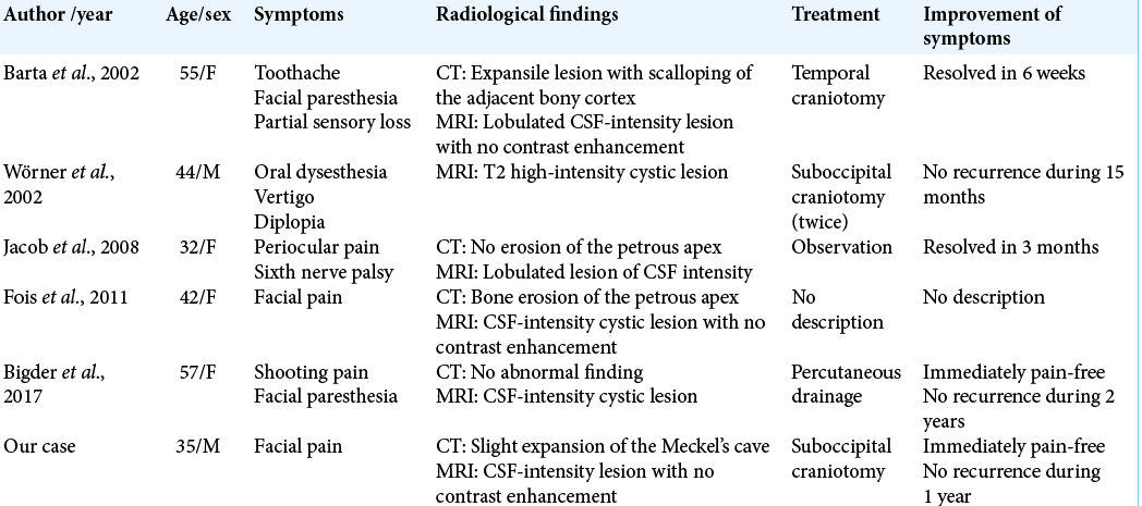
We describe a rare case of TN caused by an AC confined to MC. To date, several cases of ACs affecting MC have been reported in the literature [
CONCLUSION
Although such cases are rare, it is worth considering that TN can be caused by a small AC confined to MC. When there is subtle asymmetry of MC, the torsion or constriction of the trigeminal nerve inside MC might be indicative of this condition. Our report indicates the importance of careful evaluation of preoperative MR images for subtle evidence of compression on the trigeminal nerve in cases of TN seemingly without neurovascular compression.
Declaration of patient consent
The authors certify that they have obtained all appropriate patient consent.
Financial support and sponsorship
Nil.
Conflicts of interest
There are no conflicts of interest.
References
1. Barker FG, Jannetta PJ, Bissonette DJ, Larkins MV, Jho HD. The long-term outcome of microvascular decompression for trigeminal neuralgia. N Engl J Med. 1996. 334: 1077-84
2. Batra A, Tripathi RP, Singh AK, Tatke M. Petrous apex arachnoid cyst extending into Meckel’s cave. Australas Radiol. 2002. 46: 295-8
3. Bigder MG, Helmi A, Kaufmann AM. Trigeminal neuropathy associated with an enlarging arachnoid cyst in Meckel’s cave: Case report, management strategy and review of the literature. Acta Neurochir. 2017. 159: 2309-12
4. Fois P, Lauda L. Bilateral Meckel’s cave arachnoid cysts with extension to the petrous apex in a patient with a vestibular schwannoma. Otol Neurotol. 2011. 32: e36-7
5. Hanakita J, Kondo A. Serious complications of microvascular decompression operations for trigeminal neuralgia and hemifacial spasm. Neurosurgery. 1988. 22: 248-352
6. Ishikawa M, Nishi S, Aoki T, Takase T, Wada E, Ohwaki H. Operative findings in cases of trigeminal neuralgia without vascular compression: proposal of a different mechanism. J Clin Neurosci. 2002. 9: 200-4
7. Jacob M, Gujar S, Trobe J, Gandhi D. Spontaneous resolution of a Meckel's cave arachnoid cyst causing sixth cranial nerve palsy. J Neuroophthalmol. 2008. 28: 186-91
8. Kimura T, Sameshima T, Morita A. Trigeminal neuralgia caused by a fibrous ring around the nerve. J Neurosurg. 2012. 116: 741-2
9. Lee A, McCartney S, Burbidge C, Raslan AM, Burchiel KJ. Trigeminal neuralgia occurs and recurs in the absence of neurovascular compression. J Neurosurg. 2014. 120: 1048-54
10. Panczykowski DM, Jani RH, Hughes MA, Sekula RF. Development and evaluation of a preoperative trigeminal neuralgia scoring system to predict long-term outcome following microvascular decompression. Neurosurgery. 2019. 87: 71-9
11. Revuelta-Gutiérrez R, López-González MA, SotoHernández JL. Surgical treatment of trigeminal neuralgia without vascular compression: 20 years of experience. Surg Neurol. 2006. 66: 32-6
12. Sindou MP, Chiha M, Mertens P. Anatomical findings observed during microsurgical approaches of the cerebellopontine angle for vascular decompression in trigeminal neuralgia (350 cases). Stereotact Funct Neurosurg. 1994. 63: 203-7
13. Toda H, Iwasaki K, Yoshimoto N, Miki Y, Hashikata H, Goto M. Bridging veins and veins of the brainstem in microvascular decompression surgery for trigeminal neuralgia and hemifacial spasm. Neurosurg Focus. 2018. 45: E2
14. Wörner BA, Noll M, Rahim T, Fink U, Oeckler R. Recurrent arachnoid cyst of Meckel’s cave mimicking a brain stem ischaemia. Report of a rare case. Zentralbl Neurochir. 2003. 64: 76-9


