Tools

Aqueel Hussain Pabaney, Paul A. Mazaris, Max K. Kole, Kevin A. Reinard
  1. Department of Neurosurgery, Henry Ford Hospital, Detroit, MI, USA
  2. Department of Neurosurgery, Hartford Hospital, Hartford, CT, USA

Correspondence Address:
Aqueel Hussain Pabaney
Department of Neurosurgery, Henry Ford Hospital, Detroit, MI, USA

DOI:10.4103/2152-7806.161239

Copyright: © 2015 Pabaney AH. This is an open-access article distributed under the terms of the Creative Commons Attribution License, which permits unrestricted use, distribution, and reproduction in any medium, provided the original author and source are credited.

How to cite this article: Pabaney AH, Mazaris PA, Kole MK, Reinard KA. Endovascular management of fusiform aneurysm of anterior temporal artery: Technical report. Surg Neurol Int 20-Jul-2015;6:119

How to cite this URL: Pabaney AH, Mazaris PA, Kole MK, Reinard KA. Endovascular management of fusiform aneurysm of anterior temporal artery: Technical report. Surg Neurol Int 20-Jul-2015;6:119. Available from: http://surgicalneurologyint.com/surgicalint_articles/endovascular-management-of-fusiform-aneurysm-of-anterior-temporal-artery-technical-report/

Date of Submission
25-Jan-2015

Date of Acceptance
27-May-2015

Date of Web Publication
20-Jul-2015

Abstract

Background:The treatment of a rare, nontraumatic, fusiform aneurysm of the anterior temporal artery (ATA) via endovascular techniques is presented, and procedural nuances are highlighted.

Methods:We performed a retrospective chart review and collected demographic and clinical data on the patient presented here; procedural details were extracted from operative notes.

Results:Following successful balloon test occlusion (BTO) of the ATA, complete coil embolization of the ATA, and its associated fusiform aneurysm was performed. Postprocedurally, the patient did not suffer any adverse neurological sequelae.

Conclusion:Selective BTO of intracranial branch vessels is safe, technically feasible, and could serve as a useful technical tool in the treatment of complex, fusiform intracranial aneurysms.

Keywords: Anterior temporal artery, balloon test occlusion, coil occlusion, fusiform aneurysm

INTRODUCTION

Approximately 20% of all clinically significant intracranial aneurysms involve the middle cerebral artery (MCA).[ 7 ] While most aneurysms that originate at the MCA bifurcation or trifurcation have a saccular geometry, some MCA aneurysms may exhibit a fusiform morphology and incorporate not only the proximal MCA trunk but also major MCA branches. In contrast to saccular aneurysms, fusiform aneurysms represent a distinct subset of intracranial aneurysms with unique underlying pathological features, hemodynamic forces, anatomical distribution, as well as natural history that governs their treatment. There have been several reports of fusiform aneurysms of various MCA segments and the various modalities utilized to treat them; however, a paucity of reports exists with a focus on spontaneous, fusiform anterior temporal artery (ATA) aneurysms.[ 1 14 ] Here, we present a case of an incidental, fusiform ATA aneurysm that was coiled and highlight the endovascular techniques that resulted in excellent radiologic and clinical outcome.

CASE PRESENTATION

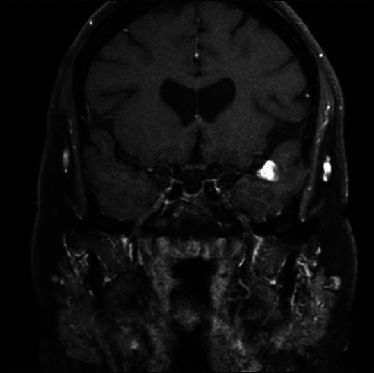
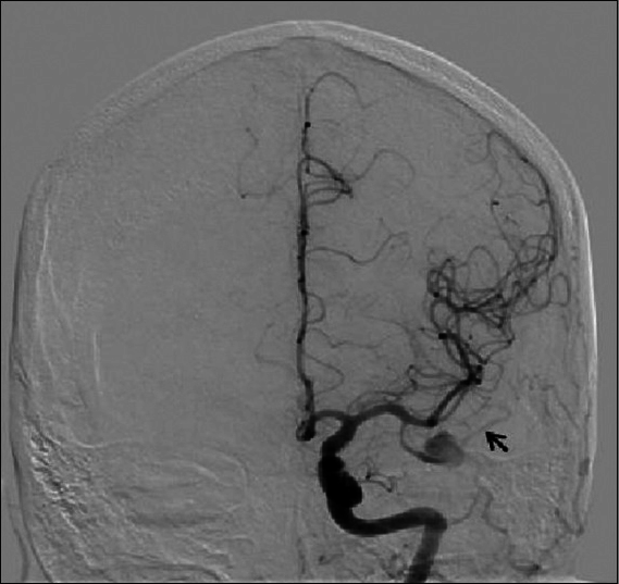
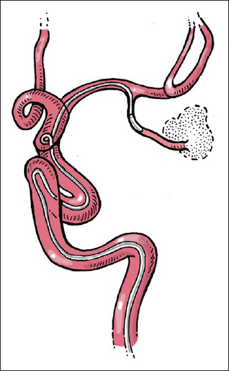
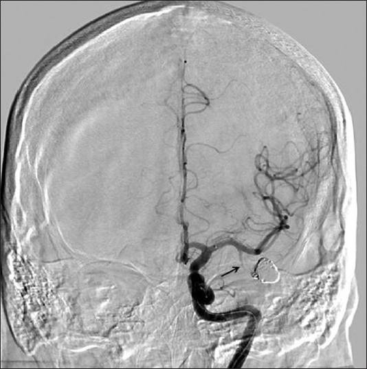
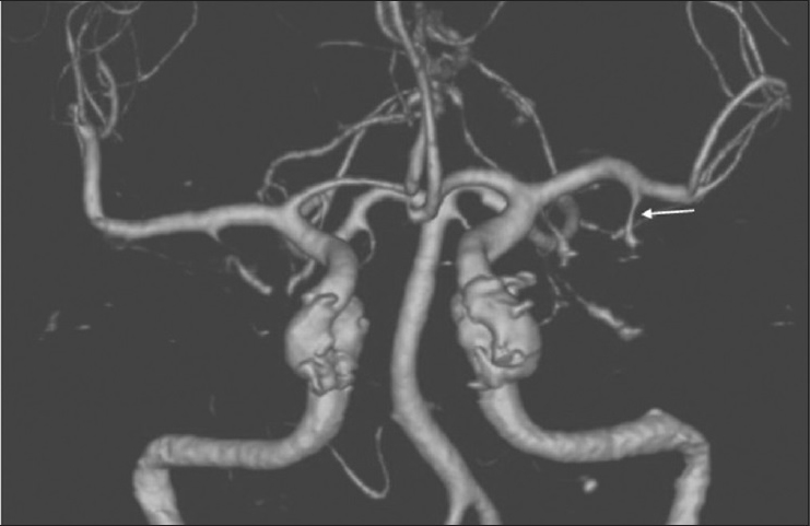
A 37-year-old, right-handed female with no significant past medical history presented to our Emergency Department with several days of holocranial headaches. Her neurological examination was benign with no evidence of meningismus. Computed tomography (CT) of the head revealed a hyperdense mass in the left temporal lobe immediately inferior to the Sylvian fissure. Magnetic resonance imaging/arteriography demonstrated a partially thrombosed aneurysm in an atypical location [ Figure 1 ]. Digital subtraction angiography with conscious sedation revealed a large fusiform and partially thrombosed ATA aneurysm from which emanated a small vessel [ Figure 2 ]. Balloon occlusion of the ATA was performed using Hyperform Occlusion balloon system (ev3/Covidien, Irvine, California, USA) for approximately 30 min and the patient's systolic blood pressure was lowered by 10%, from 130 to 115 mmHg [ Figure 3 ]. The neurological evaluation was conducted every 2–3 min for the duration of the balloon test occlusion (BTO). Because the patient tolerated the BTO of the ATA without any changes in her neurological status, the senior author (M.K.K.) proceeded and completely occluded the fusiform ATA aneurysm, as well as the distal portion of the ATA by deploying multiple Guglielmi detachable coils in the aneurysm while the patient remained awake using protocols described by Qureshi et al. [ Figure 4 ].[ 9 ] Postprocedurally, the patient was observed in the neurosurgical Intensive Care Unit overnight and was discharged home the following day with no new neurological deficits. A CT angiogram performed 12 months after the coiling procedure confirmed stable aneurysm and parent vessel occlusion with no evidence of de novo aneurysms [ Figure 5 ].


Figure 1

(original): Pretreatment magnetic resonance imaging (MRI). Coronal view of T1-weighted, postcontrast MRI brain, demonstrating contrast enhancement of partially thrombosed left anterior temporal artery aneurysm, just inferior to the sylvian fissure


Figure 2

(original): Precoiling angiogram. An anterior-posterior view of left internal carotid artery injection demonstrates the fusiform aneurysm of left anterior temporal artery. Vessel exiting the aneurysm is also visualized (black arrow)


Figure 3

(original): Balloon test occlusion. Artist's depiction of the placement and inflation of balloon in the anterior temporal artery while performing BTO


Figure 4

(original): Postcoiling angiogram. Left internal carotid artery injection is demonstrating complete occlusion of the aneurysm and the distal anterior temporal artery (ATA) following balloon occlusion test. Contrast stasis is seen in proximal ATA (black arrow)


Figure 5

(original): Follow-up imaging. Computed tomography angiogram performed 1-year postoperatively displays stable aneurysm occlusion with persistent filling of proximal segment of anterior temporal artery (arrow) with no evidence of recanalization or new aneurysms


DISCUSSION

The MCA is the largest and most complex of the three major cerebral arteries.[ 10 ] It is generally divided into four segments: M1 – The segment between the internal carotid artery (ICA) bifurcation and the genu; M2 – The segments that run over the deep insular surface; M3 – The segments that traverse the opercular surface of the sylvian fissure; and M4 – The cortical branches. The M1 segment gives rise to multiple lenticulostriate vessels from its posterior-superior surface while an anterior temporal branch often takes off from its anterior-interior surface. According to cadaveric studies, the ATA arises from the proximal M1 in a common trunk shared with the temporal-polar artery approximately 79% of the times and could have a variable course.[ 12 ] In the past, the ATA has been utilized for bypassing complex MCA bifurcation aneurysms[ 2 ] and has been successfully embolized for the treatment of skull base meningiomas.[ 15 ] More recently, functional studies have demonstrated that strokes in the ATA or temporal-polar artery could produce semantic errors in auditory comprehension and object naming tasks.[ 13 ] Therefore, the selective sacrifice of the ATA may not be as inconsequential as once thought.

With the advent of sophisticated neuroimaging modalities, intracranial fusiform aneurysms are reported with increasing frequency. To date, most studies have focused on fusiform aneurysms of the vertebrobasilar system. As such, the pathogenesis, clinical features, radiographic features, and treatment options available for fusiform aneurysms of the posterior circulation are well documented.[ 6 ] Fusiform aneurysms involving the anterior circulation, however, are rare. Most tend to arise from the MCA and predominantly occur in children and adolescents.[ 16 ] Various etiologies such as cystic medial necrosis, fibromuscular dysplasia, moya-moya disease, atherosclerosis, homocystinuria, and intimal fibroelastic abnormal have been implicated in the pathogenesis of similar non-traumatic aneurysms of ICA. Curiously, all of the aforementioned pathological processes were absent in our patient.

It has been hypothesized that spontaneous fusiform MCA aneurysms may develop as a result of arterial dissection with intramural hemorrhage between the layers of the intima and the media.[ 4 ] Patients can present either with ischemic symptoms or with subarachnoid hemorrhage with the latter group demonstrating better recovery and long-term neurological outcomes, according to some reports.[ 3 ] No specific treatment guidelines exist for the management of fusiform aneurysms of the anterior circulation. However, various authors have reported outcomes based on a wide spectrum of treatment options that includes observation, surgical treatment (clip reconstruction, ligation and excision, revascularization), and endovascular treatment (stent or balloon assisted coiling or parent vessel occlusion). Only two reports that focus on the surgical treatment of a single fusiform[ 14 ] and a saccular[ 1 ] aneurysm of ATA are available. To date, endovascular treatment of an ATA aneurysm has not been reported. Our patient was a young woman with an incidental, partially thrombosed, fusiform, left ATA aneurysm. Given her young age and the left-sided location, we recommended endovascular treatment to minimize the surgical morbidity associated with lesions involving the dominant hemisphere.

Advancements in endovascular neurosurgery and the development of modern stents and coil material have provided clinicians with innovative and minimally invasive treatment modalities for the management of disparate and complex intracranial vascular pathologies. Given the small caliber and fragile nature of the dissected vessel involved, endovascular reconstruction techniques using flow-diverting stents were deemed not feasible. In this case, the senior author (M.K.K.) performed a BTO of the ATA prior to aneurysm occlusion and parent vessel sacrifice. BTO of the ICA has a reported positive predictive value that approaches 90%[ 8 ] and could help endovascular neurosurgeons determine whether or not a patient is able to tolerate permanent occlusion for an extra- or intracranial vessel. Superselective BTO of intracranial vessels has been performed in the past prior to parent vessel sacrifice with satisfying results.[ 11 ] However, great care should be taken when selecting a diseased vessel for BTO as the fragile nature of the vessel wall may increase the risk of rupture and catastrophic neurologic compromise. Eckard et al. made use of selective injection of 30 mg of Amytal prior to performing parent vessel occlusion in the management of peripheral intracranial aneurysms.[ 5 ] However, we elected not to use Amytal injection because the technique could grossly overestimate the risk of neurological deficit following vessel occlusion. In contrast, BTO may accurately evaluate the efficacy of collateral circulation and the likelihood of neurologic compromise in the event of parent vessel sacrifice. In addition, contrast stasis– which is a reliable indicator of sluggish distal flow that may promote the pial collateral formation and, therefore, decrease the likelihood of infarction following vessel occlusion was noted on angiography. Since our patient tolerated the test for 30 min with no signs of neurological deterioration, the fusiform ATA aneurysm and the parent vessel were occluded with coils. Due to proximity to the main trunk of the MCA, we elected to use detachable coils for precise embolization instead of embolic glue, which carries a higher complication risk secondary to glue reflux into the parent vessel. If the patient failed the BTO, both the patient and the senior author (M.K.K.) were prepared for microsurgical trapping of the aneurysm and bypass.

CONCLUSION

We present a rare case of an incidental, fusiform aneurysm of ATA that was treated by endovascular coil occlusion and resulted in no neurological sequelae after patient displayed tolerance for balloon occlusion. Superselective BTO could serve as a powerful assessment tool prior to parent vessel sacrifice. Large-scale studies are required to determine the long-term safety and applicability of superselective, intracranial BTO to diverse vascular pathologies.

References

1. Asakura K, Tasaki T, Okada K. A case of unruptured anterior temporal artery aneurysm showing pupil-sparing oculomotor palsy. No Shinkei Geka. 1986. 14: 777-82

2. Bederson JB, Spetzler RF. Anastomosis of the anterior temporal artery to a secondary trunk of the middle cerebral artery for treatment of a giant M1 segment aneurysm. Case report. J Neurosurg. 1992. 76: 863-6

3. Chuang MJ, Lu CH, Cheng MH. Management of middle cerebral artery dissecting aneurysm. Asian J Surg. 2012. 35: 42-8

4. Day AL, Gaposchkin CG, Yu CJ, Rivet DJ, Dacey RG. Spontaneous fusiform middle cerebral artery aneurysms: Characteristics and a proposed mechanism of formation. J Neurosurg. 2003. 99: 228-40

5. Eckard DA, O’Boynick PL, McPherson CM, Eckard VR, Han P, Arnold P. Coil occlusion of the parent artery for treatment of symptomatic peripheral intracranial aneurysms. AJNR Am J Neuroradiol. 2000. 21: 137-42

6. Hosoya T, Adachi M, Yamaguchi K, Haku T, Kayama T, Kato T. Clinical and neuroradiological features of intracranial vertebrobasilar artery dissection. Stroke. 1999. 30: 1083-90

7. Locksley HB, Sahs AL, Knowler L. Report on the cooperative study of intracranial aneurysms and subarachnoid hemorrhage. Section II. General survey of cases in the central registry and characteristics of the sample population. J Neurosurg. 1966. 24: 922-32

8. Narayanan S, Singer R, Abruzzo TA, Hussain MS, Powers CJ, Prestigiacomo CJ. Reporting standards for balloon test occlusion. J Neurointerv Surg. 2013. 5: 503-5

9. Qureshi AI, Suri MF, Khan J, Kim SH, Fessler RD, Ringer AJ. Endovascular treatment of intracranial aneurysms by using Guglielmi detachable coils in awake patients: Safety and feasibility. J Neurosurg. 2001. 94: 880-5

10. Rhoton AL. The supratentorial arteries. Neurosurgery. 2002. 51: 53-120

11. Senbokuya N, Kanemaru K, Kinouchi H, Horikoshi T. Giant serpentine aneurysm of the distal anterior cerebral artery. J Stroke Cerebrovasc Dis. 2012. 21: e7-11

12. Tanriover N, Kawashima M, Rhoton AL Jr, Ulm AJ, Mericle RA. Microsurgical anatomy of the early branches of the middle cerebral artery: Morphometric analysis and classification with angiographic correlation. J Neurosurg. 2003. 98: 1277-90

13. Tsapkini K, Frangakis CE, Hillis AE. The function of the left anterior temporal pole: Evidence from acute stroke and infarct volume. Brain. 2011. 134: 3094-105

14. Umeoka K, Shirokane K, Mizunari T, Kobayashi S, Teramoto A. Dissecting aneurysm of the anterior temporal artery: Case report. Neurol Med Chir (Tokyo). 2011. 51: 777-80

15. Waldron JS, Sughrue ME, Hetts SW, Wilson SP, Mills SA, McDermott MW. Embolization of skull base meningiomas and feeding vessels arising from the internal carotid circulation. Neurosurgery. 2011. 68: 162-9

16. Yonas H, Agamanolis D, Takaoka Y, White RJ. Dissecting intracranial aneurysms. Surg Neurol. 1977. 8: 407-15

Leave a Reply

Your email address will not be published. Required fields are marked *