- Department of Anesthesiology, Montefiore Medical Center, NY, United States
- Department of Neurological Surgery, Montefiore Medical Center, NY, United States
- Department of Radiology, Montefiore Medical Center, NY, United States
Correspondence Address:
Merritt D. Kinon
Department of Neurological Surgery, Montefiore Medical Center, NY, United States
DOI:10.25259/SNI-148-2019
Copyright: © 2019 Surgical Neurology International This is an open-access article distributed under the terms of the Creative Commons Attribution-Non Commercial-Share Alike 4.0 License, which allows others to remix, tweak, and build upon the work non-commercially, as long as the author is credited and the new creations are licensed under the identical terms.How to cite this article: David C. Kramer, Adela Aguirre-Alarcon, Reza Yassari, Allan L. Brook, Merritt D. Kinon. Spinal cord ischemia/infarct after cauda equina syndrome from disc herniation – A case study and literature review. 10-May-2019;10:80
How to cite this URL: David C. Kramer, Adela Aguirre-Alarcon, Reza Yassari, Allan L. Brook, Merritt D. Kinon. Spinal cord ischemia/infarct after cauda equina syndrome from disc herniation – A case study and literature review. 10-May-2019;10:80. Available from: https://surgicalneurologyint.com/?post_type=surgicalint_articles&p=9313
Abstract
Background:Spinal cord infarction is rare and occurs in 12/100,000; it represents 0.3%–2% of central nervous system infarcts. Here, we present a patient who developed recurrent bilateral lower extremity paraplegia secondary to spinal cord infarction 1 day after a successful L4-5 microdiscectomy in a patient who originally presented with a cauda equina syndrome.
Case Description:A 56-year-old patient presented with an acute cauda equina syndrome characterized by severe lower back pain, a right foot drop, saddle anesthesia, and acute urinary retention. When the lumbar magnetic resonance imaging (MRI) revealed a large right paracentral lumbar disc herniation at the L4-L5 level, the patient underwent an emergency minimally invasive right-sided L4-5 discectomy. Immediately, postoperatively, the patient regained normal function. However, 1 day later, while having a bowel movement, he immediately developed the recurrent paraplegia. The new lumbar MRI revealed acute ischemia and an infarct involving the distal conus medullaris. Further, workup was negative for a spinal cord vascular malformation, thus leaving an inflammatory postsurgical vasculitis as the primary etiology of delayed the conus medullaris infarction.
Conclusions:Acute neurologic deterioration after spinal surgery which does not neurologically correlate with the operative level or procedure performed should prompt the performance of follow-up MR studies of the neuraxis to rule out other etiologies, including vascular lesions versus infarctions, as causes of new neurological deficits.
Keywords: Complication spine surgery, Disc herniation, Spinal cord infarct, Spinal cord ischemia
INTRODUCTION
Spinal cord infarctions are rare and occur in approximately 12/100,000 patients,[
CASE DESCRIPTION
A 56-year-old patient presented with an acute cauda equina syndrome characterized by severe lower back pain, saddle anesthesia, and acute urinary retention. Neurologic examination showed diffuse weakness in the right leg, more so in the distal leg muscle groups and poor rectal tone.
Medical history was significant for obesity (body mass index = 34.5), type 2 diabetes mellitus, hyperlipidemia, hypertension, peripheral vascular disease (unspecified), obstructive sleep apnea, chronic kidney disease (Stage II thought to be secondary to diabetic nephropathy), and polyarticular arthritis.
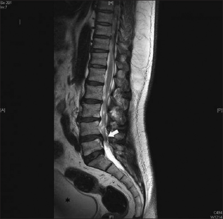
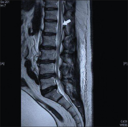
The lumbar magnetic resonance imaging (MRI) demonstrated an acute right L4-L5 paracentral disc herniation contributing to the canal and right lateral recess stenosis [
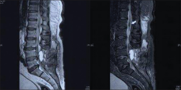
The patient tolerated the procedure well, and postoperatively, his right lower extremity weakness markedly improved. However, 1 day postoperatively, when moving his bowels (e.g., Valsalva maneuver), he acutely developed sudden bilateral lower extremity paralysis. The emergent postoperative lumbar MR showed a subtle increased T2 signal at the L1-L2 level involving the conus medullaris; autoimmune, inflammatory, and hypercoagulation workups proved negative, but the remaining differential diagnoses included ischemia versus posttraumatic/inflammatory changes [
DISCUSSION
Our patient initially presented with a cauda equina syndrome due to an L4-L5 disc herniation. Postoperatively, he acutely neurologically improved. Nevertheless, when having a bowel movement (e.g., acute Valsalva maneuver), he suddenly became paraparetic. When the postoperative MR documented ischemia involving the conus medullaris and reexploration of the prior laminectomy site prove nondiagnostic, the conclusion was that the patient had sustained a conus medullaris infarct. Etiologies of a conus medullaris syndrome variously include neoplasm, autoimmune/inflammatory, degenerative/compressive pathology, arteriovenous malformation (AVM), dural AVF, and infarct.
Demographic parameters for infarcts resulting in conus medullaris syndrome
Typically, patients with spinal/conus medullaris infarctions are males, in their early sixties, whom present with significant motor deficits and dissociated sensory loss.[
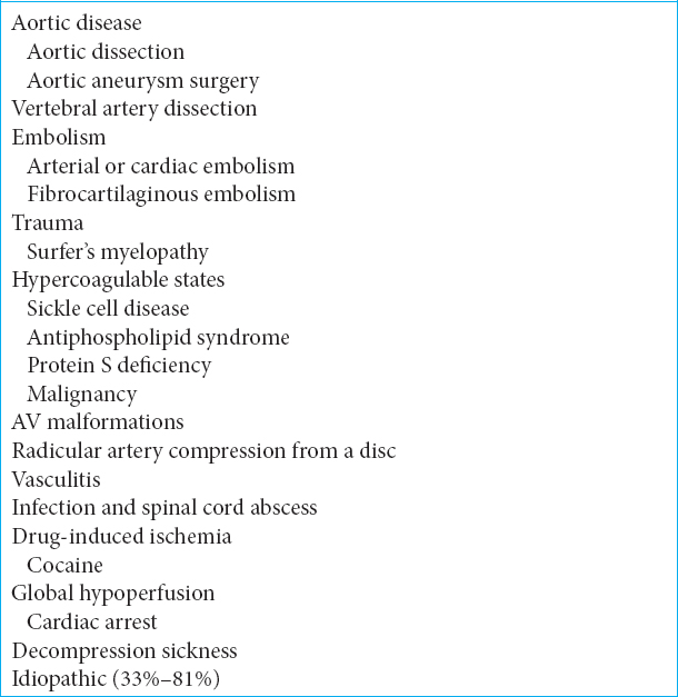
While the cervical spinal cord is the very well vascularized, supply to the midthoracic portion is most fragile. In this section, blood supply is merely provided through the artery of Adamkiewicz. Hypoperfusion, atherosclerosis, iatrogenic occlusion, vasculitis/vasculopathies, thromboembolic occlusion, trauma, infection, or inflammatory conditions have also been associated with spinal cord infarction [
Etiology of spinal cord infarction
The most frequent identifiable etiology of spinal cord infarction is occult spinal vascular malformations including AVM (most commonly found at the level of the conus medullaris) and dural AVF.[
Imaging of conus medullaris infarction
The imaging findings here were classic for spinal infarction; they included early nonspecific edema, cord swelling, and enhancement of a hemorrhagic lesion.[
Postoperative infarction of the conus medullaris
Postoperative conus medullaris infarction is even more rare than de novo infarction. There were just two reports in literature by Stevens and Iovtchev that focused on occult dural AVF acutely diagnosed following lumbar discectomy resulting in acute spinal cord infarction at a site not associated with initial surgery.[
CONCLUSIONS
The patient’s initial symptoms correlated with his lumbar disc herniation. However, his secondary acute onset of paraparesis attributed to a Valsalva maneuver, pointed to an acute vascular event involving the conus medullaris ultimately attributed to an occult AVM with resultant acute ischemia/hemorrhage or inflammatory postsurgical vasculitis.[
Financial support and sponsorship
Nil.
Conflicts of interest
There are no conflicts of interest.
References
1. Atkinson JL, Miller GM, Krauss WE, Marsh WR, Piepgras DG, Atkinson PP. Clinical and radiographic features of dural arteriovenous fistula, a treatable cause of myelopathy. Mayo Clin Proc. 2001. 76: 1120-30
2. Banit DM, Wheeler AH, Darden BV. Recurrent transverse myelitis after lumbar spine surgery:A case report. Spine (Phila Pa 1976). 2003. 28: E165-8
3. Iovtchev I, Hiller N, Ofran Y, Schwartz I, Cohen J, Rubin SA. Late diagnosis of spinal dural arteriovenous fistulas resulting in severe lower-extremity weakness:A case series. Spine J. 2015. 15: e39-44
4. Masson C, Pruvo JP, Meder JF, Cordonnier C, Touzé E, De La Sayette V. Spinal cord infarction:Clinical and magnetic resonance imaging findings and short term outcome. J Neurol Neurosurg Psychiatry. 2004. 75: 1431-5
5. Mull M, Nijenhuis RJ, Backes WH, Krings T, Wilmink JT, Thron A. Value and limitations of contrast-enhanced MR angiography in spinal arteriovenous malformations and dural arteriovenous fistulas. AJNR Am J Neuroradiol. 2007. 28: 1249-58
6. Nasr DM, Rabinstein A. Spinal cord infarcts:Risk factors, management, and prognosis. Curr Treat Options Neurol. 2017. 19: 28-
7. Novy J, Carruzzo A, Maeder P, Bogousslavsky J. Spinal cord ischemia:Clinical and imaging patterns, pathogenesis, and outcomes in 27 patients. Arch Neurol. 2006. 63: 1113-20
8. Ropper AH, Samuels MA, Klein JP.editors. Diseases of the spinal cord. Adams and Victor's Principles of Neurology. New York: McGraw-Hill Education; 2014. p.
9. Stevens EA, Powers AK, Morris PP, Wilson JA. Occult dural arteriovenous fistula causing rapidly progressive conus medullaris syndrome and paraplegia after lumbar microdiscectomy. Spine J. 2009. 9: e8-12
10. Vongveeranonchai N, Zawahreh M, Strbian D, Sundararajan S. Evaluation of a patient with spinal cord infarction after a hypotensive episode. Stroke. 2014. 45: e203-5
11. Zalewski NL, Rabinstein AA, Krecke KN, Brown RD, Wijdicks EF, Weinshenker BG. Characteristics of spontaneous spinal cord infarction and proposed diagnostic criteria. JAMA Neurol. 2019. 76: 56-63


