- Department of Neurosurgery, Karolinska University Hospital, Stockholm, Sweden,
- Department of Neurosurgery, Bezmialem Vakif University Medical School, İstanbul, Turkey,
- Department of Oncology, Royal Berkshire NHS Foundation Trust, Reading, United Kingdom.
- Department of Neuropathology, Karolinska University Hospital, Stockholm, Sweden,
Correspondence Address:
Georges Sinclair
Department of Neuropathology, Karolinska University Hospital, Stockholm, Sweden,
DOI:10.25259/SNI_176_2019
Copyright: © 2019 Surgical Neurology International This is an open-access article distributed under the terms of the Creative Commons Attribution-Non Commercial-Share Alike 4.0 License, which allows others to remix, tweak, and build upon the work non-commercially, as long as the author is credited and the new creations are licensed under the identical terms.How to cite this article: Georges Sinclair, Yahya Al-saffar, Philippa Johnstone, Mustafa Aziz Hatiboglu, Alia Shamikh. A challenging case of concurrent multiple sclerosis and anaplastic astrocytoma. 23-Aug-2019;10:166
How to cite this URL: Georges Sinclair, Yahya Al-saffar, Philippa Johnstone, Mustafa Aziz Hatiboglu, Alia Shamikh. A challenging case of concurrent multiple sclerosis and anaplastic astrocytoma. 23-Aug-2019;10:166. Available from: http://surgicalneurologyint.com/surgicalint-articles/9591/
Abstract
Background: Cases of gliomas coexisting with multiple sclerosis (MS) have been described over the past few decades. However, due to the complex clinical and radiological traits inherent to both entities, this concurrent phenomenon remains difficult to diagnose. Much has been debated about whether this coexistence is incidental or mirrors a poorly understood neoplastic phenomenon engaging glial cells in the regions of demyelination.
Case Description: We present the case of a 41-year-old patient diagnosed with a left-sided frontal contrast enhancing lesion initially assessed as a tumefactive MS. Despite systemic treatment, the patient gradually developed signs of mass effect, which led to decompressive surgery. The initial microscopic evaluation demonstrated the presence of MS and oligodendroglioma; the postoperative evolution proved complex due to a series of MS-relapses and tumor recurrence. An ulterior revaluation of the samples for the purpose of this report showed an MS-concurrent anaplastic astrocytoma. We describe all relevant clinical aspects of this case and review the medical literature for possible causal mechanisms.
Conclusion: Although cases of concurrent glioma and MS remain rare, we present a case illustrating this phenomenon and explore a number of theories behind a potential causal relationship.
Keywords: Anaplastic astrocytoma, Gamma knife radiosurgery, Magnetic resonance imaging, Multiple sclerosis, Oligodendroglioma, Tumefactive multiple sclerosis
INTRODUCTION
Multiple sclerosis (MS) is a demyelinating disease with complex clinical and radiological features. Although 85% of MS-cases develop a clinically isolated syndrome involving the optic nerves, brainstem, or spinal cord,[
CASE DESCRIPTION
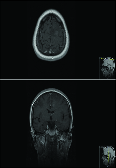
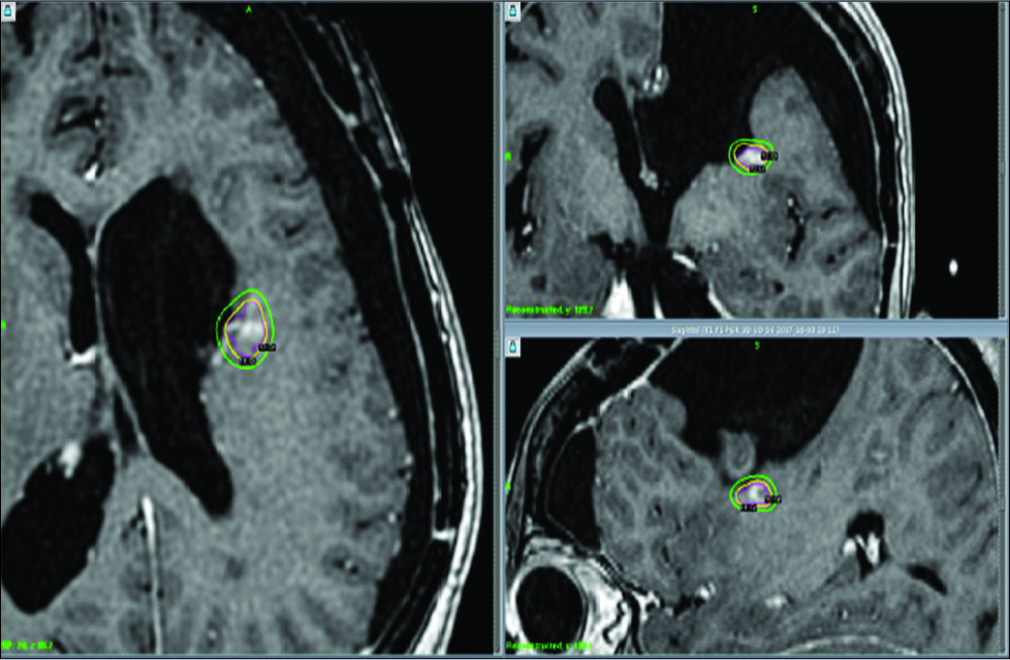
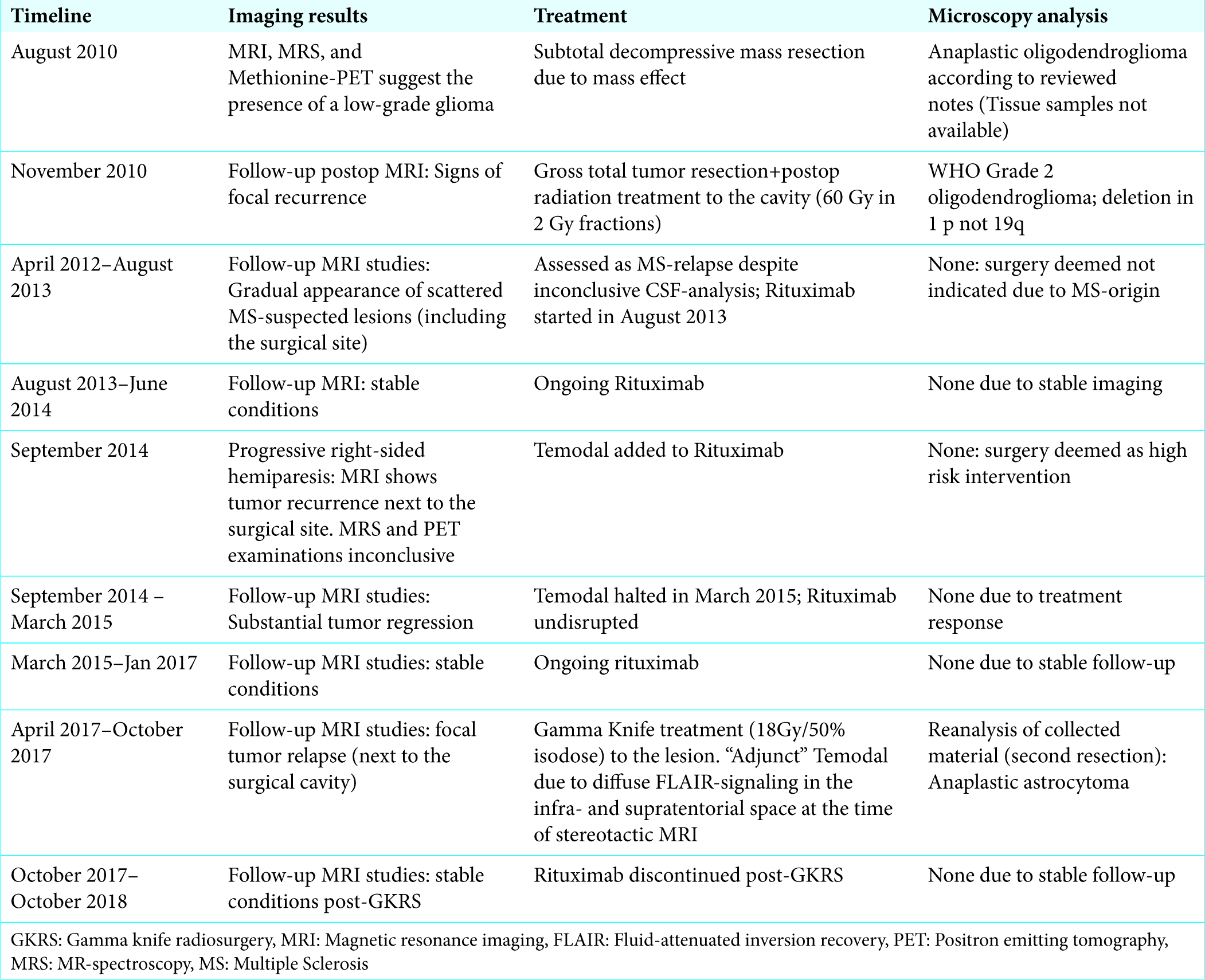
A 41-year-old female patient developed epileptic seizures in 2008. Further investigation with magnetic resonance imaging (MRI) revealed a glioma-suspect tumor within the boundaries of the left frontal lobe as well as multiple supratentorial MS-like lesions scattered in both hemispheres. The routine cerebrospinal fluid (CSF)-analysis showed evidence of MS-activity, while the complementary MR spectroscopy (MRS) study suggested the presence of a concurrent oligodendroglioma. The ensuing needle biopsy on the left frontal lobe lesion showed solely inflammatory signs of demyelination, suggesting an evolving tumefactive MS-plaque [
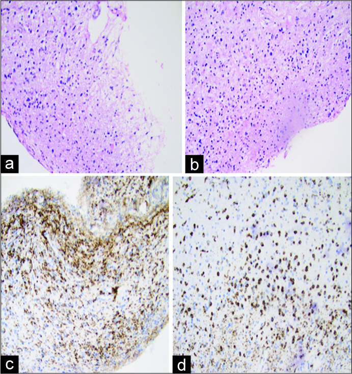
Figure 1:
Re-evaluation (2017) of available samples from needle biopsy performed in 2008 (2 samples of <4 mm). (a) Hematoxylin and Eosin glial biopsy with a mild increase in astrocytes. (b) Inflammatory process dominated by foamy macrophages (confirmed by immunohistochemistry). (c+d) CD68 immunohistochemistry: presence of a large number of macrophages.
DISCUSSION
General aspects
MS is a chronic inflammatory demyelinating disease with contentious evolution. MS-plaques are not uncommonly misdiagnosed as malignant processes, a phenomenon known as tumefactive MS; in fact, some groups have reported that up to 6% of all MS-cases are confused with other diseases, including primary brain neoplasms.[
Radiology
Glioma: In the context of glial tumors (including astrocytomas), preoperative MRI-examinations may provide key data as to the plausible grade of malignancy.[ MS: Lesions are commonly found within the confinements of the posterior fossa, periventricular areas, spinal cord, and optic nerves. Dual-echo and fluid-attenuated inversion recovery (FLAIR) imaging reveal focal areas of hyperintensity while T1-weighted MRI (with Gd) help discerns between active and inactive MS-lesions, as enhancement results from an increase in blood-brain barrier permeability in areas of “active” inflammation.[
Microscopic assessment
CSF-analysis and MS
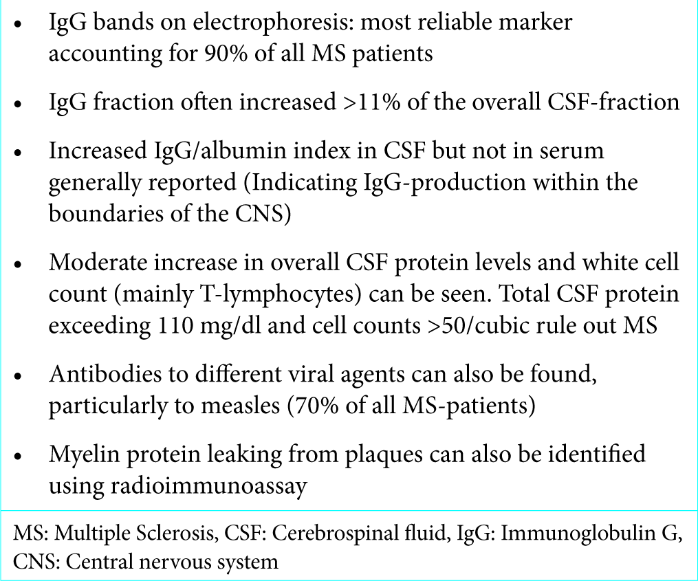
From a general perspective, the presence of demyelinating activity can be confirmed through CSF-analyses; common traits of MS-disease in CSF-analysis can be found in
Histopathologic assessment
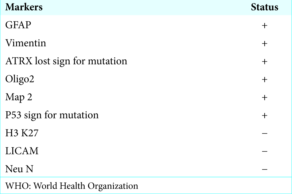
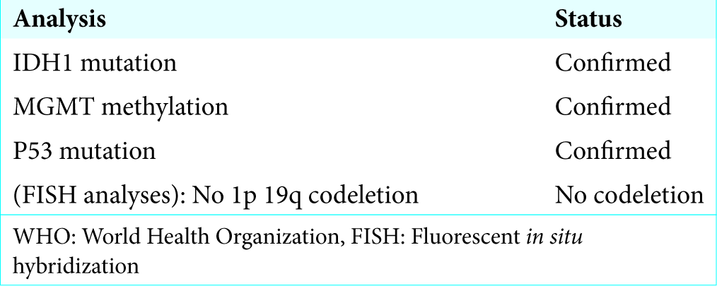
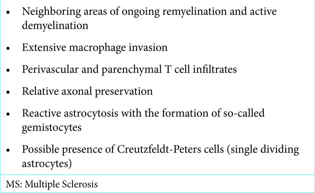
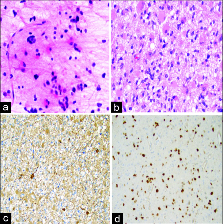
MS: Histological characteristics of chronic MS lesions are well known to neuropathologists; however, diagnosing MS at early stages may prove complex, particularly in the context of needle biopsy where meager samples frequently represent small areas of a larger lesion. Although larger samples collected from “wider” surgical margins increase diagnostic accuracy, a complete microscopic interpretation of this type of lesion may prove difficult. In this aspect, key microscopic features of early MS lesions are described in Glial tumors: The revaluation made for the purpose of this article exposed the presence of a diffuse, infiltrating isocitrate dehydrogenase (IDH) 1-mutant AA [
Table 5:
Key microscopic features of early MS disease.[
Figure 6:
Re-evaluation (2017) of 25 mm sample collected from the second resection (November 2010). (a) Hematoxylin and Eosin glial biopsy with moderately increased atypical astrocytes with gemistocytic features and with few mitoses. (b) Smear from the same biopsy: moderate increase in astrocytes with variation in size and shape. (c) Glial fibrillary acidic protein immunohistochemistry: positive in glial cells with astrocytic features. (d) KI-67 proliferation index about 25%. Samples from the first resection were not made available.
Causal relation?
Reports of concurrent MS and glial cell tumors can be traced back as early as 1912; however, despite significant advances in the field of neuro-diagnostics, the number of reported cases of coexistent MS and gliomas remains limited (<100 cases). This is probably due to the condition’s rareness and entangled histopathological characteristics, the repertoire of diagnostic tools used during disease evolution, and the degree of multidisciplinary experience. Despite the latter, our case has a striking resemblance to a number of reports.[
John Cunningham (JC)-virus
As previously described in this paper, different groups seem to share common thoughts concerning plausible environmental- modulated mechanisms and malignant transformation of glial cells during the process of remyelination; the role of the human polyoma JCV remains interesting in this context.[
CONCLUSION
Although cases of concurrent glioma and MS remain rare, their entangled evolution might pose a challenge in terms of diagnosis and treatment. Modern MRI-protocols are crucial in the diagnosis of MS and gliomas; however, further studies are warranted to overcome issues concerning MR-signal specificity, particularly in the face of a concurrent condition. In this context, microscopic evaluation remains largely dependent on reliable diagnostic markers and multidisciplinary expertise. At least, in theory, MS and CNS malignancies might synergistically concur through a set of shared micro-environmental and immune-modulated mechanisms. From that perspective, a casual origin cannot be fully ruled out. More studies on the subject are warranted and encouraged.
Declaration of patient consent
The authors certify that they have obtained all appropriate patient consent forms. In the form, the patient has given his consent for his images and other clinical information to be reported in the journal. The patients understand that their names and initials will not be published and due efforts will be made to conceal their identity, but anonymity cannot be guaranteed.
Financial support and sponsorship
Nil.
Conflicts of interest
None of the authors has any conflicts of interest to disclose.
References
1. Aarli JA, Mørk SJ, Myrseth E, Larsen JL. Glioblastoma associated with multiple sclerosis: Coincidence or induction. ? Eur Neurol. 1989. 29: 312-6
2. Abrishamchi F, Khorvash F. Coexistence of multiple sclerosis and brain tumor: An uncommon diagnostic challenge. Adv Biomed Res. 2017. 6: 101-
3. Acqui M, Caroli E, Di Stefano D, Ferrante L. Cerebral ependymoma in a patient with multiple sclerosis case report and critical review of the literature. Surg Neurol. 2008. 70: 414-20
4. Brendle C, Hempel JM, Schittenhelm J, Skardelly M, Reischl G, Bender B. Glioma grading by dynamic susceptibility contrast perfusion and 11C-methionine positron emission tomography using different regions of interest. Neuroradiology. 2018. 60: 381-9
5. Butteriss DJ, Ismail A, Ellison DW, Birchall D. Use of serial proton magnetic resonance spectroscopy to differentiate low grade glioma from tumefactive plaque in a patient with multiple sclerosis. Br J Radiol. 2003. 76: 662-5
6. Carvalho AT, Linhares P, Castro L, Sá MJ. Multiple sclerosis and oligodendroglioma: An exceptional association. Case Rep Neurol Med. 2014. 2014: 546817-
7. de la Lama A, Gómez PA, Boto GR, Lagares A, Ricoy JR, Alén JF. Oligodendroglioma and multiple sclerosis. A case report. Neurocirugia (Astur). 2004. 15: 378-83
8. Diamandis E, Gabriel CPS, Würtemberger U, Guggenberger K, Urbach H, Staszewski O. MR-spectroscopic imaging of glial tumors in the spotlight of the 2016 WHO classification. J Neurooncol. 2018. 139: 431-40
9. Ferenczy MW, Marshall LJ, Nelson CD, Atwood WJ, Nath A, Khalili K. Molecular biology, epidemiology, and pathogenesis of progressive multifocal leukoencephalopathy, the JC virus-induced demyelinating disease of the human brain. Clin Microbiol Rev. 2012. 25: 471-506
10. Filippi M, Rocca MA. MR imaging of multiple sclerosis. Radiology. 2011. 259: 659-81
11. Golombievski EE, McCoyd MA, Lee JM, Schneck MJ. Biopsy proven tumefactive multiple sclerosis with concomitant glioma: Case report and review of the literature. Front Neurol. 2015. 6: 150-
12. Green AJ, Bollen AW, Berger MS, Oksenberg JR, Hauser SL. Multiple sclerosis and oligodendroglioma. Mult Scler. 2001. 7: 269-73
13. Ho KL, Wolfe DE. Concurrence of multiple sclerosis and primary intracranial neoplasms. Cancer. 1981. 47: 2913-9
14. Imitola J, Wagoner J, Khurana JS. Rapid dissemination of granular cell astrocytoma arising from periventricular stem cell regions in chronic multiple sclerosis. J Neurooncol. 2015. 124: 147-9
15. Khalil A, Serracino H, Damek DM, Ney D, Lillehei KO, Kleinschmidt-DeMasters BK. Genetic characterization of gliomas arising in patients with multiple sclerosis. J Neurooncol. 2012. 109: 261-72
16. Khan OA, Bauserman SC, Rothman MI, Aldrich EF, Panitch HS. Concurrence of multiple sclerosis and brain tumor: Clinical considerations. Neurology. 1997. 48: 1330-3
17. Kuhlmann T, Lassmann H, Brück W. Diagnosis of inflammatory demyelination in biopsy specimens: A practical approach. Acta Neuropathol. 2008. 115: 275-87
18. Lin M, Reid P, Bakhsheshian J. Tumefactive multiple sclerosis masquerading as high grade glioma. World Neurosurg. 2018. 112: 37-8
19. Malmgren RM, Detels R, Verity MA. Co-occurrence of multiple sclerosis and glioma case report and neuropathologic and epidemiologic review. Clin Neuropathol. 1984. 3: 1-9
20. Mandel JJ, Goethe EA, Patel AJ, Heck K, Hutton GJ. Newly diagnosed ganglioglioma in an adult patient with multiple sclerosis. J Neurol Sci. 2016. 369: 51-2
21. Mantero V, Balgera R, Bianchi G, Rossi G, Rigamonti A, Fiumani A. Brainstem glioblastoma in a patient with secondary progressive multiple sclerosis. Neurol Sci. 2015. 36: 1733-5
22. Mathews T, Moossy J. Mixed glioma, multiple sclerosis, and charcot-marie-tooth disease. Arch Neurol. 1972. 27: 263-8
23. Plantone D, Renna R, Sbardella E, Koudriavtseva T. Concurrence of multiple sclerosis and brain tumors. Front Neurol. 2015. 6: 40-
24. Podbielska M, Szulc ZM, Kurowska E, Hogan EL, Bielawski J, Bielawska A. Cytokine-induced release of ceramide-enriched exosomes as a mediator of cell death signaling in an oligodendroglioma cell line. J Lipid Res. 2016. 57: 2028-39
25. Preziosa P, Sangalli F, Esposito F, Moiola L, Martinelli V, Falini A. Clinical deterioration due to co-occurrence of multiple sclerosis and glioblastoma: Report of two cases. Neurol Sci. 2017. 38: 361-4
26. Rathbone E, Durant L, Kinsella J, Parker AR, Hassan-Smith G, Douglas MR. Cerebrospinal fluid immunoglobulin light chain ratios predict disease progression in multiple sclerosis. J Neurol Neurosurg Psychiatry. 2018. 89: 1044-9
27. Rencic A, Gordon J, Otte J, Curtis M, Kovatich A, Zoltick P. Detection of JC virus DNA sequence and expression of the viral oncoprotein, tumor antigen, in brain of immunocompetent patient with oligoastrocytoma. Proc Natl Acad Sci U S A. 1996. 93: 7352-7
28. Rovira À, Wattjes MP, Tintoré M, Tur C, Yousry TA, Sormani MP. Evidence-based guidelines: MAGNIMS consensus guidelines on the use of MRI in multiple sclerosis-clinical implementation in the diagnostic process. Nat Rev Neurol. 2015. 11: 471-82
29. Sega S, Horvat A, Popovic M. Anaplastic oligodendroglioma and gliomatosis Type 2 in interferon-beta treated multiple sclerosis patients. Report of two cases. Clin Neurol Neurosurg. 2006. 108: 259-65
30. Shankar SK, Rao TV, Srivastav VK, Narula S, Asha T, Das S. Balo’s concentric sclerosis: A variant of multiple sclerosis associated with oligodendroglioma. Neurosurgery. 1989. 25: 982-6
31. Sharim J, Tashjian R, Golzy N, Pouratian N. Glioblastoma following treatment with fingolimod for relapsing-remitting multiple sclerosis. J Clin Neurosci. 2016. 30: 166-8
32. Upadhyay N, Waldman AD. Conventional MRI evaluation of gliomas. Br J Radiol. 2011. 84 Spec No 2: S107-11
33. Vrenken H, Jenkinson M, Horsfield MA, Battaglini M, van Schijndel RA, Rostrup E. Recommendations to improve imaging and analysis of brain lesion load and atrophy in longitudinal studies of multiple sclerosis. J Neurol. 2013. 260: 2458-71
34. Wattjes MP, Rovira À, Miller D, Yousry TA, Sormani MP, de Stefano MP. Evidence-based guidelines: MAGNIMS consensus guidelines on the use of MRI in multiple sclerosis establishing disease prognosis and monitoring patients. Nat Rev Neurol. 2015. 11: 597-606
35. Vieregge P, Nahser HC, Gerhard L, Reinhardt V, Nau HE. Multiple sclerosis and cerebral tumor. Clin Neuropathol. 1984. 3: 10-21
36. Von Deimling A, Louis DN, Aldape K, Brat D, Collins VP, Eberhart C.editorsWHO classification of tumours of the Central nervous system. Lyon: IARC; 2016. p. 24-7


