- Department of Neurosurgery, PGIMER, Chandigarh, India
Correspondence Address:
Ashish Aggarwal
Department of Neurosurgery, PGIMER, Chandigarh, India
DOI:10.4103/sni.sni_97_18
Copyright: © 2018 Surgical Neurology International This is an open access journal, and articles are distributed under the terms of the Creative Commons Attribution-NonCommercial-ShareAlike 4.0 License, which allows others to remix, tweak, and build upon the work non-commercially, as long as appropriate credit is given and the new creations are licensed under the identical terms.How to cite this article: Singla N, Aggarwal A. A giant partially thrombosed, partially filling, and partially calcified intracranial aneurysm. Surg Neurol Int 24-Jul-2018;9:139
How to cite this URL: Singla N, Aggarwal A. A giant partially thrombosed, partially filling, and partially calcified intracranial aneurysm. Surg Neurol Int 24-Jul-2018;9:139. Available from: http://surgicalneurologyint.com/surgicalint-articles/a-giant-partially-thrombosed-partially-filling-and-partially-calcified-intracranial-aneurysm/
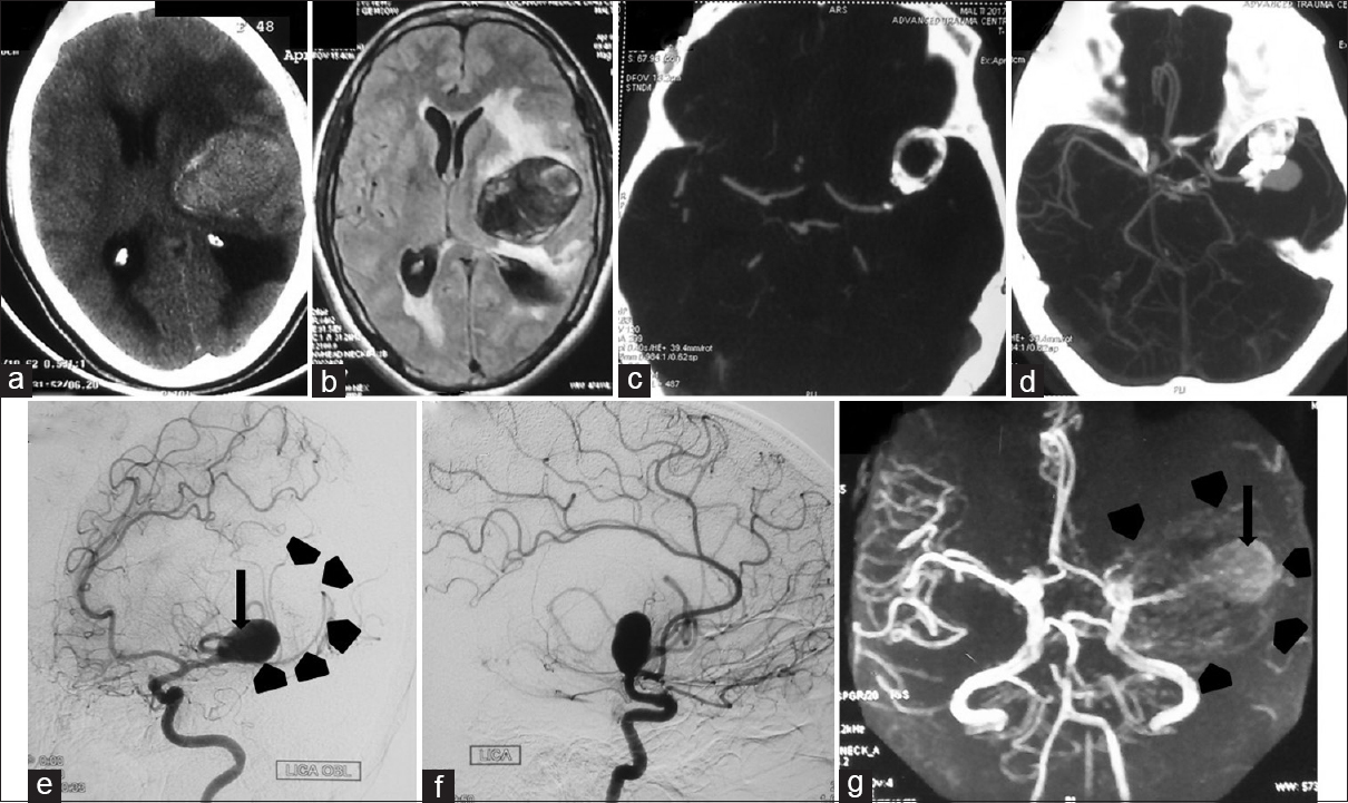
A 48-year-old female presented with intermittent headache and right hemiparesis for 6 years. Non-contrast computed tomography of head revealed a space-occupying lesion (SOL) in left frontotemporal region which was partially calcified [
Figure 1
(a) NCCT reveals SOL in left fronto temporal region which was partially calcified. (b) MRI axial flair image shows layered thrombus inside the SOL. (c and d) CT angiography shows an aneurysm arising from MCA with significant calcification. DSA of Left ICA (e) oblique view and (f) lateral view showing a large filling aneurysm arising from left MCA(arrow). In addition, there is a large non filling portion (?thrombosed ) of aneurysm shown by arrow heads. (g) MR angiography reveals central filling portion (Arrow) while the rest of the mass was not taking up contrast (Arrow heads)
Declaration of patient consent
The authors certify that they have obtained all appropriate patient consent forms. In the form the patient(s) has/have given his/her/their consent for his/her/their images and other clinical information to be reported in the journal. The patients understand that their names and initials will not be published and due efforts will be made to conceal their identity, but anonymity cannot be guaranteed.
Financial support and sponsorship
Nil.
Conflicts of interest
There are no conflicts of interest.


