- Department of Anaesthesia, All India Institute of Medical Sciences, Rishikesh, Uttarakhand, India.
- Department of Neurosurgery, All India Institute of Medical Sciences, Rishikesh, Uttarakhand, India.
Correspondence Address:
Sanjay Agrawal, Department of Anaesthesia, All India Institute of Medical Sciences, Rishikesh, Uttarakhand, India.
DOI:10.25259/SNI_186_2023
Copyright: © 2023 Surgical Neurology International This is an open-access article distributed under the terms of the Creative Commons Attribution-Non Commercial-Share Alike 4.0 License, which allows others to remix, transform, and build upon the work non-commercially, as long as the author is credited and the new creations are licensed under the identical terms.How to cite this article: Nirupa Ramakumar1, Priyanka Gupta1, Rajnish Arora2, Sanjay Agrawal1. A prospective exploratory study to assess echocardiographic changes in patients with supratentorial tumors – Effect of craniotomy and tumor decompression. 05-May-2023;14:166
How to cite this URL: Nirupa Ramakumar1, Priyanka Gupta1, Rajnish Arora2, Sanjay Agrawal1. A prospective exploratory study to assess echocardiographic changes in patients with supratentorial tumors – Effect of craniotomy and tumor decompression. 05-May-2023;14:166. Available from: https://surgicalneurologyint.com/surgicalint-articles/12309/
Abstract
Background: Functional changes in the myocardium secondary to increased intracranial pressure (ICP) are studied sparingly. Direct echocardiographic changes in patients with supratentorial tumors have not been documented. The primary aim was to assess and compare the transthoracic echocardiography changes in patients with supratentorial tumors presenting with and without raised intracranial pressure for neurosurgery.
Methods: Patients were divided into two groups based on preoperative radiological and clinical evidence of midline shift of
Results: Ninety patients were assessed, 88 were included for analysis. Two were excluded based on a poor echocardiographic window (1) and change in the operative plan (1). Demographic variables were comparable. About 27% of the patients in Group 2 had ejection fraction
Conclusion: The study demonstrated that in patients with supratentorial tumors with ICP, cardiac dysfunction might be present in the preoperative period.
Keywords: Echocardiography, Intracranial hypertension, Left, Supratentorial neoplasms, Optic nerve sheath diameter, Ventricular dysfunction
INTRODUCTION
Brain-heart interactions in patients with brain insult hold a leading place in the developing field of organ cross-talk, with clinically significant implications throughout the perioperative period.[
Although presumably less frequent, cardiac alterations following brain tumors are comparable to the site-specific harm brought on by brain lesions when the regions responsible for controlling circulatory function are injured.[
There is scant evidence to support the hypothesis that cerebral lesions with mass effect cause elevated intracranial pressure (ICP).[
Only a few case reports have investigated a persistent increase in ICP in primary brain tumors that causes cardiac dysfunction.[
With this background, this study was done to assess and compare the cardiac function using transthoracic echocardiography in patients with supratentorial tumor who underwent craniotomy and tumor excision, both before and after surgery (primary objective) and to find any correlation of ONSD USG with midline intracranial shift and cardiac parameters (secondary objective).
MATERIALS AND METHODS
This prospective observational study was conducted on 90 patients with supratentorial tumor of either sex, aged between the age group 18 and 60 years with revised cardiac risk index less than two scheduled for elective craniotomy and tumor excision over a period of 2 years. The patients were divided into two groups based on midline shift seen on the preoperative computed tomography (CT) scan/magnetic resonance imaging (a) Group 1: Midline shift of ≤6 mm and (b) Group 2: Midline shift of more than 6 mm. Patients with pregnancy, known case of cardiac illness, body mass index >35 kg/m2, lesions in the prefrontal cortex, insula, hypothalamus, amygdala, hippocampus, and previous eye surgery and tumor involving optic nerve were excluded from the study. The Institutional Ethics Committee approval (AIIMS/IEC/20/504) was sought, and written informed consent was obtained from either patients or next of the kin. The study was registered with Clinical Trials Registry India (CTRI/2020/10/028313).
Radiological evaluation
Preoperative radiological variables including size, location and number of the tumor, laterality, presence, or absence of cerebral edema and midline shift was noted. The cerebral edema was graded as minimal (maximum thickness of edema was less than the radius of mass), moderate (between the radius and diameter of the mass), or severe (more than the diameter of the mass).
Cardiac evaluation
Transthoracic echocardiography was performed by NR using P21x, 1–5 MHz, phased array probe (Sonosite, Bothell, USA) at preoperative period (24 h) and postoperatively at 48 h. Chamber quantification was performed as per the guidelines of the American Society of Echocardiography.[
ONSD
Patients’ eyes were scanned in supine position using a high resolution 7-MHz linear array transducer (Sonosite, Inc., Bothell, WA, USA) on closed eyelids. The structure of the eyes was visualized to align with the optic nerve directly opposite the probe with the ONSD width perpendicular to the vertical axis of the scanning plane. ONSD bilaterally was measured 3 mm behind the globe and an average of three readings from each eye for calculation. The ONSD measurements were classified as ipsilateral ONSD (craniotomy side, ONSD-Ipsilateral [ONSD-IP]) and contralateral ONSD (side opposite the craniectomy, ONSD-Contralateral [ONSD-CL]) according to the side of the lesion. Transorbital ONSD >5.5 mm was considered as indicative of raised ICP.[
Perioperative management
A written informed written consent was taken. All patients were kept nil per oral 6 h for solids and 2 h for clear liquids, premedicated with Tablet Ranitidine 150 mg early morning at 6 am. General anesthesia was given to all patients as per institutional protocol. Standard American Society of Anesthesiologist intraoperative monitors were applied. Anesthesia was induced with intravenous (i.v) propofol 1.5–2 mg/kg and fentanyl 2 μg/kg, while i.v Vecuronium 0.1 mg/kg was used to facilitate orotracheal intubation. Maintenance of Anesthesia was achieved with 50% Oxygen: Air mixture propofol infusion@ 100–150 μg/kg/min and fentanyl infusion @ 1 μg/kg/h and intermittent vecuronium boluses. Following surgical intervention, patients were shifted to the intensive care unit.
Statistical analysis
As it was an exploratory study, power analysis for Paired t-test was conducted in G-POWER to determine a sufficient sample size using an alpha of 0.05, a power of 0.80, an effect size of 0.3 (Using Cohen’s Convention), and two tails. Based on the assumptions, the desired sample size total of 90 patients was taken. Categorical data were presented as percentages (%). Pearson’s Chi-square test and Fishers exact test were used to evaluate differences between groups for categorized variables. Normally distributed data were presented as means and standard deviation, or 95% confidence intervals (CI). Student’s t-test paired and unpaired and analysis of variance were used for comparison between various quantitative parameters. Pearson’s correlation test was used for correlation between various quantitative parameters. All tests were performed at a 5% level of significance; thus, an association was significant if P < 0.05. Analysis was carried out using the Statistical Package for the Social Studies for Windows version 23.0 and online GraphPad software (Prism 5 for Windows) version 5.01.
RESULTS
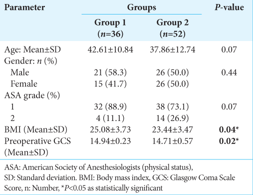
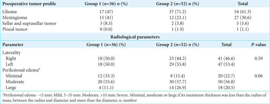
Demographic and lesion characteristics
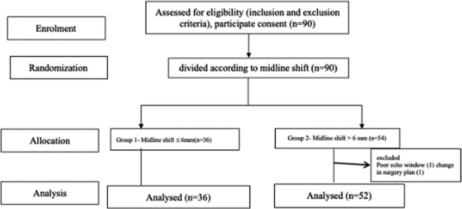
Ninety patients were assessed for eligibility; data of 88 were included for analysis [
Intraoperative and hemodynamic changes
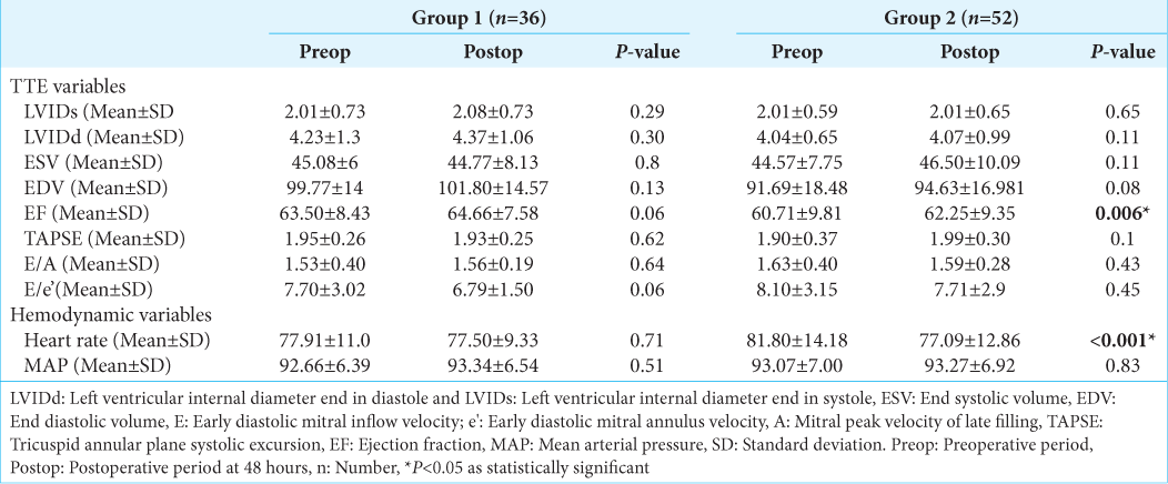
Intraoperative events like hypotension were seen in 8 (22%) patients (group 1) and 18 patients (34%) (group 2) (P = 0.21). The heart rate was significantly higher in preoperative period as compared to postoperative period in group 2 (P < 0.001) [
Echocardiographic changes
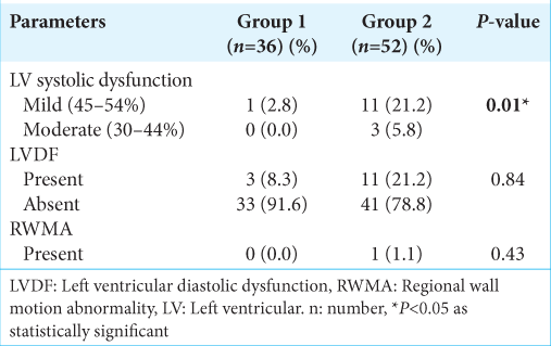
Echocardiographic changes between the groups are shown in [
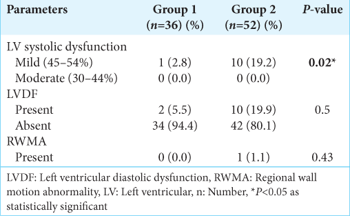
The echocardiographic changes before and after surgery in both the groups are shown in
ONSD-USG changes
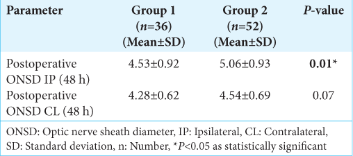
Intergroup and intragroup analysis of ONSD variables depicted in [
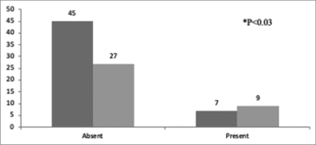
The preoperative ONSD-IP and ONSD-CL value shows a correlation of 0.31 to mid-line shift (MLS) values which were found to be statistically significant (P = 0.003). The patients with ONSD-ipsilateral (IL) >5.5 mm had more perilesional edema (56% – moderate and 26% – severe) than in patients with ONSD < 5.5 mm. (P = 0.024). Nine (25%) patients with ONSD >5.5 mm in the preoperative period had LV diastolic dysfunction which was statistically significant (P = 0.02) [
DISCUSSION
Our study demonstrated that left ventricular ejection fraction (LVEF) is affected in patients with intracranial space occupying lesion and it may show improvement in few within first 24–48 h of tumor excision. Cross-talk between the brain pathology and the cardiac functions is focusing on wide range of cardiac-functional alterations secondary to brain pathology. Modest cardiac dysfunction secondary to intracranial pathology may have ramifications in the perioperative period. Association of increased ICP and cardiac dysfunction is sparsely reported.[
Preoperative hypertension, hyperlipidaemia, increasing age, and asymptomatic coronary artery disease can cause diastolic dysfunction. Intraoperative direct effects of anesthetics on cardiac inotropy, lusitropy, and peripheral vasodilatation cannot be underestimated in unmasking any subclinical systolic cardiac dysfunction. The absence of a regional wall motion abnormality rules out systolic failure due to an acute myocardial infarction. Increased sympathetic surge with resultant increased levels of circulating endogenous catecholamines consequent to increased ICP is described in the literature.[
To determine diastolic function, we employed the streamlined measurement technique recommended by Greenstein and Mayo under the aegis of the American College of Chest Physicians, employing e and E/e’ using echocardiography.[
In our study, diastolic dysfunction was present in three patient (8.3%) in patients with midline shift of <6 mm and in 11 (21.2) % of patients with MLS of >6 mm preoperatively. Chronic sympathetic activity and myocardial norepinephrine discharge maybe postulated as a cause of LV diastolic dysfunction. Chronic hypertension could be the cause of diastolic dysfunction in the group with MLS <6 mm. Grassi et al., in their study, discovered a correlation between elevated sympathetic nerve activity as detected by microneurography and the existence of asymptomatic LV diastolic failure.[
Group 2 patients (increased ICP) had a higher heart rate in the preoperative period than group 1 (P < 0.05). It can be because ICP returned to normal after the tumor removal. This finding contrasts the classic Cushing’s reflex causing bradycardia primarily found in these patients. The Koszewicz et al. study, which examined the profile of autonomic dysfunctions in 30 patients with the primary brain tumors, is consistent with this conclusion. The subjects had increased heart rates and elevated blood pressure with low heart rate variability.[
There was statistically significant improvement in EF in the postoperative period at 48 h in few patients. This concurs with study by Srinivasaiah and Praveen et al. who found significant improvement in systolic dysfunction after surgery.[
Interestingly, ONSD values in our patients resemble those found in patients with elevated ICP. Montorfano et al. demonstrated ONSD value of 5.82 mm (95% CI 5.58–6.06 mm) in patients with elevated ICP.[
There was a significant decrease in ONSD- IP and ONSD-CL after the surgery in both the groups (P < 0.001). These results are consistent with these studies.[
A recently published study by Bäuerle et al. revealed that the ultrasonic measurement of ONSD could not accurately estimate ICP in patients with SAH.[
We found positive correlation between ONSD and radiological findings of raised ICP. Robba et al. demonstrated that ONSD was the best non-invasive way to measure ICP and recommended the following formula to do so: nICP ONSD = 5.00 × ONSD–13.92 mmHg.[
Cardiovascular disease is the second largest cause of death in a retrospective study patients with brain tumors, 30–50% comprised patients with meningioma.[
Our study had few limitations. The study included a small sample size, long-term effects at 6 months/1 year were not evaluated, biochemical markers of cardiac dysfunction, or catecholamine levels measuring the sympathetic activity were not observed, and quantitative measurement of ICP was not done. We also did not have previous transthoracic echocardiography reports of the patients as it is not routinely advised when they visit the preanesthetic clinic. Prospective multicenter randomized trials are essential to evaluate the impact of intraoperative TTE. Further studies are required to investigate the temporal course of the changes in the echocardiographic variables in chronically raised ICP scenarios and to see the long-term effects at 6 months/1 year.
CONCLUSION
Our study demonstrated that in patients with supratentorial tumors with features of increased ICP, cardiac dysfunction maybe present in the preoperative period; hence, 2D-echocardiography must be included in preoperative assessment for patients presenting for craniotomy and tumor excision.
Declaration of patient consent
The Institutional Review Board (IRB) permission obtained for the study.
Financial support and sponsorship
Nil.
Conflicts of interest
There are no conflicts of interest.
Disclaimer
The views and opinions expressed in this article are those of the authors and do not necessarily reflect the official policy or position of the Journal or its management. The information contained in this article should not be considered to be medical advice; patients should consult their own physicians for advice as to their specific medical needs.
Acknowledgment
I would like to thank the anesthesia technical team for the help extended throughout the study period.
References
1. Bäuerle J, Niesen WD, Egger K, Buttler KJ, Reinhard M. Enlarged optic nerve sheath in aneurysmal subarachnoid hemorrhage despite normal intracranial pressure. J Neuroimaging. 2016. 26: 194-6
2. Cuisinier A, Maufrais C, Payen JF, Nottin S, Walther G, Bouzat P. Myocardial function at the early phase of traumatic brain injury: A prospective controlled study. Scand J Trauma Resusc Emerg Med. 2016. 24: 129
3. Dubourg J, Javouhey E, Geeraerts T, Messerer M, Kassai B. Ultrasonography of optic nerve sheath diameter for detection of raised intracranial pressure: A systematic review and meta-analysis. Intensive Care Med. 2011. 37: 1059-68
4. Fedriga M, Czigler A, Nasr N, Zeiler FA, Park S, Donnelly J. Autonomic nervous system activity during refractory rise in intracranial pressure. J Neurotrauma. 2021. 38: 1662-9
5. Grassi G, Quarti-Trevano F, Seravalle G, Arenare F, Volpe M, Furiani S. Early sympathetic activation in the initial clinical stages of chronic renal failure. Hypertension. 2011. 57: 846-51
6. Geeraerts T, Launey Y, Martin L, Pottecher J, Vigué B, Duranteau J. Ultrasonography of the optic nerve sheath may be useful for detecting raised intracranial pressure after severe brain injury. Intensive Care Med. 2007. 33: 1704-11
7. Greenstein YY, Mayo PH. Evaluation of left ventricular diastolic function by the intensivist. Chest. 2018. 153: 723-32
8. Jin K, Brennan PM, Poon MT, Sudlow CL, Figueroa JD. Raised cardiovascular disease mortality after central nervous system tumor diagnosis: Analysis of 171,926 patients from UK and USA. Neurooncol Adv. 2021. 3: vdab136
9. Kalim Z, Siddiqui OA, Nadeem A, Hasan M, Rashid H. Assessment of optic nerve sheath diameter and its postoperative regression among patients undergoing brain tumor resection in a tertiary care center. J Neurosci Rural Pract. 2022. 13: 270-5
10. Kazdal H, Kanat A, Findik H, Sen A, Ozdemir B, Batcik OE. Transorbital ultrasonographic measurement of optic nerve sheath diameter for intracranial midline shift in patients with head trauma. World Neurosurg. 2016. 85: 292-7
11. Kienzler JC, Zakelis R, Marbacher S, Bäbler S, Schwyzer L, Remonda E. Changing the paradigm of intracranial hypertension in brain tumor patients: A study based on noninvasive ICP measurements. BMC Neurol. 2020. 20: 268
12. Kopelnik A, Fisher L, Miss JC, Banki N, Tung P, Lawton MT. Prevalence and implications of diastolic dysfunction after subarachnoid hemorrhage. Neurocrit Care. 2005. 3: 132-8
13. Koszewicz M, Michalak S, Bilinska M, Budrewicz S, Zaborowski M, Slotwinski K. Profile of autonomic dysfunctions in patients with primary brain tumor and possible autoimmunity. Clin Neurol Neurosurg. 2016. 151: 51-4
14. Krishnamoorthy V, Prathep S, Sharma D, Gibbons E, Vavilala MS. Association between electrocardiographic findings and cardiac dysfunction in adult isolated traumatic brain injury. Indian J Crit Care Med. 2014. 18: 570-4
15. Krishnamoorthy V, Chaikittisilpa N, Lee J, Mackensen GB, Gibbons EF, Laskowitz D. Speckle tracking analysis of left ventricular systolic function following traumatic brain injury: A pilot prospective observational cohort study. J Neurosurg Anesthesiol. 2020. 32: 156-61
16. Lang RM, Badano LP, Mor-Avi V, Afilalo J, Armstrong A, Ernande L. Recommendations for cardiac chamber quantification by echocardiography in adults: An update from the American Society of Echocardiography and the European Association of Cardiovascular Imaging. J Am Soc Echocardiogr. 2015. 28: 1-39.e14
17. Lechowicz-Glogowska E, Rojek A, Duszynska W. The irreversible neurogenic stress cardiomyopathy during large supratentorial brain tumor resection. Neurocrit Care. 2019. 31: 587-91
18. Lee SH, Kim HS, Yun SJ. Optic nerve sheath diameter measurement for predicting raised intracranial pressure in adult patients with severe traumatic brain injury: A meta-analysis. J Crit Care. 2020. 56: 182-7
19. Mazzeo AT, Micalizzi A, Mascia L, Scicolone A, Siracusano L. Brain-heart crosstalk: The many faces of stress-related cardiomyopathy syndromes in anaesthesia and intensive care. Br J Anaesth. 2014. 112: 803-15
20. Montorfano L, Yu Q, Bordes SJ, Sivanushanthan S, Rosenthal RJ, Montorfano M. Mean value of B-mode optic nerve sheath diameter as an indicator of increased intracranial pressure: A systematic review and meta-analysis. Ultrasound J. 2021. 13: 35
21. Moretti R, Pizzi B, Cassini F, Vivaldi N. Reliability of optic nerve ultrasound for the evaluation of patients with spontaneous intracranial hemorrhage. Neurocrit Care. 2009. 11: 406-10
22. Mrozek S, Gobin J, Constantin JM, Fourcade O, Geeraerts T. Crosstalk between brain, lung and heart in critical care. Anaesth Crit Care Pain Med. 2020. 39: 519-30
23. Nagueh SF, Smiseth OA, Appleton CP, Byrd BF, Dokainish H, Edvardsen T. Recommendations for the evaluation of left ventricular diastolic function by echocardiography: An update from the American Society of Echocardiography and the European Association of Cardiovascular Imaging. J Am Soc Echocardiogr. 2016. 29: 277-314
24. Prasad Hrishi AP, Lionel KR, Prathapadas U. Head rules over the heart: Cardiac manifestations of cerebral disorders. Indian J Crit Care Med. 2019. 23: 329-35
25. Prathep S, Sharma D, Hallman M, Joffe A, Krishnamoorthy V, Mackensen GB. Preliminary report on cardiac dysfunction after isolated traumatic brain injury. Crit Care Med. 2014. 42: 142-7
26. Praveen R, Jayant A, Mahajan S, Jangra K, Panda NB, Grover VK. Perioperative cardiovascular changes in patients with traumatic brain injury: A prospective observational study. Surg Neurol Int. 2021. 12: 174
27. Rajajee V, Fletcher JJ, Rochlen LR, Jacobs TL. Comparison of accuracy of optic nerve ultrasound for the detection of intracranial hypertension in the setting of acutely fluctuating vs stable intracranial pressure: Post-hoc analysis of data from a prospective, blinded single center study. Crit Care. 2012. 16: R79
28. Ragauskas A, Bartusis L, Piper I, Zakelis R, Matijosaitis V, Petrikonis K. Improved diagnostic value of a TCD-based non-invasive ICP measurement method compared with the sonographic ONSD method for detecting elevated intracranial pressure. Neurol Res. 2014. 36: 607-14
29. Robba C, Cardim D, Tajsic T, Pietersen J, Bulman M, Donnelly J. Ultrasound non-invasive measurement of intracranial pressure in neurointensive care: A prospective observational study. PLoS Med. 2017. 14: e1002356
30. Robba C, Santori G, Czosnyka M, Corradi F, Bragazzi N, Padayachy L. Optic nerve sheath diameter measured sonographically as non-invasive estimator of intracranial pressure: A systematic review and meta-analysis. Intensive Care Med. 2018. 44: 1284-94
31. Ruggieri F, Gemma M, Calvi MR, Nicelli E, Agarossi A, Beretta L. Perioperative serum brain natriuretic peptide and cardiac troponin in elective intracranial surgery. Neurocrit Care. 2012. 17: 395-400
32. Santos S, Castro M, Raimundo A, Ribeiro A. Anaesthetic management of large meningioma excision complicated by Takotsubo and posterior reversible encephalopathy. BMJ Case Rep. 2022. 15: e246690
33. Schmidt EA, Despas F, Traon AP, Czosnyka Z, Pickard JD, Rahmouni K. Intracranial pressure is a determinant of sympathetic activity. Front Physiol. 2018. 9: 11
34. Serri K, El Rayes M, Giraldeau G, Williamson D, Bernard F. Traumatic brain injury is not associated with significant myocardial dysfunction: An observational pilot study. Scand J Trauma Resusc Emerg Med. 2016. 24: 31
35. Shirodkar CG, Rao SM, Mutkule DP, Harde YR, Venkategowda PM, Mahesh MU. Optic nerve sheath diameter as a marker for evaluation and prognostication of intracranial pressure in Indian patients. Indian J Crit Care Med. 2014. 18: 728-34
36. Srinivasaiah B, Muthuchellappan R, Sesha UR. A prospective observational study of electrocardiographic and echocardiographic changes in traumatic brain injury-effect of surgical decompression. Br J Neurosurg. 2022. p. 1-6
37. Wang LJ, Chen LM, Chen Y, Bao LY, Zheng NN, Wang YZ. Ultrasonography assessments of optic nerve sheath diameter as a non-invasive and dynamic method of detecting changes in intracranial pressure. JAMA Ophthalmol. 2018. 136: 250-6
38. Wu GB, Tian J, Liu XB, Wang ZY, Guo JY. Can optic nerve sheath diameter assessment be used as a non-invasive tool to dynamically monitor intracranial pressure?. J Integr Neurosci. 2022. 21: 54
39. Yogendranathan N, Herath HM, Pahalagamage SP, Kulatunga A. Electrocardiographic changes mimicking acute coronary syndrome in a large intracranial tumour: A diagnostic dilemma. BMC Cardiovasc Disord. 2017. 17: 91


