- Department of Neurosurgical, SAID Hilmani, Casablanca, Morocco.
- Department of Anesthesiology, Clinic Dar Salam, UHC Ibn Rusch, Casablanca, Morocco.
DOI:10.25259/SNI_57_2020
Copyright: © 2020 Surgical Neurology International This is an open-access article distributed under the terms of the Creative Commons Attribution-Non Commercial-Share Alike 4.0 License, which allows others to remix, tweak, and build upon the work non-commercially, as long as the author is credited and the new creations are licensed under the identical terms.How to cite this article: Said Hilmani1, Tarek Mesbahi1, Abderrahman Bouaggad2, Abdelhakim Lakhdar1. A rare complication of ventriculoperitoneal shunt: Pleural effusion without intrathoracic ventriculoperitoneal shunt catheter. Surg Neurol Int 18-Sep-2020;11:291
How to cite this URL: Said Hilmani1, Tarek Mesbahi1, Abderrahman Bouaggad2, Abdelhakim Lakhdar1. A rare complication of ventriculoperitoneal shunt: Pleural effusion without intrathoracic ventriculoperitoneal shunt catheter. Surg Neurol Int 18-Sep-2020;11:291. Available from: https://surgicalneurologyint.com/surgicalint-articles/10271/
Abstract
Background: Symptomatic pleural effusion following ventriculoperitoneal shunt (VPS) insertion is very rare and poorly understood in the literature in contrary to other mechanical complications.
Case Description: We report a case of 15 month-year-old girl who had VP shunt for congenital hydrocephalus. Twelve months after surgery, she was diagnosed with massive hydrothorax. Chest X-ray and thoracoabdominal CT scan confirmed the right pleurisy and showed the tip of the peritoneal catheter in the general peritoneal cavity. We made thoracic drainage of the transudative pleural effusion. When we released the chest tube, 24 h after, the girl showed a respiratory distress again and the effusion resumed at the X-ray control. Her symptoms abated after the realization of a ventriculoatrial shunt “VAS.” Repeat chest X-ray confirmed the resolution of the hydrothorax.
Conclusion: Despite the not yet well-understood mechanism of this rare and important VPS complication, management is simple based on X-ray confirmation, thoracentesis with biological analysis, and catheter replacement, especially in atrium “VAS.”
Keywords: Hydrothorax, Ventriculoatrial shunt, Ventriculoperitoneal shunt complication
INTRODUCTION
Mechanical and shunt infection are the most common complications of ventriculoperitoneal shunt (VPS), especially in the pediatric patients treated for hydrocephalus. Pleural effusion complicating VPS is a very rare condition. Most cases described with hydrothorax are due to the migration of the catheter tip into the pleural space.[
CASE REPORT
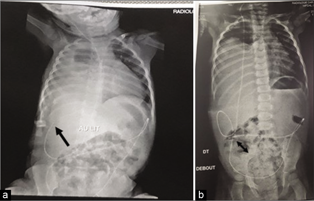
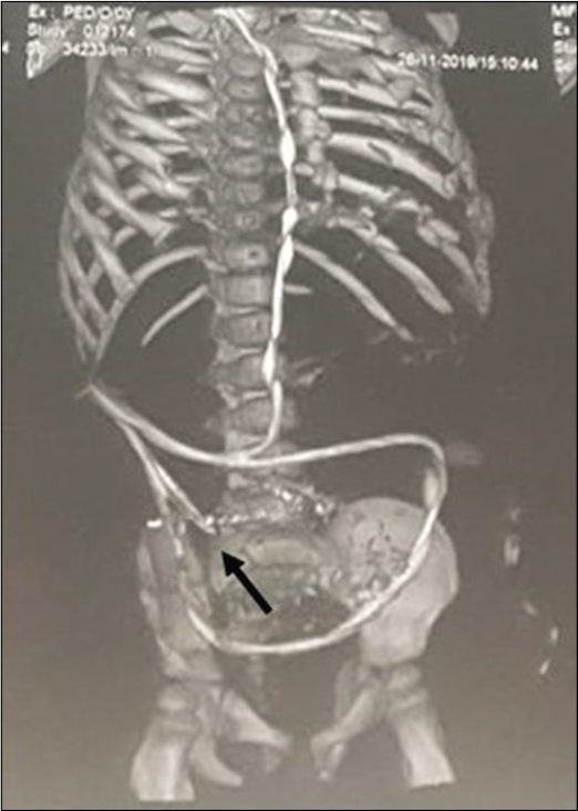
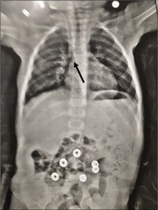
A 15-month-old girl presenting a congenital hydrocephalus, diagnosed at uterine life. At the age of 3 months, a right VAS was made due to the progressive character of her congenital hydrocephalus. Immediate and short time follow-up was simple. One year later, the infant goes to the emergency department for a progressive respiratory distress with polypnea and diminution of oxygen saturation. She was afebrile with pulmonary auscultation evidencing hypophony of the right hemithorax and abdomen not distended and depressible without pain. The blood analysis did not show significant alterations. A thoracoabdominal X-ray shows complete opacity of the right hemithorax, without evidence of intestinal obstruction or intraperitoneal air and with a well-positioned catheter tip but in contact of diaphragm on sleep position and in pelvic cavity on up position [
DISCUSSION
Mechanical failures and infections are the common complications of VP shunts.[
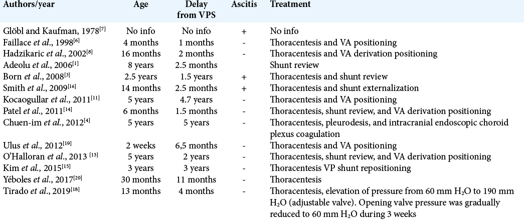
In our case, the pleural effusion is right, ipsilateral to the position of the VP shunt, in which as the majority of cases described in the literature. The imaging allowed us to confirm the existence of the distal tip of the catheter in intra-abdominal and showed a minimum amount of free peritoneal fluid, with no clear ascites. The cause of CSF passage from the abdominal cavity to the thorax is not clear. We suggest that diaphragmatic catheter tip microtrauma is the most cause. However, we cannot rule out the existence of a continuity diaphragm solution. Finally, there are still questions that remain unanswered. Why hydrothorax occurs on the right side, without concomitant CSF ascites and many months or years after VPS in the majority of cases?
CONCLUSION
Hydrothorax following a VPS without catheter migration is an uncommon and serious complication. Contrary to the unclear mechanism, management is simple based on imaging, thoracentesis with biologic analysis, and shunt revisions. Different types of CSF shunting (VA shunt) or endoscopic treatment (third ventriculostomy with choroid plexus coagulation) may be considered as alternative therapeutic approaches.
Declaration of patient consent
Patient’s consent not required as patients identity is not disclosed or compromised.
Financial support and sponsorship
Nil.
Conflicts of interest
There are no conflicts of interest.
References
1. Adeolu AA, Komolafe EO, Abiodun AA, Adetiloye VA. Symptomatic pleural effusion without intrathoracic migration of ventriculoperitoneal shunt catheter. Childs Nerv Syst. 2006. 22: 186-8
2. Akyüz M, Uçar T, Göksu E. A thoracic complication of ventriculoperitoneal shunt: Symptomatic hydrothorax from intrathoracic migration of a ventriculoperitoneal shunt catheter. Br J Neurosurg. 2004. 18: 171-3
3. Born M, Reichling S, Schirrmeister J. Pleural effusion: Beta-Trace protein in diagnosing ventriculoperitoneal shunt complications. J Child Neurol. 2008. 23: 810-2
4. Chuen-Im P, Smyth MD, Segura B, Ferkol T, Rivera-Spoljaric K. Recurrent pleural effusion without intrathoracic migration of ventriculoperitoneal shunt catheter: A case report. Pediatr Pulmonol. 2012. 47: 91-5
5. Drake JM, Kestle JR, Milner R, Cinalli G, Boop F, Piatt J. Randomized trial of cerebrospinal fluid shunt valve design in pediatric hydrocephalus. Neurosurgery. 1998. 43: 294-303
6. Faillace WJ, Garrison RD. Hydrothorax after ventriculoperitoneal shunt placement in a premature infant: An iatrogenic postoperative complication. Case report. J Neurosurg. 1998. 88: 594-7
7. Glöbl H, Kaufmann H. Shunts and complications. Prog Pediatr Radiol. 1978. 6: 231-71
8. Hadzikaric N, Nasser M, Mashani A, Ammar A. CSF hydrothorax-VP shunt complication without displacement of a peritoneal catheter. Childs Nerv Syst. 2002. 18: 179-82
9. Kahle KT, Kulkarni AV, Limbrick DD, Warf BC. Hydrocephalus in children. Lancet. 2016. 387: 788-99
10. Kim JH, Roberts DW, Bauer DF. CSF hydrothorax without intrathoracic catheter migration in children with ventriculoperitoneal shunt. Surg Neurol Int. 2015. 6: S330-3
11. Kocaogullar Y, Güney O, Kaya B, Erdi F. CSF hydrothorax after ventriculoperitoneal shunt without catheter migration: A case report. Neurol Sci. 2011. 32: 949-52
12. Mishra RK, Chaturvedi A, Jena BR, Rath GP. Anesthetic considerations for ventriculoatrial shunt insertion in a child with cerebrospinal fluid ascites. J Pediatr Neurosci. 2018. 13: 249-51
13. O’Halloran PJ, Kaliaperumal C, Caird J. Chemotherapy-induced cerebrospinal fluid malabsorption in a shunted child: Case report and review of the literature. BMJ Case Rep. 2013. 2013: bcr2012008255
14. Patel AP, Dorantes-Argandar A, Raja AI. Cerebrospinal fluid hydrothorax without ventriculoperitoneal shunt migration in an infant. Pediatr Neurosurg. 2011. 47: 74-7
15. Porcaro F, Procaccini E, Paglietti MG, Schiavino A, Petreschi F, Cutrera R. Pleural effusion from intrathoracic migration of a ventriculo-peritoneal shunt catheter: Pediatric case report and review of the literature. Ital J Pediatr. 2018. 44: 42
16. Smith JC, Cohen E. Beta-2-transferrin to detect cerebrospinal fluid pleural effusion: A case report. J Med Case Rep. 2009. 3: 6495
17. Taub E, Lavyne MH. Thoracic complications of ventriculoperitoneal shunts: Case report and review of the literature. Neurosurgery. 1994. 34: 181-3
18. Tirado CA, Giménez-Pando J, Aguirre AM, Grande-Tejada A, Gilete-Tejero IJ, Botana-Fernández M. Pleural effusion in a child with a correctly placed ventricle-peritoneal shunt. Br J Neurosurg. 2019. 1: 1-5
19. Ulus A, Kuruoglu E, Ozdemir SM, Yapici O, Sensoy G, Yarar E. CSF hydrothorax: Neither migration of peritoneal catheter into the chest nor ascites. Case report and review of the literature. Childs Nerv Syst. 2012. 28: 1843-8
20. Yéboles RM, Vázquez L, Seoane M, Castro S, Ruiz B. Hydrothorax as a complication of a ventricle peritoneal shunt. A case report. Neurocirugia (Astur). 2017. 28: 202-6


