- Departments of Neurosurgery, All India Institute of Medical Sciences, Jodhpur, Rajasthan, India.
- Departments of Radiology, All India Institute of Medical Sciences, Jodhpur, Rajasthan, India.
- Departments of Pathology, All India Institute of Medical Sciences, Jodhpur, Rajasthan, India.
Correspondence Address:
Jaskaran Singh Gosal
Departments of Pathology, All India Institute of Medical Sciences, Jodhpur, Rajasthan, India.
DOI:10.25259/SNI_164_2020
Copyright: © 2020 Surgical Neurology International This is an open-access article distributed under the terms of the Creative Commons Attribution-Non Commercial-Share Alike 4.0 License, which allows others to remix, tweak, and build upon the work non-commercially, as long as the author is credited and the new creations are licensed under the identical terms.How to cite this article: Jaskaran Singh Gosal1, Kartikeya Shukla1, Kokkula Praneeth1, Sarbesh Tiwari2, Mayank Garg1, Suryanarayanan Bhaskar1, Poonam Elhence3, Deepak Kumar Jha1, Sudeep Khera3. Coexistent pituitary adenoma and frontal convexity meningioma with frontal sinus invasion: A rare association. 05-Sep-2020;11:270
How to cite this URL: Jaskaran Singh Gosal1, Kartikeya Shukla1, Kokkula Praneeth1, Sarbesh Tiwari2, Mayank Garg1, Suryanarayanan Bhaskar1, Poonam Elhence3, Deepak Kumar Jha1, Sudeep Khera3. Coexistent pituitary adenoma and frontal convexity meningioma with frontal sinus invasion: A rare association. 05-Sep-2020;11:270. Available from: https://surgicalneurologyint.com/surgicalint-articles/10247/
Abstract
The coexistence of pituitary adenoma (PA) and meningioma in the same patient is rare, after excluding radiotherapy-induced meningiomas. Most of the literature on their coexistence describes meningiomas located in the close vicinity to PA, that is, in the sellar/parasellar region. We describe a case of a 65-year-old lady with a nonfunctioning PA and an associated frontal convexity meningioma with frontal sinus invasion. The imaging was nonspecific for the meningioma, and its association with concomitant PA has not been reported before.
Keywords: Convexity, Frontal sinus invasion, Meningioma, Pituitary adenoma
A 65-year-old lady presented with painless progressively decreasing vision both eyes for 2 years and worsening frontal headache for the past 1 year. On examination, she was oriented, cooperative. Her vision was finger counting (FC) at 1 foot in the left eye and FC at 3 feet right eye. Visual field examination was suggestive of bilateral temporal hemianopsia. The fundus examination revealed bilateral early primary optic atrophy. The rest of the examination, including her speech, was normal. Clinical localization was suprasellar mass and the clinical diagnosis of nonfunctioning pituitary adenoma (PA) was made.[
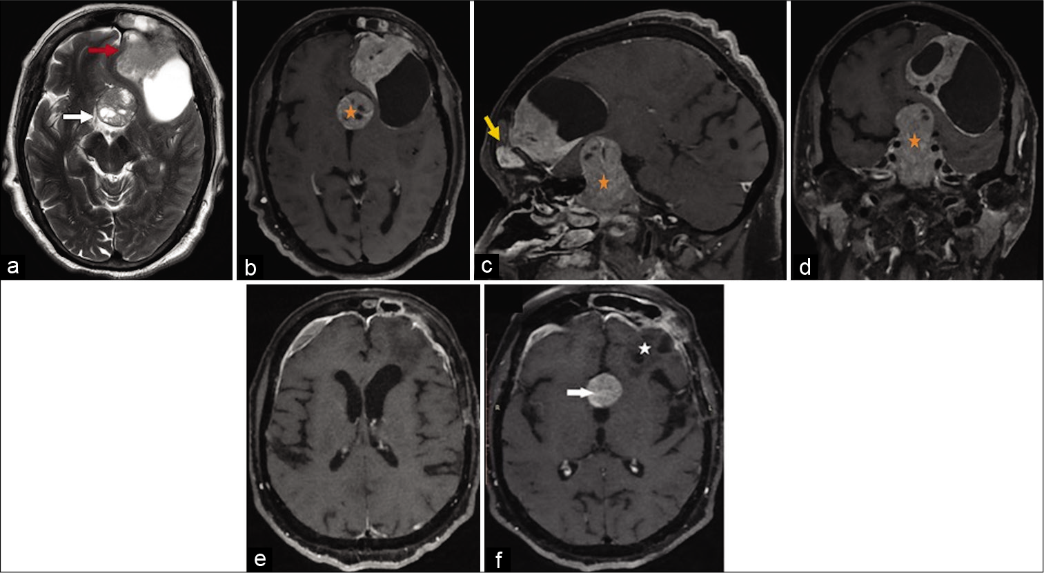
However, magnetic resonance imaging of the brain surprisingly showed two separate lesions: a well-defined homogenously enhancing sellar-suprasellar mass lesion consistent with a pituitary macroadenoma and an extra-axial left frontal convexity mass lesion with a cystic component with dural enhancement and no perilesional edema. Interestingly, the frontal convexity mass lesion was invading the frontal sinus [
Figure 1:
The axial T2 image (a) shows two discrete heterogeneous extra-axial lesions at suprasellar cistern (white arrow) and left frontal convexity (red arrow). The axial (b), sagittal, (c) and oblique coronal (d) contrast-enhanced T1 scan shows a sellar lesion with suprasellar extension extending till the floor of third ventricle (asterisk) consistent with pituitary macroadenoma. The heterogeneous solid cystic enhancing lesion at the left frontal convexity is dural based, shows extension into the left frontal sinus (yellow arrow). Note that both the lesions are discrete with normal brain parenchyma between them. Postoperative scan (e and f): the postcontrast T1 axial image shows complete resection of the left frontal convexity meningioma (asterisk in Figure f) with postoperative dural thickening at operative site. Persistent enhancing pituitary macroadenoma (white arrow) is seen in the axial (f) image.
We planned to tackle both the lesions simultaneously using the left frontal craniotomy for frontal convexity lesion and subsequently to use a subfrontal approach for the pituitary macroadenoma.
An intraoperative impression of the convexity lesion was that of meningioma; it invaded the frontal sinus with the parenchymal invasion at places. Gross total excision of meningioma done, and frontal sinus was repaired using muscle, bone wax, and exteriorized using the pericranium. Using the subfrontal corridor, the suprasellar part of the PA was decompressed. However, due to significantly high vascularity of PA, an adequate decompression could not be achieved, and a significant residual was left. We planned to tackle the PA through the endoscopic endonasal transsphenoidal approach, but the patient did not give consent for the second surgery. Despite this, the patient had subjective improvement in her vision in the immediate postoperative period. Postoperative imaging showed operative changes in the frontal convexity region and the residual sellar mass lesion [
Figure 2:
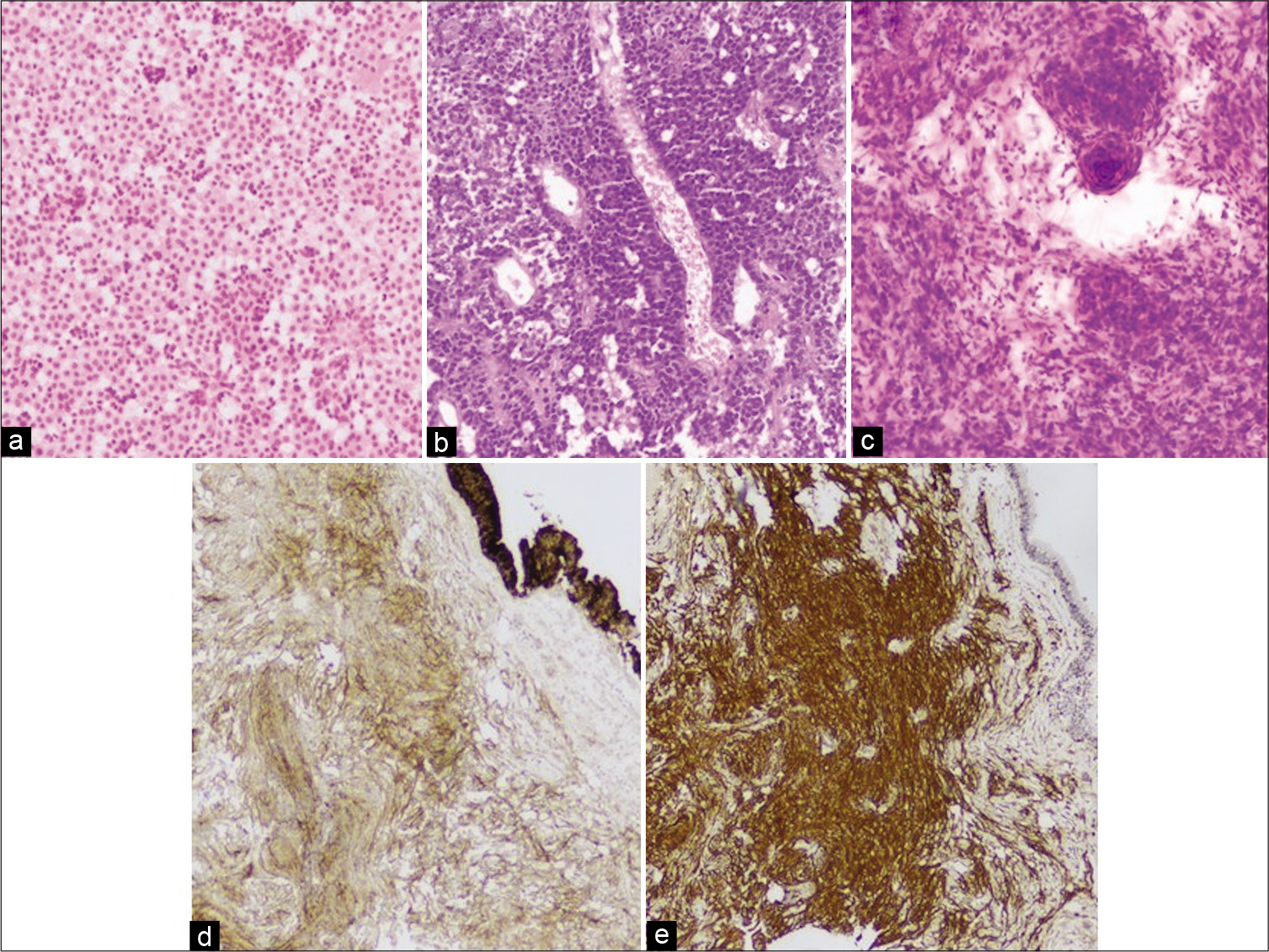
(a and b) Hematoxylin and eosin (H&E) staining, ×10, shows sheets of monomorphic cells with round nuclei, salt and pepper chromatin consistent with pituitary adenoma (c). H&E, ×10, whorl, suggestive of meningioma confirmed with immunohistochemistry (IHC) (d). IHC for vimentin, ×4 and (e). IHC for EMA, ×4.
There is a well-known association of meningiomas with neurofibromas and gliomas (neurofibromatosis, NF Type I) and schwannoma (NF Type II).[
Declaration of patient consent
The authors certify that they have obtained all appropriate patient consent.
Financial support and sponsorship
Nil.
Conflicts of interest
There are no conflicts of interest.
References
1. Abs R, Parizel PM, Willems PJ, Van De Kelft E, Verlooy J, Mahler C. The association of meningioma and pituitary adenoma: Report of seven cases and review of the literature. Eur Neurol. 1993. 33: 416-22
2. Amirjamshidi A, Mortazavi SA, Shirani M, Saeedinia S, Hanif H. Coexisting pituitary adenoma and suprasellar meningioma-a coincidence or causation effect: Report of two cases and review of the literature. J Surg Case Rep. 2017. 2017: rjx039
3. Ben Nsir A, Khalfaoui S, Hattab N. Simultaneous occurrence of a pituitary adenoma and a foramen magnum meningioma: Case report. World Neurosurg. 2017. 97: 748
4. Bhaskar S, Gosal JS, Garg M, Jha DK. Letter: The neurological examination. Oper Neurosurg (Hagerstown). 2020. 18: E262
5. Cannavò S, Curtò L, Fazio R, Paterniti S, Blandino A, Marafioti T. Coexistence of growth hormone-secreting pituitary adenoma and intracranial meningioma: A case report and review of the literature. J Endocrinol Invest. 1993. 16: 703-8
6. Da Costa LB, Riva-Cambrin J, Tandon A, Tymianski M. Pituitary adenoma associated with intraventricular meningioma: Case report. Skull Base. 2007. 17: 347-51
7. Das KK, Gosal JS, Sharma P, Mehrotra A, Bhaisora K, Sardhara J. Falcine meningiomas: Analysis of the impact of radiologic tumor extensions and proposal of a modified preoperative radiologic classification scheme. World Neurosurg. 2017. 104: 248-58
8. De Vries F, Lobatto DJ, Zamanipoor Najafabadi AH, Kleijwegt MC, Verstegen MJ, Schutte PJ. Unexpected concomitant pituitary adenoma and suprasellar meningioma: A case report and review of the literature. Br J Neurosurg. 2019. 17: 1-5
9. Furtado SV, Venkatesh PK, Ghosal N, Hegde AS. Coexisting intracranial tumors with pituitary adenomas: Genetic association or coincidence?. J Cancer Res Ther. 2010. 6: 221-3
10. Gajbhiye S, Gosal JS, Pandey S, Das KK. Apoplexy inside a giant medial sphenoid wing meningothelial (Grade I) meningioma: An extremely rare but a potentially dangerous complication. Asian J Neurosurg. 2019. 14: 961-3
11. Gosal JS, Behari S, Joseph J, Jaiswal AK, Sardhara JC, Iqbal M. Surgical excision of large-to-giant petroclival meningiomas focusing on the middle fossa approaches: The lessons learnt. Neurol India. 2018. 66: 1434-46
12. Gosal JS, Shukla K, Garg M, Bhaskar S, Jha D. Collision tumors in the spinal cord: Co-existent intramedullary dermoid with spinal lipoma. Interdiscip Neurosurg. 2020. 21: 100751
13. Honegger J, Buchfelder M, Schrell U, Adams EF, Fahlbusch R. The coexistence of pituitary adenomas and meningiomas: Three case reports and a review of the literature. Br J Neurosurg. 1989. 3: 59-69
14. Hyodo A, Nose T, Maki Y, Enomoto T. Pituitary adenoma and meningioma in the same patient (author’s transl). Neurochirurgia (Stuttg). 1982. 25: 66-7
15. Mathuriya SN, Vasishta RK, Dash RJ, Kak VK. Pituitary adenoma and parasagittal meningioma: An unusual association. Neurol India. 2000. 48: 72-4
16. Ravisankar M, Khatri D, Gosal JS, Arulalan M, Jaiswal AK, Das KK. Surgical excision of trigeminal (V3) schwannoma through endoscopic transpterygoid approach. Surg Neurol Int. 2019. 10: 259


