- Department of Neurosurgery, King Edward VII Memorial Hospital, Mumbai, Maharashtra, India.
DOI:10.25259/SNI_106_2020
Copyright: © 2020 Surgical Neurology International This is an open-access article distributed under the terms of the Creative Commons Attribution-Non Commercial-Share Alike 4.0 License, which allows others to remix, tweak, and build upon the work non-commercially, as long as the author is credited and the new creations are licensed under the identical terms.How to cite this article: Aimee Goel, Abhidha Harshad Shah, Ravikiran Vutha, Atul Goel. External syringomyelia in longstanding benign foramen magnum tumors. 02-May-2020;11:92
How to cite this URL: Aimee Goel, Abhidha Harshad Shah, Ravikiran Vutha, Atul Goel. External syringomyelia in longstanding benign foramen magnum tumors. 02-May-2020;11:92. Available from: https://surgicalneurologyint.com/surgicalint-articles/9989/
Abstract
Background: The effect of benign foramen magnum tumours on cranial and spinal dimensions and cerebrospinal fluid (CSF) spaces is unclear. In this study, we measured alterations in cerebrospinal fluid (CSF) spaces in the spinal canal and in the posterior cranial fossa distant from the site of benign foramen magnum tumors.
Methods: Twenty-nine magnetic resonance imaging scans of patients with foramen magnum tumors (8 meningiomas and 21 C2 neurinomas) were identified for radiological morphometric analysis and compared with normal control scans. The anterior-posterior distance between the pontomedullary junction and the clivus, the spinal canal diameter, spinal cord diameter, and cord-canal ratios were measured at the C6 and T2 levels.
Results: The mean spinal canal diameter was significantly higher in tumor scans at both the C6 and T2 spinal levels than in controls (13.8 mm vs. 11.4 mm at C6; pP=0.01). Further, the mean cord:canal ratio was significantly lower in tumor scans at both levels (0.49 vs. 0.64 at C6; PP=0.0009). There was no significant difference in mean anteroposterior distance from the clivus to the pontomedullary junction (10.4 mm vs. 10.3 mm; P=0.91).
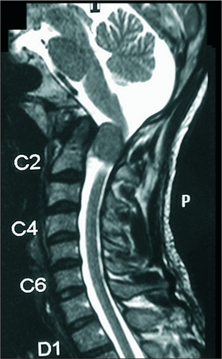
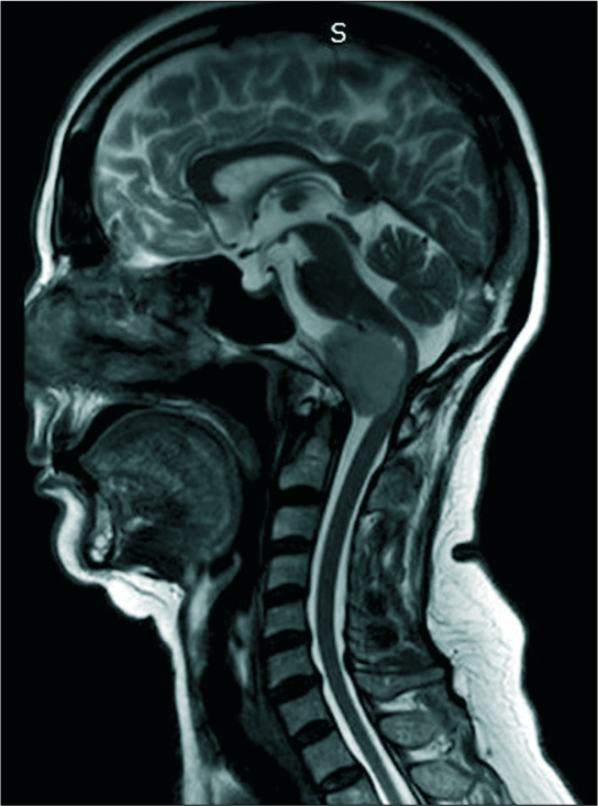
Conclusion: In the presence of benign foramen magnum tumors, the spinal canal diameter and CSF volume in the spinal canal increased at the C6 and T2 levels, distant from the tumor site, a phenomenon we describe as “external syringomyelia”.
Keywords: C2-neurinoma, External syringobulbia, External syringomyelia, Foramen magnum meningioma
INTRODUCTION
In 2016, we introduced the term “external syringomyelia” and “external syringobulbia” to describe the presence of excessive cerebrospinal fluid (CSF) volume in the spinal canal outside the spinal cord substance.[
MATERIALS AND METHODS
We retrieved preoperative T2-weighted magnetic resonance imaging (MRI) scans of patients with C2 neurinomas and foramen magnum meningiomas that were treated from 2010 to 2017. We evaluated 29 patients (21 patients with C2 neurinoma and 8 patients with foramen magnum meningioma) 17 male:12 female, mean age 45.8 years, range 27 - 68 years with cervical MRI images extending to the T2 level [
Comparison of MR studies for tumor versus control patients
For both the tumor and control groups, on sagittal cervical MR studies, we measured the anteroposterior spinal canal diameter and spinal cord width at the C6 and T2 mid- vertebral body index sites. The spinal cord: canal ratio was then calculated at each level along with the maximum anteroposterior dimensions of the CSF column anterior to the pontomedullary junction and posterior to the clivus.
Statistical analysis
Statistical analysis was performed using GraphPad Prism software. The unpaired t-test results are outlined in
RESULTS
At both the C6 and T2 levels, the spinal canal diameter in tumor scans was significantly higher, and the cord:canal ratio was significantly lower than in control scans. At the T2 level, the spinal cord girth was significantly lower in patients with benign foramen magnum tumors versus controls, but there was no significant difference at the C6 level. Further, there was no statistically significant difference in CSF width between the pontomedullary junction and the clivus between tumor patients versus controls.
DISCUSSION
Our analysis revealed that in the presence of benign tumors of the foramen magnum such as C2 neurinomas or meningiomas, the spinal canal diameter increases, and the cord: canal ratio decreases distal to the tumor. We have described this finding as external syringomyelia in the previous papers.[
Low-grade or benign spinal tumors, including neurinomas and meningiomas, are generally associated with chronic local spinal changes, such as an increase in spinal canal dimensions, erosion of bones of the spinal canal, reduction of extradural fat, thinning of the dural tube, and a host of other alterations. Although spinal alterations in the vicinity of the benign spinal tumors have been previously described in the literature, alterations distant from the site of the tumor have not been elucidated.[
Here, we described changes in the bony cervical (C6) and thoracic spinal (T2) canal width distal to the site of benign foramen magnum tumors. These changes are remarkably similar to those seen in long-standing atlantoaxial instability with odontoid process spinal cord compression at the craniovertebral junction.[
CONCLUSION
Utilizing cervical MR studies, we documented an increase in C6 and T2 spinal canal diameter and CSF spaces remote from the site of benign foramen magnum tumors.
Declaration of patient consent
Patient’s consent not obtained as patients identity is not disclosed or compromised.
Financial support and sponsorship
Nil.
Conflicts of interest
There are no conflicts of interest.
References
1. Goel A, Desai K, Muzumdar D. Surgery on anterior foramen magnum meningiomas using a conventional posterior suboccipital approach: a report on an experience with 17 cases. Neurosurgery. 2001. 49: 102-6
2. Goel A, Jain S, Shah A. Radiological evaluation of 510 cases of basilar invagination with evidence of atlantoaxial instability (Group A). World Neurosurg. 2018. 110: 533-43
3. Goel A, Kaswa A, Shah A, Rai S, Gore S, Dharurkar P. Extraspinal-interdural surgical approach for C2 neurinomas-report of an experience with 50 cases. World Neurosurg. 2018. 110: 575-82
4. Goel A, Nadkarni T, Shah A, Sathe P, Patil M. Radiological evaluation of Basilar invagination without obvious atlantoaxial instability (Group B-basilar invagination): An analysis based on a study of 75 patients. World Neurosurg. 2016. 95: 375-82
5. Goel A, Sathe P, Shah A. Atlantoaxial fixation for Basilar invagination without obvious atlantoaxial instability (Group B-basilar invagination): Outcome analysis of 63 surgically treated cases. World Neurosurg. 2017. 99: 164-70
6. Goel A. Chiari malformation is atlantoaxial instability the cause? Outcome analysis of 65 patients with Chiari malformation treated by atlantoaxial fixation. J Neurosurg Spine. 2015. 22: 116-27
7. Goel A. Is Chiari malformation nature’s protective “air-bag”? Is its presence diagnostic of atlantoaxial instability?. J Craniovertebr Junction Spine. 2014. 5: 107-9
8. Goel A. Is syringomyelia pathology or a natural protective phenomenon?. J Postgrad Med. 2001. 47: 87-8


