- Skull Base Research Center, Loghman Hakim Hospital, Shahid Beheshti University of Medical Sciences, Tehran, Iran,
- Medius Klinik Kirchheim, University of Tübingen, Kirchheim, Germany,
- Division of Vascular and Endovascular Neurosurgery, Firoozgar Hospital, Iran University of Medical Sciences, Tehran, Iran.
Correspondence Address:
Mahmoud Omidbeigi
Skull Base Research Center, Loghman Hakim Hospital, Shahid Beheshti University of Medical Sciences, Tehran, Iran,
DOI:10.25259/SNI_312_2020
Copyright: © 2020 Surgical Neurology International This is an open-access article distributed under the terms of the Creative Commons Attribution-Non Commercial-Share Alike 4.0 License, which allows others to remix, tweak, and build upon the work non-commercially, as long as the author is credited and the new creations are licensed under the identical terms.How to cite this article: Guive Sharifi1, Mahmoud Lotfinia2, Mahmoud Omidbeigi1, Sina Asaadi3, Farahnaz Bidari Zerehpoosh1. Meningothelial meningioma of the oculomotor nerve: A case report and review of the literature. 02-Oct-2020;11:314
How to cite this URL: Guive Sharifi1, Mahmoud Lotfinia2, Mahmoud Omidbeigi1, Sina Asaadi3, Farahnaz Bidari Zerehpoosh1. Meningothelial meningioma of the oculomotor nerve: A case report and review of the literature. 02-Oct-2020;11:314. Available from: https://surgicalneurologyint.com/surgicalint-articles/10308/
Abstract
Background: The origin of meningioma tumors is known as the meningothelial or arachnoid cap cells. The arachnoid granulations or villi are concentrated along with the dural venous sinuses in the cerebral convexity, parasagittally, and sphenoid wing regions. The majority of meningiomas are found in these locations with dural attachment. Infrequently, meningiomas develop without dural attachment but in dural adjacent. There are numerous reports of patients with cranial nerve involvement as a result of the compressive effect of the sinus cavernous or adjacent structures meningioma tumor on the cranial nerve.
Case Description: In this study, we reviewed all reports of patients with third nerve involvement as a result of meningioma tumors in addition to the introduction of a new case. We present a 47-year-old woman presented with headache, diplopia, and ptosis. A gadolinium-enhanced mass on anterolateral of the left cerebral peduncle with no dural attachment was suggesting for Schwannoma at preoperative imaging. An adhesive 10 × 5 × 4 mm meningothelial meningioma arising from the oculomotor nerve was resected.
Conclusion: The findings of this review suggest that there may be other mechanisms as the origin of meningiomas tumors. It is crucial to take into account origination mechanisms of meningioma using ectopic meningiomas due to the increasing prevalence of meningioma.
Keywords: Meningioma, Meningothelial, Oculomotor nerve
INTRODUCTION
Meningiomas are the most common primary brain tumors.[
In this report, we present a 47-year-old woman with oculomotor nerve originating meningioma without any dural connection.
CASE REPORT
A 47-year-old woman referred to our center with a history of gradually worsening symptoms of headache, diplopia, and left-sided ptosis (eyelid drooping) for 1 year ago that has progressed intensely during the past 4 months.
Magnetic resonance imagining that performed in another center revealed a localized midbrain lesion and referred to our center 9 months after her initial symptoms.
Examination
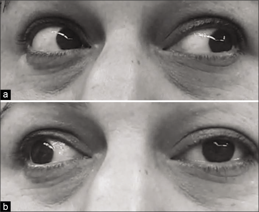
The neurological examination revealed left eye hypotropia and ptosis. The medial and upward gaze of the left eye was impaired and deviated slightly out and down in the primary position [
Imaging
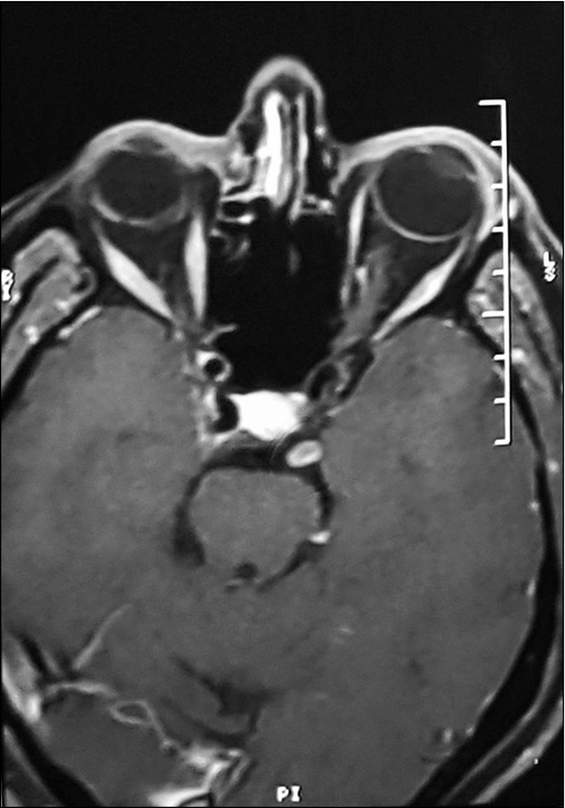
Multiplanar images at different MRI sequences with and without intravenous contrast showed small spherical tumor (10 × 5 × 5 mm) abutting anterolateral of the left cerebral peduncle. The tumor appeared isointense on T1-weighted images, hyperintense on T2-weighted images. Imaging confirmed a gadolinium-enhanced mass with no dural attachment, suggesting for Schwannoma [
Operation
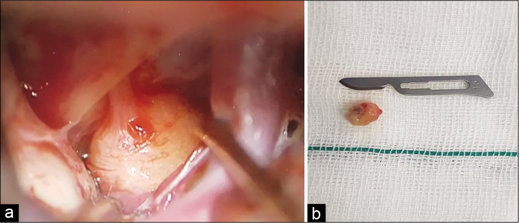
After general anesthesia, lateral subfrontal craniotomy was performed. Dura opened on curly Lina Fashion. The carotid artery was found. By opening the liliequist membrane, the posterior communicating artery was followed to the interpeduncular fossa. Firm dark pink global tumor with a compressive effect on the mesencephalic area appeared. The third nerve ran into the tumor [
Histology
The histopathological study of the 10 × 5 × 4 mm dark pink mass revealed round to oval centrally located nuclei with dispersed chromatin and eosinophilic cytoplasmic. Lobules of the tumor are separated from each other with collagen sheets and contained whorls and psammoma bodies. Necrosis was not a feature. The final diagnosis was meningothelial meningioma (WHO Grade I).[
Postoperative
On postoperative examination, all preoperative signs were found as complete paralysis of the left oculomotor nerve without slight responses and reflexes, which was expected due to the tumor and nerve resection. At a 6-month follow-up, she continued to have diplopia and eyelid drooping. In the brain MRI finding, there was no evidence of recurrence during 1 year follow-up.
DISCUSSION
The origin of meningioma tumors is known as the meningothelial or arachnoid cap cells. The arachnoid villi have large numbers of arachnoid cap cells.[
There are numerous reports of patients with third nerve involvement as a result of meningioma tumors, but in almost all of them, the compressive effect of the sinus cavernous or adjacent structures meningioma tumor on the oculomotor nerve has caused these symptoms.[
In this report, we present a middle-aged woman with oculomotor nerve paralysis. Imaging and examination findings were suggesting for oculomotor schwannoma. Histology of the tumor revealed a meningothelial meningioma tumor.
As our knowledge, there is only one similar case to ours that reported by Hart et al.[
Mohri et al.[
All cranial nerve meningioma tumors have been reported in the middle-aged woman and located on the left side. In all three cases, the tumor had caused paralysis symptoms of the involved nerve and had not invaded the surrounding structures. Reported trigeminal and accessory origin tumors were a meningothelial meningioma, like our case.
Fujimoto et al. explained the origin of meningioma without dural attachment using the number of mechanisms.[
Due to the increasing prevalence of meningioma,[
CONCLUSION
The findings of this review suggest that there may be other mechanisms as the origin of meningiomas tumors. It is crucial to take into account origination mechanisms of meningioma using ectopic meningiomas due to the increasing prevalence of meningioma.
Declaration of patient consent
Patient’s consent not required as patients identity is not disclosed or compromised.
Financial support and sponsorship
Publication of this article was made possible by the James I. and Carolyn R. Ausman Educational Foundation.
Conflicts of interest
There are no conflicts of interest.
References
1. Buetow M, Buetow P, Smirniotopoulos J. Typical, atypical, and misleading features in meningioma. Radiographics. 1991. 11: 1087-106
2. Cahill KS, Claus EB. Treatment and survival of patients with nonmalignant intracranial meningioma: Results from the Surveillance, Epidemiology, and end results program of the national cancer institute. Clinical article. J Neurosurg. 2011. 115: 259-67
3. Duong LM, McCarthy BJ, McLendon RE, Dolecek TA, Kruchko C, Douglas LL. Descriptive epidemiology of malignant and nonmalignant primary spinal cord, spinal meninges, and cauda equina tumors, United States, 2004-2007. Cancer. 2012. 118: 4220-7
4. Fujimoto Y, Kato A, Taniguchi M, Maruno M, Yoshimine T. Meningioma arising from the trigeminal nerve: A case report and literature review. J Neurooncol. 2004. 68: 185-7
5. Gokce G, Ceylan OM, Altinsoy HI. Recurrent alternating oculomotor nerve palsy: An unusual presentation of parasagittal meningioma. Neuroophthalmology. 2013. 37: 82-5
6. Hart AJ, Allibone J, Casey A, Thomas D. Malignant meningioma of the oculomotor nerve without dural attachment. J Neurosurg. 1998. 88: 1104-6
7. Kunimatsu A, Kunimatsu N, Kamiya K, Katsura M, Mori H, Ohtomo K. Variants of meningiomas: A review of imaging findings and clinical features. Jpn J Radiol. 2016. 34: 459-69
8. Louis DN, Perry A, Reifenberger G, von Deimling A, FigarellaBranger D, Cavenee WK. The 2016 World Health Organization classification of tumors of the central nervous system: A summary. Acta Neuropathol. 2016. 131: 803-20
9. Mohri M, Yamano J, Saito K, Nakada M. Spinal accessory nerve meningioma at the foramen magnum with medullar compression: A case report and literature review. World Neurosurg. 2019. 128: 158-61
10. Piatt JH, Campbell GA, Oakes WJ. Papillary meningioma involving the oculomotor nerve in an infant: Case report. J Neurosurg. 1986. 64: 808-12
11. Shechtman DL, Woods AD, Tyler JA. Pupil sparing incomplete third nerve palsy secondary to a cavernous sinus meningioma: Challenges in management. Clin Exp Optom. 2007. 90: 132-8
12. Sughrue ME, Rutkowski MJ, Aranda D, Barani IJ, McDermott MW, Parsa AT. Factors affecting outcome following treatment of patients with cavernous sinus meningiomas. J Neurosurg. 2010. 113: 1087-92
13. Tomaru U, Hasegawa T, Hasegawa F, Kito M, Hirose T, Shimoda T. Primary extracranial meningioma of the foot: A case report. Jpn J Clin Oncol. 2000. 30: 313-7
14. Wiemels J, Wrensch M, Claus EB. Epidemiology and etiology of meningioma. J Neurooncol. 2010. 99: 307-14
15. Winterkorn JM, Bruno M. Relative pupil-sparing oculomotor nerve palsy as the presenting sign of posterior fossa meningioma. J Neuroophthalmol. 2001. 21: 207-9
16. Zhang J, Chi LY, Meng B, Li F, Zhu SG. Meningioma without dural attachment: Case report, classification, and review of the literature. Surg Neurol. 2007. 67: 535-9


