- Department of Orthopaedic Surgery, Henry Ford Health System, Detroit, Michigan, USA
Correspondence Address:
Charles C. Yu
Department of Orthopaedic Surgery, Henry Ford Health System, Detroit, Michigan, USA
DOI:10.4103/sni.sni_324_17
Copyright: © 2017 Surgical Neurology International This is an open access article distributed under the terms of the Creative Commons Attribution-NonCommercial-ShareAlike 3.0 License, which allows others to remix, tweak, and build upon the work non-commercially, as long as the author is credited and the new creations are licensed under the identical terms.How to cite this article: Morenikeji A. Buraimoh, Charles C. Yu, Michael P. Mott, Gregory P. Graziano. Sacroiliac stabilization for sacral metastasis: A case series. 06-Dec-2017;8:287
How to cite this URL: Morenikeji A. Buraimoh, Charles C. Yu, Michael P. Mott, Gregory P. Graziano. Sacroiliac stabilization for sacral metastasis: A case series. 06-Dec-2017;8:287. Available from: http://surgicalneurologyint.com/surgicalint-articles/sacroiliac-stabilization-for-sacral-metastasis-a-case-series/
Abstract
Background:The sacrum is a rare location for spinal metastasis. These lesions are typically large and destructive by the time of diagnosis, making treatment difficult. When indicated, surgical stabilization offers pain relief and preserves independence in patients with impending and acute pathological sacral fractures.
Case Description:Three consecutive patients presented with sacral metastases. After either failing radiation therapy or presenting with acute fracture and instability, the patients underwent intralesional excision, bilateral L4 to ilium fusion with instrumentation, and sacroiliac (SI) screw fixation. Pain improved after surgery, and there were no wound healing complications. Two patients could continue walking without any assistive device, while one patient required a walker.
Conclusion:Stabilization with combined modified Galveston fixation and SI screw fixation relieves pain and allows maintenance of independence in patients with sacral metastasis.
Keywords: Iliosacral screw, sacral metastasis, sacroiliac fixation
INTRODUCTION
Metastatic tumors comprise the majority of malignant sacral tumors.[
Sacral metastatic disease
Metastatic disease of the sacral spine is rare and occurs in approximately 5–10% of cancer patients. Meanwhile, the prevalence of asymptomatic and symptomatic metastatic lesions throughout the entire spine may be as high as 70% and 14%, respectively.[
Surgical management of sacral metastatic disease
The management of metastatic sacral lesions is complicated as they are often large and destructive by the time they are diagnosed [
CASE REPORT
Data from three consecutive patients with sacral malignancies undergoing lumbopelvic fixation were evaluated [
Imaging and surgical fixation
All patients underwent computed tomography (CT) and magnetic resonance imaging (MRI) scans preoperatively. They underwent decompression and instrumented fusion utilizing the modified Galveston/iliosacral screw technique [
Case 1
Clinical presentation
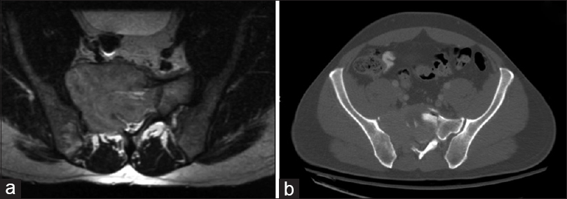
A 53-year-old female with remote history of granulosa cell ovarian cancer presented with increasing sciatic/radicular pain and gluteal numbness. CT showed a very large expansile lytic lesion involving the entire right sacral ala, eroding through the right SI joint and dorsal cortex, with accompanying visceral and other bony metastases. Two months later, following biopsy confirmation of recurrent ovarian cancer and after failed nonoperative management [e.g., including and palliative radiation (30 gray)], she underwent surgery performed by a multidisciplinary team.
Lumbosacral/Lumbopelvic surgery
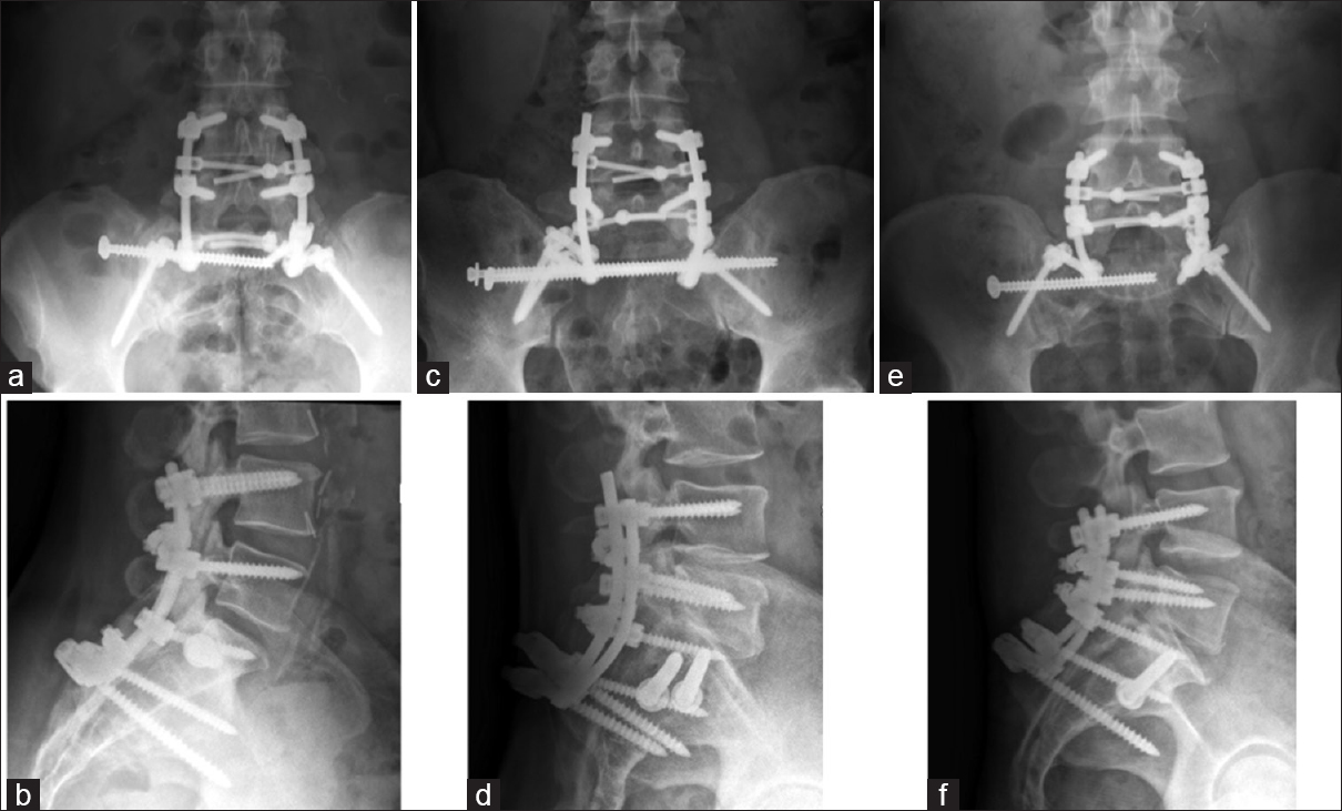
The lumbosacral spine was approached using a standard posterior midline exposure from L4 to the sacrum. The tumor was partially debulked and pedicle screws were inserted bilaterally at L4 and L5 (under fluoroscopic guidance). As the right S1 pedicle was destroyed, only a left S1 pedicle was inserted. Bilateral iliac bolts were inserted at the posterior superior iliac spine. Two rods were applied, and cross-connectors were used to connect the rods to the iliac bolts. Two cross-clamps were used to increase the strength of the construct. The orthopedic oncologist performed an intralesional excision followed by the percutaneous right SI screw placement.[
The patient sustained immediate pain relief, stayed 4 postoperative days, and was discharged home. The only complication was left iliac bolt irritation. At 82 postoperative weeks, the patient had occasional back pain, was on no pain medication, and ambulated without difficulty. The follow-up CT confirmed solid lumbar and iliolumbar fusion with partial SI fusion. Cases 2 and 3 are summarized in
DISCUSSION
The goals of surgery for symptomatic sacral metastases include relief of pain/radiculopathy and the preservation of function. This typically requires decompressing the neural elements and attendant lumbopelvic stabilization. The literature demonstrates that intralesional excision accompanied by instrumented fusion meets the intended goals and allows patients to maintain or improve their ambulatory status postoperatively.[
Technical aspects of sacropelvic surgery
Ideally, instrumentation of the sacrum and pelvis should occur prior to impending fracture.[
Multidisciplinary approach
The multidisciplinary approach, including medical oncology, radiation oncology, surgical oncology, and spinal surgeons is essential to optimize the management/outcomes of spinal surgery dealing with metastatic disease to the sacrum.[
CONCLUSION
Decompression and instrumented fusion of symptomatic sacral metastatic disease utilizing the modified Galveston and SI fixation system is both safe and effective. This technique provided excellent pain relief, helped maintain postoperative mobility, and independence.
Financial support and sponsorship
Nil.
Conflicts of interest
There are no conflicts of interest.
References
1. Chip Routt ML, Meier MC, Kregor PJ, Mayo KA. Percutaneous iliosacral screws with the patient supine technique. Oper Tech Orthop. 1993. 3: 35-45
2. Fujibayashi S, Neo M, Nakamura T. Palliative dual iliac screw fixation for lumbosacral metastasis. Technical note. J Neurosurg Spine. 2007. 7: 99-102
3. Gunterberg B, Romanus B, Stener B. Pelvic strength after major amputation of the sacrum. An exerimental study. Acta Orthop Scand. 1976. 47: 635-42
4. Kollender Y, Meller I, Bickels J, Flusser G, Issakov J, Merimsky O. Role of adjuvant cryosurgery in intralesional treatment of sacral tumors. Cancer. 2003. 97: 2830-8
5. McGee AM, Bache CE, Spilsbury J, Marks DS, Stirling AJ, Thompson AG. A simplified Galveston technique for the stabilisation of pathological fractures of the sacrum. Eur Spine J. 2000. 9: 451-4
6. Quraishi NA, Giannoulis KE, Edwards KL, Boszczyk BM. Management of metastatic sacral tumours. Eur Spine J. 2012. 21: 1984-93
7. Rose PS, Buchowski JM. Metastatic disease in the thoracic and lumbar spine: Evaluation and management. J Am Acad Orthop Surg. 2011. 19: 37-48
8. Vrionis FD, Small J. Surgical management of metastatic spinal neoplasms. Neurosurg Focus. 2003. 15: E12-


